Single-cell dissection of chronic lung allograft dysfunction and native lung fibrosis reveal convergent and unique mechanisms
Yan et al. report an integrative single-cell transcriptomic analysis of fibrosis-associated lung diseases that reveals cellular and molecular signatures in chronic lung allograft dysfunction. Image credit: Ellen Weiss.
-
Research Letter
×
Abstract
Authors
Nadir Yehya, Jacob E. Till, Nishi Srivastava, Donglan Zhang, Jason D. Christie, Erica L. Carpenter, Nilam S. Mangalmurti, Wanding Zhou

-
Review
×
Abstract
Bronchopulmonary dysplasia (BPD) remains a debilitating disease in premature infants. The chronic pathogenesis of BPD with complex prenatal and postnatal programming challenges attempts at precisely defining or treating disease. While existing BPD definitions categorize disease severity, a lack of consideration of disease heterogeneity and endotypes has contributed to the failure of clinical trials to improve BPD outcomes. Recent studies have used advanced lung imaging techniques, echocardiography, and lung function tests to identify airway, parenchymal, and vascular BPD endotypes. These endotypes carry different prognoses and require endotype-specific treatment strategies to optimize infant outcomes. In this Review, we focus on the pathogenic mechanisms that specify individual BPD endotypes and discuss how combining biomarkers, functional studies, and artificial intelligence–based characterization of endotypes can inform precision therapies for BPD.
Authors
Megha Sharma, Gangaram Akangire, Noah H. Hillman, Winston M. Manimtim, Mark Ivan Attard, Venkatesh Sampath
-
Research Articles
×
Abstract
Apoptosis and necroptosis are 2 distinct destinies of cells stimulated with TNF-α; however, it remains unclear how apoptosis and necroptosis are differentially regulated. This study validates the key regulatory role of speckle-type POZ protein (SPOP) in balancing apoptosis and necroptosis. SPOP promotes the polyubiquitination and degradation of receptor-interacting serine/threonine-protein kinase 3 (RIPK3), thereby inhibiting necrosome formation and decreasing cellular sensitivity to necroptosis. Conversely, SPOP interacted with RIPK1 independently of its E3 ubiquitin ligase activity, protecting it from ubiquitination and degradation, thereby enhancing RIPK1 expression and cellular sensitivity to apoptosis. Inhibiting RIPK1 kinase activity with 7-Cl-O-Nec-1 impeded both SPOP-mediated apoptosis and SPOP deficiency–mediated necroptosis. Besides, inhibition or loss of RIPK3 rescued SPOP deficiency–mediated necroptosis. Pancancer analyses indicated that the SPOP/RIPK1/RIPK3 axis is dysfunctional in a variety of tumors. In 3 representative tumor types with high expression of SPOP and RIPK1, kidney renal clear cell carcinoma, liver hepatocellular carcinoma, and breast invasive carcinoma, this regulatory mechanism remains applicable. Based on these findings, a combination therapy using the second mitochondria-derived activator of caspases (Smac) mimetic SM164 and sunitinib was developed, demonstrating a more pronounced efficacy than sunitinib monotherapy, and this sensitizing effect was dependent on the expression level of RIPK1. These results suggest that the combination of Smac mimetics with tyrosine kinase inhibitors holds potential clinical value for tumors with dysregulated SPOP/RIPK1/RIPK3 signaling.
Authors
Yuzhong Ye, Changjie Yue, Zaosong Zheng, Hailong Ruan, Yuanpeng Zhang, Qi Miao, Xiaoping Zhang, Wen Xiao, Lei Liu
×Abstract
Acute lower respiratory infections are the primary cause of global mortality in postneonatal children. Most respiratory viruses primarily involve upper airway infection and inflammation, yet nasal responses are poorly characterized. Using a mouse model of human metapneumovirus (HMPV), we found viral burden was higher in nasal airways and exhibited delayed clearance. Despite high burden, there was low nasal expression of type I and III interferon (IFN). Single-cell RNA-sequencing (scRNA-Seq) from HMPV-infected mice showed lower nasal IFN-stimulated gene (ISG) expression and nasal enrichment of genes negatively regulating IFN. scRNA-Seq of patients with COVID-19 verified lower ISG expression in upper airways. HMPV infection downregulated nasal expression of IFN regulatory factor 3, suggesting a mechanism for limited response. To rescue the quiescent environment, we administered type I or III IFN to upper airways early postinfection, leading to lower nasal HMPV titer and virus-specific CD8+ T cell upregulation. Intranasal immunization adjuvanted with type I or III IFN improved immune response, reduced clinical disease, and enhanced viral clearance in HMPV and influenza infection. IFN adjuvant increased recruitment of dendritic cells, recruitment of resident memory T cells, and neutralizing antibodies. These findings reveal locally suppressed IFN production contributes to a quiescent nasal immune landscape that delays viral clearance and impairs mucosal vaccine responses.
Authors
Jorna Sojati, Olivia B. Parks, Taylor Eddens, Jie Lan, Monika Johnson, John V. Williams
×Abstract
Dravet syndrome (DS) is an early-onset epilepsy caused by loss-of-function mutations in the SCN1A gene, which encodes Nav1.1 channels that preferentially regulate activity of inhibitory neurons early in development. DS is associated with a high incidence of sudden unexpected death in epilepsy (SUDEP) by a mechanism that may involve respiratory failure. Evidence also shows that loss of Scn1a impaired activity of neurons in the retrotrapezoid nucleus (RTN) that regulate breathing in response to CO2/H+, suggesting breathing problems precede seizures and serve as a biomarker of SUDEP. Consistent with this, we showed that Scn1a+/– mice exhibited a blunted ventilatory response to CO2/H+ prior to overt seizure activity that worsened with disease progression. Later in development, some Scn1a+/– mice also showed a blunted ventilatory response to hypoxia. Importantly, the severity of respiratory problems correlated with mortality. We also found that pharmacological activation of Nav1.1 rescued activity deficits of RTN neurons in Scn1a+/– mice. We conclude that disordered breathing may be an early biomarker of SUDEP in DS, and at the cellular level, loss of Scn1a disrupts RTN neurons by mechanisms involving disinhibition and pharmacological activation of Nav1.1 to reestablish inhibitory control of RTN neurons rescues activity deficits.
Authors
Brenda M. Milla, Eliandra N. da Silva, Cleyton R. Sobrinho, Monica L. Strain, Daniel K. Mulkey
×Abstract
Wiedemann-Steiner syndrome (WDSTS) is a rare genetic cause of intellectual disability that is primarily caused by heterozygous loss-of-function variants in the gene encoding the histone lysine methyltransferase 2A (KMT2A). Prior studies have shown successful postnatal amelioration of disease phenotypes for Rett, Rubinstein-Taybi, and Kabuki syndromes, which are related Mendelian disorders of the epigenetic machinery. To explore whether the neurological phenotype in WDSTS is treatable in utero, we created a mouse model carrying a loss-of-function variant placed between 2 loxP sites. Kmt2a+/LSL mice demonstrated core features of WDSTS including growth retardation, craniofacial abnormalities, and hypertrichosis as well as hippocampal memory defects. The neurological phenotypes were rescued upon restoration of KMT2A in utero following breeding to a nestin-Cre. Together, our data provide a mouse model to explore the potential therapeutic window in WDSTS. Our work suggests that WDSTS has a window of opportunity extending at least until the midpoint of in utero development, making WDSTS an ideal candidate for future therapeutic strategies.
Authors
Tinna Reynisdottir, Kimberley J. Anderson, Katrin Möller, Stefán Pétursson, Andrew Brinn, Katheryn P. Franklin, Juan Ouyang, Asbjorg O. Snorradottir, Cathleen M. Lutz, Aamir R. Zuberi, Valerie B. DeLeon, Hans T. Bjornsson
×Abstract
Recent evidence suggests that cellular metabolism, including glycolysis and fatty acid synthesis in lymphatic endothelial cells (LECs), plays essential roles in developing functional lymphatic systems. Site-1 protease (S1P) proteolytically activates membrane-bound latent transcription factor sterol regulatory element-binding proteins (SREBPs), which are required to induce lipid biosynthesis. In this study, we generated mice with pan-endothelial or LEC-specific deficiency of either S1P or SREBP2. Mouse embryos with pan-endothelial deletion of S1P showed defective lymphatic vessel migration in skin and lymphedema, while their blood vasculature formation was relatively normal. Mice lacking S1P in LECs or SREBP2 in LECs exhibited chylous ascites, reduced lipogenic gene expression, and reduced VEGFR3 expression and progressively developed wasting, resulting in postnatal death by approximately 8 weeks of age. Additionally, mice with SREBP2 deletion in LECs exhibited dilated lacteal and mesenteric lymphatics and accumulation of lipids in the lacteal before weaning age, indicating apparent lymphatic malfunctioning. These data indicate that S1P-SREBP2–mediated cholesterol biosynthesis is pivotal in lymphatic vascular development. We also found that treating human dermal LECs with VEGF-C induced proteolytic activation of SREBP2 with concomitant phosphorylation of Akt and the expression of genes involved in cholesterol biosynthesis. Those effects were canceled out by treating the cells with an S1P inhibitor or SREBP inhibitor. These data demonstrate that the S1P/SREBP2 axis is critical in VEGF-C/VEGFR3 mitogenic signaling in LECs.
Authors
Yuji Kondo, Yizhi Jiang, Xin Geng, Jianhua Song, Summer Simeroth, J. Michael McDaniel, Pengchun Yu, R. Sathish Srinivasan, Lijun Xia
×Abstract
Bowel smooth muscle experiences mechanical stress constantly during normal function and pathologic mechanical stressors in disease states. We tested the hypothesis that pathologic mechanical stress could alter transcription to induce smooth muscle phenotypic class switching. To test this hypothesis, primary human intestinal smooth muscle cells (HISMCs), seeded on electrospun aligned poly-ε-caprolactone nano-fibrous scaffolds, were subjected to pathologic, high-frequency (1 Hz) uniaxial 3% cyclic stretch (loaded) or kept unloaded in culture for 6 hours. RNA-Seq, quantitative PCR (qPCR), and quantitative IHC defined loading-induced changes in gene expression. NicheNet predicted how differentially expressed genes might affect HISMCs and other bowel cells. These studies show loading induced differential expression of 4,537 HISMC genes. Loaded HISMCs had a less contractile phenotype, with increased expression of synthetic SMC genes, proinflammatory cytokines, and altered expression of axon guidance molecules, growth factors, and morphogens. Many differentially expressed genes encode secreted ligands that could act cell autonomously on smooth muscle and on other cells in the bowel wall. These data show that HISMCs undergo remarkably rapid phenotypic plasticity in response to mechanical stress that may convert contractile HISMCs into proliferative fibroblast-like cells or proinflammatory cells. These mechanical stress–induced changes in HISMC gene expression may be relevant for human bowel disease.
Authors
Sharon M. Wolfson, Katherine Beigel, Sierra E. Anderson, Brooke Deal, Molly Weiner, Se-Hwan Lee, Deanne M. Taylor, Su Chin Heo, Robert O. Heuckeroth, Sohaib K. Hashmi
×Bcl6 expression is associated with a distinct immune landscape and spatial transcriptome in COVID-19
Abstract
The regulation of follicular (F) and germinal center (GC) immune reactivity in human lymph nodes (LNs), particularly during the acute stages of viral infection, remains poorly understood. We have analyzed lung-draining lymph nodes (LD-LNs) from COVID-19 autopsies using multiplex imaging and spatial transcriptomics to examine the immune landscape with respect to follicular immune reactivity. We identified 3 groups of donors based on the Bcl6 prevalence of their reactive follicles (RFs): RF-Bcl6no/lo, RF-Bcl6int, and RF-Bcl6hi. A distinct B/Tfh immune landscape, associated with increased prevalence of proliferating B cell and Tfh cell subsets, was found in RF-Bcl6hi LD-LNs. The comparison between LD-LNs and subdiaphragmatic (SD) LNs from the same donor revealed a divergent Bcl6 expression between the 2 anatomical sites. LD-LN Bcl6 expression was also associated with a distinct spatial transcriptomic profile. TH1-associated genes/pathways (e.g., CXCR3, STAT5, TNF signaling) were significantly upregulated in RF-Bcl6no/lo tissues, while the RF-Bcl6hi tissues exhibited significant upregulation of GC-promoting genes/pathways (e.g., CXCL13, B-cell receptor signaling). Our findings reveal a heterogeneous F/GC landscape in COVID-19 LD-LNs, highlighting specific molecular targets and pathways that could regulate human F/GC immune dynamics during acute viral infections.
Authors
Cloé Brenna, Bernat Bramon Mora, Kalliopi Ioannidou, Julien Bodelet, Mia L. Siebmanns, Simon Burgermeister, Spiros Georgakis, Michail Orfanakis, Yannick D. Muller, Nazanin Sédille, Matthew J. Feinstein, Jon W. Lomasney, Oliver Y. Chén, Giuseppe Pantaleo, Sabina Berezowska, Laurence de Leval, Raphael Gottardo, Constantinos Petrovas
×Abstract
The intestinal mucosal epithelium forms a barrier between luminal contents and the body. MicroRNAs (miRNAs) regulate mucosal homeostasis by controlling inflammatory responses and structural integrity. Here, we discovered a protective role for miR147 in intestinal inflammation using a miR147tdTomato reporter mouse. miR147 was enriched in the intestines, with the highest expression in the colonic epithelial cells at the luminal surface, with prominent expression in differentiated enterocytes. Mice with general or intestinal epithelial deletion of miR147 showed increased intestinal inflammation and diminished mucosal healing during colitis. RNA sequencing of miR147-deficient cells showed dysregulated immune signaling, with upregulated proinflammatory cytokine pathways and reduced type I interferon responses and revealed Ndufa4 as a likely miR147 target. Ndufa4, a mitochondrial protein regulating energy metabolism and inflammation, was elevated at the crypt base, inversely correlating with miR147. Mice lacking the miR147 binding site in Ndufa4’s 3′-UTR phenocopied miR147-deficient mice during colitis. Spatial and single-cell transcriptomic analyses in murine and human colons showed mutually exclusive miR147 and Ndufa4 expression, consistent with a regulatory relationship in epithelial differentiation and metabolism. These findings underscore miR147’s role in intestinal homeostasis and mucosal healing, suggesting it as a therapeutic target for inflammatory bowel disease.
Authors
Agnieszka K. Czopik, Arash Dabiri, Chia-Hao Tung, Victoria Vaughn, Xiangsheng Huang, Jinlian Wang, Hui Li, Nicolas F. Moreno, Natalia V. Piwko, Katherine Figarella, Hongfang Liu, Zhongming Zhao, Xiaoyi Yuan, Holger K. Eltzschig
×Abstract
The mechanisms underlying cyst growth and progression in Autosomal Dominant Polycystic Kidney Disease (ADPKD) remain unresolved. Since cyst expansion requires epithelial salt and water secretion likely involving basolateral membrane K+ recycling, we investigated the role of KCNN4-encoded K+ channel KCa3.1, inhibited by the potent, pharmacospecific, well-tolerated antagonist, senicapoc. We hypothesized that genetic and/or pharmacological inactivation of KCNN4/KCa3.1 would slow PKD progression. KCNN4 was upregulated in kidneys of patients with ADPKD and of mechanistically distinct PKD mouse models. Cyst expansion in Pkd1–/– murine metanephroi was stimulated by KCa3.1 agonist and was prevented/reversed by senicapoc. In rapidly and/or slowly progressive mouse Pkd1 models, Kcnn4 inactivation slowed renal cyst growth; attenuated PKD-stimulated cAMP and ERK/Myc signaling pathways; reduced PKD-associated ciliary elongation, cell proliferation, and fibrosis; improved renal function; and prolonged survival. Importantly, senicapoc treatment of Pkd1 mouse models phenocopied most effects of Kcnn4 inactivation. This first study on the efficacy of KCa3.1 inhibition in PKD progression recommends senicapoc as a clinical trial candidate for ADPKD.
Authors
Guanhan Yao, Almira Kurbegovic, Camila Parrot, William Foley, William Roman, Seth L. Alper, Marie Trudel
×Abstract
Macrophage migration inhibitory factor (MIF) is an upstream regulatory cytokine that is associated with advanced disease and poor outcomes in multiple cancer types, including melanoma. We investigated whether anti-MIF therapy could enhance the antitumor effects of the immune checkpoint inhibitor anti–programmed cell death 1 (anti–PD-1) in 2 murine tumor models. The therapeutic efficacy of anti-MIF, alone or combined with anti–PD-1, was tested in the YUMMER1.7 melanoma and MC38 colorectal cancer models. Tumor growth and survival were assessed in untreated Mif-knockout (KO) and low-expression human MIF allele (CATT5) mice and compared with wild-type (WT) or high-expression MIF allele (CATT7) mice. Tumor-bearing animals underwent cytokine profiling, tumor immunohistochemistry, flow cytometry, and scRNA-Seq. We also correlated functional variant MIF alleles with melanoma incidence and progression in patients. Our results showed that combined anti-MIF and anti–PD-1 significantly reduced tumor growth, improved survival, and promoted tumor regression, accompanied by enhanced TH1 cytokine levels, increased macrophage activation–related cytokines, and increased type 1 conventional dendritic cells. scRNA-Seq analysis revealed an expansion of intratumor Cd74/C1q/Aif1-expressing macrophages, which exhibited an antitumor phenotype, in response to anti-MIF therapy. MIF-KO and CATT5 mice exhibited reduced tumor burdens compared with WT or CATT7 mice alone and in the presence of anti–PD-1. In patients with melanoma, the high-MIF expression genotype (-173C/C) occurred at higher frequencies compared with healthy controls. These findings highlight that the addition of anti-MIF to anti–PD-1 reduces tumor growth, enhances antitumor responses, prolongs survival, and augments key intratumor immune cell populations involved in immune activation against tumors. This approach merits further consideration for clinical trial development.
Authors
Thuy T. Tran, Gabriela Athziri Sánchez-Zuno, Lais Osmani, Jasmine Caulfield, Caroline Naomi Valdez, Marta Piecychna, Lin Leng, Michelle E. Armstrong, Seamas C. Donnelly, Carlo B. Bifulco, Terri Clister, Rajan P. Kulkarni, Lin Zhang, Mario Sznol, Lucia Jilaveanu, Harriet M. Kluger, Insoo Kang, Richard Bucala
×Abstract
BACKGROUND SARS-CoV-2 has evolved subvariants since the emergence of the Omicron variant in 2021. Whether these changes impact viral shedding and transmissibility is not known.METHODS POSITIVES is a prospective longitudinal cohort of individuals with mild SARS-CoV-2 infection. Ambulatory, immunocompetent participants who did not receive antivirals self-administered 6 anterior nasal swabs over 15 days. Samples were analyzed by qPCR to quantify viral RNA, semiquantitative viral culture to detect shedding of replication-competent virus, and whole-genome sequencing to classify subvariants. Our predictor of interest was Omicron subvariants: BA.1x, BA.2x, BA.4/5x, XBB.x, and JN.x. Outcomes included RNA levels and duration of shedding replication-competent virus. We additionally explored whether symptoms are a valid marker for ending isolation.RESULTS The median peak nasal SARS-CoV-2 RNA (6.0–6.3 log10 RNA copies/mL), median days to peak RNA (4–5 days), median days to undetectable viral RNA (12–14 days), and median days to negative viral culture (4–8 days) were similar across Omicron subvariants. Number and duration of symptoms were also similar. For all subvariants, a sizeable percentage (range 27.5%–56.0%) shed replication-competent virus after fever resolution and improvement of symptoms.CONCLUSION Despite ongoing viral evolution, key aspects of viral dynamics of SARS-CoV-2 infection, including the duration of shedding replication-competent virus, have not substantially changed across Omicron subvariants. Replication-competent shedding of these subvariants is detected for a large proportion of people who meet criteria for ending isolation.FUNDING NIH (U19 AI110818, R01 AI176287, K24 HL166024), the Massachusetts Consortium on Pathogen Readiness, and the Massachusetts General Hospital Department of Medicine.
Authors
Julie Boucau, Owen T. Glover, Caitlin Marino, Gregory E. Edelstein, Manish C. Choudhary, Yijia Li, Brooke M. Leeman, Zahra Reynolds, Karry Su, Dessie Tien, Chase B. Mandell, Eliza Passell, Andrew Alexandrescu, Emory Abar, Mamadou Barry, Dibya Ghimire, Tammy D. Vyas, Jatin M. Vyas, Jacob E. Lemieux, Jonathan Z. Li, Mark J. Siedner, Amy K. Barczak
×Abstract
Spinal muscular atrophy (SMA) is a neuromuscular disease caused by low levels of survival motor neuron (SMN) protein. Several therapeutic approaches boosting SMN are approved for human patients, delivering remarkable improvements in lifespan and symptoms. However, emerging phenotypes, including neurodevelopmental comorbidities, are being reported in some treated patients with SMA, indicative of alterations in brain development. Here, using a mouse model of severe SMA, we revealed an underlying neurodevelopmental phenotype in SMA where prenatal SMN-dependent defects in translation drove disruptions in nonmotile primary cilia across the central nervous system (CNS). Low levels of SMN caused widespread perturbations in translation at E14.5 targeting genes associated with primary cilia. The density of primary cilia in vivo, as well as cilial length in vitro, was significantly decreased in prenatal SMA mice. Proteomic analysis revealed downstream perturbations in primary cilia-regulated signaling pathways, including Wnt signaling. Cell proliferation was concomitantly reduced in the hippocampus of SMA mice. Prenatal transplacental therapeutic intervention with SMN-restoring risdiplam rescued primary cilia defects in SMA mouse embryos. Thus, SMN protein is required for normal cellular and molecular development of primary cilia in the CNS. Early, systemic treatment with SMN-restoring therapies can successfully target neurodevelopmental comorbidities in SMA.
Authors
Federica Genovese, Yu-Ting Huang, Anna A.L. Motyl, Martina Paganin, Gaurav Sharma, Ilaria Signoria, Deborah Donzel, Nicole C.H. Lai, Marie Pronot, Rachel A. Kline, Helena Chaytow, Kimberley J. Morris, Kiterie M.E. Faller, Thomas M. Wishart, Ewout J.N. Groen, Michael A. Cousin, Gabriella Viero, Thomas H. Gillingwater
×Abstract
BACKGROUND Active vitamin D metabolites, including 25-hydroxyvitamin D (25D) and 1,25-dihydroxyvitamin D (1,25D), have potent immunomodulatory effects that attenuate acute kidney injury (AKI) in animal models.METHODS We conducted a phase 2, randomized, double-blind, multiple-dose, 3-arm clinical trial comparing oral calcifediol (25D), calcitriol (1,25D), and placebo among 150 critically ill adult patients at high risk of moderate to severe acute kidney injury (AKI). The primary endpoint was a hierarchical composite of death, kidney replacement therapy (KRT), and kidney injury (baseline-adjusted mean change in serum creatinine), each assessed within 7 days following enrollment using a rank-based procedure. Secondary endpoints included new or progressive AKI and a composite of KRT or death. Hypercalcemia was the key safety endpoint. We also performed RNA-Seq on circulating CD14+ monocytes collected immediately prior to randomization and 2 days later.RESULTS The global rank score for the primary endpoint was similar among calcifediol- (n = 51) versus placebo- (n = 49) treated patients (P = 0.85) and for calcitriol (n = 50) versus placebo-treated patients (P = 0.58). Secondary endpoints also occurred at similar rates across groups. Hypercalcemia occurred in 1 patient in the calcifediol group (1.7%), 1 patient in the calcitriol group (2.0%), and no patients in the placebo group. Compared with placebo, calcitriol upregulated more individual genes and pathways in circulating monocytes than did calcifediol, including pathways involving IFN-α, IFN-γ, oxidative phosphorylation, DNA repair, and heme metabolism.CONCLUSION Treatment with calcifediol or calcitriol in critically ill adults upregulated multiple genes and pathways involving immunomodulation, DNA repair, and heme metabolism, but it did not attenuate AKI.TRIAL REGISTRATION ClinicalTrials.gov (NCT02962102)FUNDING NIH/NIDDK grant K23DK106448 (to DEL) and NIH/NHLBI grant R01HL16687 (to EYK).
Authors
David E. Leaf, Tushar Shenoy, Kevin Zinchuk, Shruti Gupta, Julie-Alexia Dias, Daniel Sanchez-Almanzar, Adit A. Ginde, Humra Athar, Changde Cheng, Tomoyoshi Tamura, Edy Y. Kim, Sushrut S. Waikar
×Abstract
Cardiac hypertrophy is a common adaptation to cardiovascular stress and often a prelude to heart failure. We examined how S-palmitoylation of the small GTPase, Ras-related C3 botulinum toxin substrate 1 (Rac1), impacts cardiomyocyte stress signaling. Mutation of the Cys-178 palmitoylation site impaired activation of Rac1 when overexpressed in cardiomyocytes. Cardiomyocyte-specific Rac1 conditional knockin (Rac1cKI) mice expressing a Rac1C178S mutant protein exhibited normal cardiac structure and function but developed more severe cardiac hypertrophy in response to angiotensin II (AngII) infusion, cardiomyocyte-specific overexpression of AngII type 1 receptor (AT1R), and cardiac pressure overload. Moreover, pressure overload and AT1R overexpression evoked cardiac failure phenotypes in Rac1cKI mice not observed in controls. Mechanistically, Rac1cKI hearts and cardiomyocytes genetically resistant to Rac1 S-palmitoylation had a profound increase in protein kinase A (PKA) substrate phosphorylation in response to acute β-adrenergic stimulation, as did Rac1cKI hearts subjected to chronic AngII treatment, AT1R overexpression, or pressure overload that correlates with more advanced heart failure phenotypes. This was not associated with increased PKA enzymatic activity, suggesting potential deficits in phosphatase activity at PKA-regulated phospho-sites. Taken together, this study suggests Rac1 S-palmitoylation dampens adrenergic drive and PKA-dependent modulation of the phospho-proteome in response to cardiovascular stress, revealing essential functions for S-acylated Rac1 in cardiac adaptation.
Authors
James P. Teuber, Rachel E. Scissors, Arasakumar Subramani, Nageswara Madamanchi, Matthew J. Brody
×Abstract
In vitro studies have implicated orphan receptor GPRC5B in β cell survival, proliferation, and insulin secretion, but its relevance for glucose homeostasis in vivo is largely unknown. Using tamoxifen-inducible, β cell–specific GPRC5B-KO mice (Ins-G5b–KOs), we show here that loss of GPRC5B does not affect β cell function in the lean state but results in strongly reduced insulin secretion and disturbed glucose tolerance in mice subjected to high-fat diet for 16 weeks. Flow cytometry and single-cell expression analyses in islets from obese mice show a reduced β cell abundance and a less mature β cell phenotype in Ins-G5b–KOs. Expression of β cell–specific transcription factor MafA is reduced both on the RNA and protein level, as are transcripts of MafA target genes. Mechanistically, we show that phosphorylation of cAMP response element-binding protein (CREB), a major regulator of MafA expression, is reduced in islets of obese Ins-G5b–KOs, and we show that this phenotype precedes the downregulation of MafA and MafA target genes. Taken together, GPRC5B helps to maintain mature β cell function in obesity through cAMP/CREB-dependent regulation of MafA expression.
Authors
Tianpeng Wang, Remy Bonnavion, Janett Piesker, Stefan Günther, Nina Wettschureck
×Abstract
Impaired muscle regrowth in aging is underpinned by reduced proinflammatory macrophage function and subsequently impaired muscle cellular remodeling. Macrophage phenotype is metabolically controlled through TCA intermediate accumulation and activation of HIF1A. We hypothesized that transient hypoxia following disuse in old mice would enhance macrophage metabolic inflammatory function, thereby improving muscle cellular remodeling and recovery. Old (20 months) and young adult mice (4 months) were exposed to acute (24 hour) normobaric hypoxia immediately following 14 days of hind limb unloading and assessed during early reambulation (4 and 7 days) compared to age-matched controls. Treated aged mice had improved proinflammatory macrophage profiles, muscle cellular remodeling, and functional muscle recovery to the levels of young control mice. Likewise, young adult mice had enhanced muscle remodeling and functional recovery when treated with acute hypoxia. Treatment in aged mice restored the muscle molecular fingerprint and biochemical spectral patterns (Raman spectroscopy) observed in young mice and strongly correlated with improved collagen remodeling. Finally, intramuscular delivery of hypoxia-treated macrophages recapitulated the muscle remodeling and recovery effects of whole-body hypoxic exposure in old mice. These results emphasize the role of proinflammatory macrophages during muscle regrowth in aging and highlight immunometabolic approaches as a route to improve muscle cellular dynamics and regrowth.
Authors
Zachary J. Fennel, Negar Kosari, Paul-Emile Bourrant, Elena M. Yee, Robert J. Castro, Anu S. Kurian, Jonathan Palmer, Morgan Christensen, Katsuhiko Funai, Ryan M. O’Connell, Anhong Zhou, Micah J. Drummond
×Abstract
We conceived of a type of antitumor mechanism of action by which a soluble target in the tumor microenvironment, such as a tumor-driving growth factor, can be phagocytized along with cancer cells via antibody-dependent cellular phagocytosis (ADCP) using an antibody bispecific for the soluble target and a solid target overexpressed on the cancer cell surface. We explored this concept through engineering bispecific antibodies (BsAbs) co-targeting human epidermal growth factor receptor-2 (HER2) and vascular endothelial growth factor A (VEGFA) in an scFv-IgG format (VHS). We showed that the HER2-VEGFA BsAbs but not the parental antibodies alone or in combination induced co-phagocytosis of VEGFA and HER2-overexpressing cancer cells by tumor-associated macrophages via ADCP. In both immunocompromised and immunocompetent mice with aggressive tumors, the BsAbs demonstrated greater anti-metastasis activity and produced a greater survival benefit than the parental antibodies alone or in combination, in a manner dependent on Fcγ receptors on the macrophages. Our results provide proof of the concept that HER2-VEGFA BsAbs achieve enhanced antitumor activity by leveraging HER2 overexpressed on the cancer cell surface to induce co-phagocytosis of VEGFA. Our findings warrant clinical testing of the strategy to treat metastasis and recurrence of HER2-overexpressing solid tumors that respond to anti-VEGFA therapy.
Authors
Yang Lu, Songbo Qiu, Zhen Fan
×Abstract
BACKGROUND Understanding age-associated differences in acute and memory adaptive immunity to SARS-CoV-2 and how they contribute to more favorable outcomes in children is critically important.METHODS We evaluated SARS-CoV-2–specific T cell, B cell, and antibody responses in 329 peripheral blood samples collected from nonhospitalized children, adolescents, and adults at 3 time points, including acute and memory time points.RESULTS Most children produced robust CD4+ T cell responses during infection and developed memory CD4+ T cells; however, young children less than 4 years old often had undetectable CD4+ T cell responses compared with older children and adults. Young children also generated reduced frequencies of memory B cells; despite this, they mounted substantial and durable neutralizing antibody responses. CD4+ T cell responses in children were biased toward non-spike epitopes, especially in asymptomatic cases. Memory B cells in children were preferentially classical memory or, paradoxically, CXCR3+.CONCLUSION These findings support the concept that the kinetics and composition of T and B cell responses shift across age groups and may be associated with milder COVID-19 outcomes in children.FUNDING NIH National Institute of Allergy and Infectious Diseases (NIAID) award AI142742, the Duke University School of Medicine, and grants from the Children’s Miracle Network Hospitals, the Translating Duke Health Children’s Health and Discovery Initiative, the NIH NIAID (R01-AI161008-02), and the Defense Advanced Research Projects Agency N66001-09-C-2082. NIH Career Development Awards (K23-AI135090 and K01-AI173398). NIH contract 75N93019C00065.
Authors
L. Benjamin Hills, Numana Bhat, Jillian H. Hurst, Amber Myers, Thomas W. Burke, Micah T. McClain, Elizabeth Petzold, Alexandre T. Rotta, Nicholas A. Turner, Alba Grifoni, Daniela Weiskopf, Yvonne Dogariu, Genevieve G. Fouda, Sallie R. Permar, Alessandro Sette, Christopher W. Woods, Matthew S. Kelly, Shane Crotty
×Abstract
The phosphorylation of the ribosomal protein S6 (RPS6) was reported to be increased in myeloid cell subsets after stimulation with peanut extract in peanut-allergic individuals or with anti-IgE antibodies in both allergic and nonallergic donors. The mechanisms driving this increase of RPS6 phosphorylation (pS6) and its clinical impacts remain to be elucidated. Therefore, we investigated the mechanism of pS6 induction in plasmacytoid DCs (pDCs) and conventional DCs (cDCs) using whole blood stimulated with peanut extract or anti-IgE antibodies. This approach included in vitro basophil depletion and the application of receptor antagonists. Clinical associations with differential pS6 were performed with data from a well-defined cohort of peanut-allergic individuals participating in the food intervention trial TINA. Our findings revealed an increase of pS6 in pDCs and cDCs via histamine receptor 2 (H2R) signaling after IgE-dependent basophil degranulation and histamine release. In adults — but not in children — pS6 in cDCs was positively associated with food allergy severity, as determined by titrated oral food challenges. The association of pS6 in cDCs with food allergy severity in an age-dependent manner suggests a possibly novel functional mechanism, which may contribute to the course of food allergy, e.g., via increased antigen presentation.
Authors
Florent Fauchère, Andreas Thiel, Margitta Worm, Julian Braun
×Abstract
BACKGROUND Cisplatin is often the cytotoxic drug of choice for chemoradiation therapy (CRT) for head and neck squamous cell carcinoma (HNSCC), but it can lead to irreversible hearing loss. There may be similar oncologic outcomes but different toxicity profiles depending on whether cisplatin is given at 75–100 mg/m2 every 3 weeks or 30–40 mg/mg2 weekly. This study compares cisplatin-induced hearing loss in patients with HNSCC receiving similar cumulative doses of cisplatin administered either on higher-dose or lower-dose treatment schedules.METHODS Using the Enhancing Cancer Hearing Outcomes (ECHO) dataset from 5 academic centers, we conducted a multicenter retrospective cohort study of adults (≥18 years) with HNSCC receiving cisplatin-based CRT. Participants were grouped by cisplatin dose schedule: every 3 weeks (≥75 mg/m²) or weekly (<75 mg/m²). Hearing loss was assessed using American Speech-Language-Hearing Association (ASHA) and Common Terminology Criteria for Adverse Events (CTCAE) v5.0 threshold shift criteria based on audiograms obtained ≤120 days before and after treatment. Risk differences and predictors of hearing loss were evaluated using χ2 analyses and multivariate regression. Kaplan-Meier curves assessed overall and disease-free survival.RESULTS Among 564 participants (1,127 ears), lower-dose weekly cisplatin was associated with significantly lower incidence of hearing loss (ASHA criteria: 57% vs. 82%; CTCAE criteria: 39% vs. 69%). CTCAE grade ≥2 hearing loss occurred in 18% of the weekly group versus 50% of the 3-week group. Multivariate analysis confirmed treatment schedule as an independent predictor of ototoxicity. Two-year survival outcomes did not differ between groups.CONCLUSIONS Weekly low-dose cisplatin significantly reduced the incidence and severity of hearing loss without compromising survival, supporting its broader use in CRT for HNSCC.
Authors
Katharine A. Fernandez, Abu S. Chowdhury, Amanda Bonczkowski, Paul D. Allen, Maura H. Campbell, David S. Lee, Charvi Malhotra, Brandi R. Page, Deborah A. Mulford, Candice Evita Ortiz, Peter L. Santa Maria, Peter Kullar, Saad A. Khan, Shawn D. Newlands, Nicole C. Schmitt, Lisa L. Cunningham
×Abstract
The gastrointestinal epithelium depends on the apical junctional complex (AJC), composed of tight and adherens junctions, to regulate barrier function. Here, we identify the apical polarity protein Crumbs homolog 3 (CRB3) as an important regulator of AJC assembly and barrier function in intestinal epithelium. Using primary murine colonic epithelial cells (colonoids) from inducible, conditional Crb3-knockout (Crb3ERΔIEC) and control (Crb3fl/fl) mice, we show that CRB3 deficiency compromised barrier function that was associated with a hypercontractile perijunctional actomyosin network and impaired AJC assembly. Loss of CRB3 exacerbated proinflammatory cytokine–induced AJC remodeling, leading to increased intestinal permeability. Crb3ERΔIEC cells exhibited increased RhoA activity and junctional tension, which could be reversed by ROCK-II or myosin II inhibition, restoring junctional architecture. Mechanistically, CRB3A interacts with the actin cytoskeletal linker protein, Merlin (NF2) via its FERM-binding domain, and NF2 knockdown phenocopied CRB3 loss, suggesting their cooperative role in AJC assembly. These findings establish CRB3 and NF2 signaling as key regulators of perijunctional actomyosin contractility and AJC organization during both de novo junctional assembly and inflammation-induced remodeling. This work defines a CRB3- and NF2-dependent pathway by which epithelial cells regulate mechanical tension to coordinate barrier assembly during homeostasis and junctional remodeling under inflammatory stress.
Authors
Shuling Fan, Saranyaraajan Varadarajan, Vicky Garcia-Hernandez, Sven Flemming, Arturo Raya-Sandino, Ben Margolis, Charles A. Parkos, Asma Nusrat
×Abstract
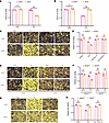
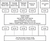
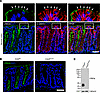
Chronic lung allograft dysfunction (CLAD) is the leading cause of mortality after lung transplantation, yet its molecular mechanisms remain poorly understood. To elucidate the pathogenesis of CLAD, we conducted a comprehensive single-cell transcriptomic analysis of CLAD lungs, integrating our generated datasets with approximately 1.6 million cells from 15 published studies of other fibrotic lung diseases. By applying pseudo-bulk approaches to mitigate batch effects, we identified molecular signatures specific to CLAD and those shared with idiopathic pulmonary fibrosis, COVID-19, and other fibrotic conditions. Our analysis revealed CLAD-specific cellular subsets including Fibro.AT2 cells, exhausted CD8+ T cells, and superactivated macrophages while suggesting that pathogenic keratin 17–positive, keratin 5–negative (KRT17+KRT5−) cells represent a common fibrotic mechanism across fibrotic lung diseases. Additionally, we performed donor-recipient cell deconvolution in lung allografts, uncovering distinct transcriptional programs and intercellular crosstalk between donor- and recipient-derived cells that drive allograft fibrosis. Recipient-derived stromal and immune cells showed enhanced pro-fibrotic and allograft rejection pathways compared with their donor counterparts. By leveraging insights from other fibrotic diseases to elucidate CLAD-specific mechanisms, our study provides a molecular framework for understanding CLAD pathogenesis and identifies potential therapeutic targets for this treatment-refractory condition.
Authors
Yuanqing Yan, Taisuke Kaihou, Emilia Lecuona, Xin Wu, Masahiko Shigemura, Haiying Sun, Chitaru Kurihara, Ruli Gao, Felix L. Nunez-Santana, G.R. Scott Budinger, Ankit Bharat