Issue published July 8, 2026 Previous issue | Next issue

- Volume 11, Issue 13
On the cover:
Fibrosis: a special issue on scientific advances and next-generation treatments
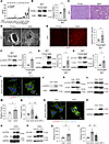
Curated by Special Issue Editors Gavin Arteel and Melanie Königshoff, this special issue on fibrosis includes a collection of original research and Reviews that spotlight fibrosis across organs, including mechanisms that underlie fibrosis onset, progression, and resolution. The cover image, from Hsiao and colleagues’ original research article, PCPE-1 promotes cardiac fibrosis with aging and obesity, is a modern art–like image showing fibrotic cardiac tissue. The image was generated using Google Gemini.
Curated by Special Issue Editors Gavin Arteel and Melanie Königshoff, this special issue on fibrosis includes a collection of original research and Reviews that spotlight fibrosis across organs, including mechanisms that underlie fibrosis onset, progression, and resolution. The cover image, from Hsiao and colleagues’ original research article, PCPE-1 promotes cardiac fibrosis with aging and obesity, is a modern art–like image showing fibrotic cardiac tissue. The image was generated using Google Gemini.
Go to section:
-
Reviews
×
Abstract
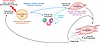
Fibrosis is a major cause of mortality and morbidity worldwide with limited therapeutic options. Our understanding of fibrosis has significantly improved and led to the identification of “core” fibrogenic mechanisms that fuel a self-sustaining vicious cycle following the initial insult. The fibrotic niche is the result of complex cellular and molecular interactions that need to be disrupted to achieve transformational therapies. In this Review, we describe the current understanding of fibrogenic mechanisms, the progress and limitations of omics approaches in the identification of novel fibrotic pathways, and advances in therapeutic modalities that all together have the potential to unleash innovative cross-organ antifibrotic therapies.
Authors
Cynthia Lebeaupin, Katelyn L. Donahue, Ken Dower, Thomas A. Wynn, Kevin M. Hart, Thomas Fabre
×Abstract
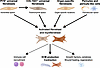
Organ fibrosis involves a complex interplay between diverse cell types and signaling pathways that ultimately leads to the pathologic accumulation of excessive extracellular matrix, subsequently resulting in organ dysfunction. In recent years, the first drugs for the treatment of idiopathic pulmonary fibrosis have been approved; however, there is a major unmet need for effective antifibrotic therapies across organs. Despite the complexity of the fibrotic process in different tissues, certain features are shared and may form the basis for future therapeutic strategies. This Review will highlight these shared characteristics, cell states, and signaling pathways across organs with the goal of highlighting potential antifibrotic strategies.
Authors
Benjamin D. Humphreys
×Abstract
Fibrosis affects almost all organ systems, resulting in a dysfunctional extracellular matrix that impairs function and can lead to failure. Crosstalk between immune cells and the stromal environment exacerbates fibrosis in all organs and is an attractive therapeutic target. Here, we discuss recent findings regarding the cellular and molecular mechanisms that underlie inflammation and fibrosis across organs. We focus on how reciprocal immune/stromal signaling maintains fibrotic niches, outline strategies for therapeutic intervention beyond current antifibrotic agents, and highlight the bone marrow fibrotic disease myelofibrosis as a model for understanding, and ultimately reversing, fibrosis in human disease.
Authors
Lucas Greven, Stijn N.R. Fuchs, Hélène F.E. Gleitz, Rebekka K. Schneider
×Abstract
The ECM is a dynamic component of the tumor microenvironment with a critical role in cancer progression, invasion, metastasis, immune exclusion, and response to therapy. Recent advances in proteomic analyses investigating the insoluble ECM fractions (termed “matrisome analysis”), along with single-cell RNA sequencing and spatial transcriptomics, have revealed cancer-specific patterns of ECM remodeling. These studies have identified a panel of recurrently upregulated ECM proteins, including annexin A1, fibrillin-1, fibronectin, periostin, and tenascin-C, actively contributing to tumor growth, invasion, angiogenesis, and immune exclusion. The expression of the cancer-associated ECM is largely driven by cancer-associated fibroblasts (CAFs), whose molecular diversity has been dissected through single-cell profiling and consolidated in emerging CAF atlases across cancers. By investigating the matrisome composition and CAF heterogeneity, these studies have unraveled the pivotal role of the stroma in shaping tumor biology. Based on these discoveries, ECM proteins and CAFs are now being explored as biomarkers and therapeutic targets. Future integration of multi-omics datasets with clinical outcomes will help to translate these insights into novel biomarkers for patient stratification and stroma-directed therapeutic interventions.
Authors
Romain Desert, Orlando Musso, Thomas F. Baumert
×Abstract
As the principal ECM-producing cell type, fibroblasts are essential regulators of tissue architecture and function in development, homeostasis, and disease. While their disease-promoting functions in fibrosis have long been the center of attention, it is increasingly recognized that fibroblasts exert critical homeostatic roles across organs, acting as sentinels that regulate the function, proliferation, and recruitment of epithelial, endothelial and immune cells in health and disease. Here, we will review the roles of fibroblasts and fibroblast-like cells in tissue maintenance, physiological wound healing, regeneration, maladaptive fibrosis, and cancer across major organs, including the skin, lung, liver, intestine, and kidney, and highlight organ-specific and shared populations and functions. We will discuss the role of PI16+ and COL15A1+ universal fibroblasts, organ-specific fibroblasts, and pericyte and pericyte-like stellate cells as cellular sources for the majority of CTHRC1+ activated fibroblasts and αSMA+ or LRRC15+ myofibroblasts and highlight the functions of specialized subpopulations, such as inflammatory fibroblasts, antigen-presenting fibroblasts, and fibroblast-like cells, including mesothelial and smooth muscle cells. A refined understanding of fibroblast heterogeneity holds promise for novel therapeutic concepts, aimed at targeting pathogenic subpopulations while preserving or enhancing homeostatic functions.
Authors
Xiaochun Yang, Marcella Steffani, Sandeep Nadella, Dean Sheppard, Florian Rieder, Yuval Rinkevich, Rafael Kramann, David A. Tuveson, Robert F. Schwabe



-
Research Articles
×
Abstract
Heart failure with preserved ejection fraction (HFpEF) is a multifactorial disease that develops in several clinical settings. Despite its complex pathogenesis, evidence indicates a central role for fibrosis in the progression of left ventricular diastolic dysfunction (LVDD). Through exploratory research into adipokines derived from brown adipose tissue (BAT), we identified a secreted-type profibrotic protein, procollagen C-endopeptidase enhancer-1 (PCPE-1), whose expression increased in BAT with aging. PCPE-1 promotes the cleavage of procollagens and is a critical initiator of fibrillogenesis. This molecule was increased in the plasma of aged mice. In addition to aging, obesity led to an increase in PCPE-1 expression in the LV of mice. Both systemic and BAT-specific PCPE-1 depletion ameliorated LV fibrosis and LVDD in the obese HFpEF model. Our data also showed that age-associated LVDD was ameliorated in the systemic PCPE-1–KO mouse fed with a normal chow diet. Conversely, the overexpression of PCPE-1 expression in BAT was shown to lead to aggravation of LV fibrosis and LVDD. Mechanistically, we found ROS/DNA damage/c-Fos/c-Jun signaling resulted in an increased production of PCPE-1 in brown adipocytes. These results indicate PCPE-1 may represent a druggable target for aging- and obesity-related HFpEF.
Authors
Yung-Ting Hsiao, Yohko Yoshida, Hirotsugu Tsuchimochi, Jingyuan Tang, Tin May Aung, Chun-Han Chang, Agian Jeffilano Barinda, Zhihong Li, Nur Syakirah Binti Othman, Tom Yoshizaki, Yiwei Ling, Shujiro Okuda, Manabu Abe, Seiya Mizuno, Satoru Takahashi, Takayuki Inomata, Hidetaka Kioka, Yasushi Sakata, Daichi Maeda, Yuya Matsue, Takaaki Furihata, Hiroshi Iwata, James T. Pearson, Kinya Otsu, Kenneth Walsh, Akihito Ishigami, Tohru Minamino, Ippei Shimizu
×Abstract
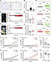
Resident cardiac fibroblast–derived (RCF-derived) cardiac myofibroblasts (CMFs) contribute to myocardial repair but also drive adverse ventricular remodeling and contractile dysfunction after myocardial infarction (MI). The sodium-activated potassium channel Slick (Slo2.1) has been described in cardiomyocyte (CM) mitochondria; however, transcriptomic analyses indicate higher Slick expression in RCFs/CMFs. Here, we investigated the role of Slick in cardiac fibroblast function and post-MI remodeling. Using live-cell imaging and whole-cell patch-clamp recordings, we found that plasma membrane Slick channels in RCFs and CMFs regulated potassium (K+) efflux and modulated store-operated calcium entry (SOCE), particularly in CMFs. Global Slick KO and conditional CMF-specific KO hearts exhibited reduced fibrosis and preserved left ventricular function after ischemia/reperfusion injury. This cardioprotection was associated with diminished CMF activation and proliferation, reduced inflammation, and improved CM survival after MI. Collectively, these findings identify fibroblast Slick channels as regulators of SOCE-dependent fibrogenesis and demonstrate that their deletion mitigates maladaptive remodeling and functional decline after MI.
Authors
Jiaqi Yang, Lin Zhu, David Spähn, Melanie Cruz Santos, Sophia Schanz, Selina Maier, Lena Birkenfeld, Helmut Bischof, Anna Roslan, Nina Wettschureck, Oliver Borst, Lucas Matt, Robert Lukowski
×Abstract
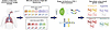
Adaptive remodeling of retrodiscal tissue following anterior disc displacement (ADD) of the temporomandibular joint (TMJ) has been recognized for decades, yet the underlying cellular dynamics and molecular mechanisms remain unclear. Using a porcine ADD model, this study investigated the cellular and molecular basis driving retrodiscal tissue adaptation. Histological staining revealed adaptive remodeling of retrodiscal tissue after ADD induction, with dense connective tissue and cartilaginous masses replacing loose connective tissue. Single-cell RNA-Seq captured pronounced fibroblast expansion during tissue remodeling, notably the FB2 subcluster with high developmental potential, and the emergence of a mural cell subcluster, MC4, associated with extracellular matrix (ECM) remodeling. CellChat analysis highlighted MC4-FB2 crosstalk via FGF2 and BMP5 signaling. The combination of pathway-aware multilayered hierarchical network (P-NET) and Seurat with drug database screening identified 5 promising compounds. Among them, zaprinast demonstrated the most robust effects by enhancing the remodeling capability of fibroblasts in vitro and alleviated TMJ deformation in vivo. Collectively, fibroblast activation is pivotal for early retrodiscal tissue adaptation after ADD, which is driven by MC4-derived FGF2/BMP5 signaling. Zaprinast treatment potentiates this remodeling process. These findings provide potentially new insights into the cellular basis of TMJ adaptation and identify potential therapeutic targets for ADD management.
Authors
Wenlin Yuan, Yilin Chen, Ruojin Yan, Wei Liu, Chenyu Wang, Ying Wang, Qiaoli Dai, Wen Li, Mengqi Zhu, Xiao Chen, Jiejun Shi
×Abstract
Survival after lung transplantation is limited by chronic, progressive graft failure, termed chronic lung allograft dysfunction (CLAD). Graft-resident mesenchymal cells (MCs) drive CLAD pathogenesis and exhibit stable dysregulated signaling, yet the transcriptomic and epigenomic drivers underlying this fibrogenic transformation remain elusive. We used single-cell multiomic profiling to characterize gene expression and chromatin accessibility in MCs isolated from bronchoalveolar lavage fluid of lung transplant recipients with and without CLAD, collected early after transplantation or after disease onset. MCs obtained after CLAD onset demonstrated a distinct transcriptomic signature compared with non-CLAD controls, enabling classification of disease status at the single-cell level with greater than 98% accuracy using signature genes. Chromatin accessibility analyses identified enrichment of CCAAT-enhancer-binding protein family transcription factors, specifically CEBPD, in CLAD MCs. MCs early after transplantation showed minimal accessibility differences, suggesting that CEBPD-associated regulatory changes emerge over time. Integration analyses identified 8 MC states and a CLAD-specific shift toward a fibrotic state. CEBPD, SOX4, and FOXP2 were identified as putative regulators of this state with substantial overlap in predicted targets. Targeting CEBPD reversed fibrotic phenotypes of CLAD MCs (decreased ECM expression, contractility, proliferation, and migration). Together, these data provide insights into transcriptomic and epigenomic changes in posttransplant MCs, facilitating the nomination of biomarkers and therapeutic targets.
Authors
Lu Lu, A. Patrick McLinden, Natalie M. Walker, Ragini Vittal, Yichen Wang, Fatemeh Fattahi, Stephen T. Russell, Michael P. Combs, Joshua D. Welch, Vibha N. Lama
×Abstract
Sustained injury to renal tubular epithelial cells (TECs), driven by excessive autophagy, is a critical mechanism underlying kidney fibrosis. Our previous work identified JLP — a TEC-expressed scaffolding protein — as an endogenous antifibrotic factor that counteracts TGF-β1–induced autophagy and fibrogenesis. However, the mechanism underlying JLP downregulation in renal fibrosis remains unclear. Here, we delineated a TGF-β1/LEF1/β-catenin/JLP axis that governs TEC autophagy through a dichotomous regulatory circuit. Under physiological conditions, low levels of β-catenin and LEF1 with minimal nuclear localization permitted normal JLP expression, which in turn maintained autophagy in check. In contrast, during renal injury, TGF-β1 promoted the expression and nuclear translocation of β-catenin and LEF1, which together suppressed JLP transcription. This loss of JLP-mediated inhibition led to unchecked autophagy and exacerbated fibrotic damage. Analyses of kidney tissues from patients with CKD, murine fibrotic kidneys, and cultured HK-2 cells confirmed consistent JLP downregulation accompanied by upregulation and nuclear accumulation of LEF1 and β-catenin. Therapeutic intervention using the β-catenin/LEF1 inhibitor iCRT3 or LEF1-targeted silencing in murine fibrosis models restored JLP expression, attenuated TEC autophagy, and ameliorated renal fibrosis. These findings revealed an autoregulatory circuit controlling TEC autophagy and fibrogenesis, and supported LEF1 and β-catenin as potential therapeutic targets in CKD.
Authors
Chen Li, Meng Zhang, Maoqing Tian, Zeyu Tang, Yuying Hu, Yuyu Long, Xiaofei Wang, Liwen Qiao, Jiefei Zeng, Yujuan Wang, Xinghua Chen, Cheng Chen, Xiaoyan Li, Lu Zhang, Huiming Wang
×Abstract
Impaired adhesion and differentiation of keratinocytes is a hallmark of several skin diseases, but only some of the factors that regulate these processes have been identified. Here, we studied the role of isoform-rich dermokine — a wound- and tumor-regulated protein — in keratinocytes using a combination of multiomics and functional approaches. CRISPR/Cas9-induced knockout of dermokine isoforms in human keratinocytes inhibited differentiation of these cells in 3-dimensional organotypic skin cultures, which was confirmed by quantitative proteomics. In 2-dimensional monocultures, dermokine deficiency affected the proteome and phosphoproteome as revealed by mass spectrometry. We found reduced abundance of differentiation-specific proteins and increased phosphorylation of the cell adhesion protein p120 (catenin δ-1). The adhesive strength of dermokine-knockout keratinocytes was impaired, which was rescued by p120 knockdown or ROCK inhibition. Finally, we verified the correlation between decreased dermokine expression and increased p120 phosphorylation in human non-healing wounds. These results identify dermokine as a regulator of keratinocyte adhesion and differentiation, involving at least in part its effect on p120 phosphorylation and ROCK. Our data point to a function of dermokine in the pathogenesis of chronic wounds.
Authors
Vahap Canbay, Till Wüstemann, Weihua Tian, Tobias A. Beyer, Camilla Reiter Elbæk, Michael Stumpe, Gaetana Restivo, Chatpakorn Christiansen, Anabel Migenda Herranz, Susanne Mailand, Jürg Hafner, Rune Busk Damgaard, Steffen Goletz, Jörn Dengjel, Ulrich auf dem Keller, Chiara Francavilla
×Abstract
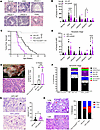
Antibody production by B cells has emerged as an important factor in regulating antitumor immunity with both suppressive and promotive roles in cancer. However, the specific effect of antibody deficiency during development of pancreatic ductal adenocarcinoma (PDAC) has not been explored. To address this question, we crossed the well-established KPC mouse model to mice lacking all circulating immunoglobulin (Ig) due to genetic ablation of both Ig secretion and Ig class switching (KPC-μSAID mice). KPC-μSAID mice exhibited a two-fold acceleration in tumor formation, a two-fold reduction in median survival, and increased liver metastases versus KPC-WT control mice. Immunofluorescence analysis of pancreatic tissues from antibody-sufficient KC- and KPC-WT mice showed that IgG was predominantly localized within the extracellular matrix (ECM). Furthermore, in both KC- and KPC-μSAID mice, ECM density and podoplanin+ cancer-associated fibroblasts (CAFs) were significantly reduced. In the KPC-μSAID tumor microenvironment (TME), intratumoral myeloid-derived suppressor cells (MDSC) were also increased, while CD4+ and CD8+ T cells decreased, relative to tumor-bearing KPC-WT mice, with macrophage exhibiting a mixed polarization phenotype. These findings were recapitulated in antibody subclass–deficient, KPC-AID mice, suggesting a potentially novel function of IgG in suppressing PDAC progression by directly or indirectly regulating pancreatic fibrosis and the density of the ECM.
Authors
Jeremy B. Foote, Sujith Sarvesh, Sameer Al Diffalha, David K. Crossman, Changde Cheng, Myung-Hee Kim, Cherlene Hardy, Julienne L. Carstens, Kyoko Kojima, Bart J. Rose, Christopher A. Klug
×Abstract
Cardiovascular disease (CVD) contributes to morbidity and mortality in people with HIV (PWH) receiving antiretroviral therapy (ART). In the REPRIEVE trial, pitavastatin reduced atherosclerotic CVD risk to a magnitude inconsistent with pitavastatin’s impact solely on LDL cholesterol and inflammation. Here, atorvastatin and ART used in REPRIEVE, including tenofovir, emtricitabine, and dolutegravir, ritonavir and darunavir were examined in 2 mouse models: transgenic HIV-Tg26 mice and HIV-PDX mice engrafted with T cells from PWH. HIV-Tg26 and HIV-PDX mice had higher cardiac fibrosis than littermate controls without HIV. Administration of tenofovir, emtricitabine, and dolutegravir or ritonavir, but not darunavir, resulted in an approximately 2-fold increase in fibrosis. Mice depleted of platelet TGF-β1 or treated with atorvastatin were partially protected from HIV- and ART-induced cardiac fibrosis, steatosis, and diastolic dysfunction. Atorvastatin’s effects were independent of changes in inflammatory cytokines, which correlated with reduced platelet activation and TGF-β signaling in cardiac endothelial cells, fibroblasts, and macrophages undergoing mesenchymal transition. Our results indicate that certain ART regimens accelerate HIV-associated CVD characterized by heart failure with preserved ejection fraction via platelet TGF-β1–dependent processes, which were mitigated by atorvastatin. Our findings provide a potential mechanism for the pleiotropic effects of statins in HIV/ART-linked CVD, which could be targeted by antiplatelet agents or inhibition of TGF-β signaling.
Authors
Kumar Subramani, Denys Babii, Brienne Cole, Tayyab A. Afzal, Thamizhiniyan Venkatesan, Trevor Word, Sandra Gostynska, Sixia Chen, Kar-Ming Fung, Ali Danesh, Itzayana G. Miller, Paul Klotman, Brad R. Jones, Jeffrey Laurence, Jasimuddin Ahamed
×Abstract
Cutaneous radiation injury is an unintended consequence of radiotherapy for many common cancers and can progress to debilitating radiation-induced skin fibrosis (RISF). Existing radiation injury models do not fully capture the skin toxicities observed in patients, contributing to the lack of efficacious therapies to mitigate RISF. To address this, we developed an ex vivo human skin model that recapitulates the temporal radiation injury and RISF response. Human skin explants (n = 12) subjected to ionizing radiation demonstrated DNA double-stranded breaks and robust p53-driven transcriptional programming of cell cycle arrest, apoptosis, and senescence compared with nonirradiated controls. Irradiated skin also exhibited induction of pro-inflammatory cytokines, epithelial-mesenchymal transition, profibrotic TGF-β1–mediated signaling, and thickened collagen over time. P53 regulators murine double minute 2 (MDM2) and miR-34a were induced after irradiation and may be leveraged to modulate injury response. Notably, RNA-sequencing of postradiotherapy breast skin from patients who had undergone mastectomy showed similar p53, inflammatory, and TGF-β1 signatures as the ex vivo model, supporting its translational relevance. Together, this model provides a platform for identifying biomarkers and testing therapies to prevent or mitigate cutaneous radiation toxicities. Targeting the dynamic p53-driven profibrotic radiation response represents a potentially new therapeutic avenue to improve quality of life for patients after radiotherapy.
Authors
Caroline Dodson, Sophie M. Bilik, Gabrielle DiBartolomeo, Hannah Pachalis, Lindsey G. Siegfried, Jordan A.K. Johnson, Seth R. Thaller, Irena Pastar, Marjana Tomic-Canic, Anthony J. Griswold, Rivka C. Stone
×Abstract
Fontan-associated liver disease (FALD) is a frequent complication in single-ventricle patients palliated with the Fontan operation. FALD severity can impact clinical decisions; however, the pathophysiology of FALD progression is unknown. Single-cell spatial transcriptomics (ST) was performed on liver explant tissue sections from FALD patients with early (n = 1) and advanced fibrosis (n = 1) using CosMx Spatial Molecular Imaging with in situ hybridization of 6000 genes. Immunofluorescence for liver zonation and cellular stress markers was performed to confirm protein expression based on ST analysis in additional FALD tissues (n = 18). Unbiased clustering yielded 12 liver cell types, comprising 6 subtypes of hepatocytes. FALD with advanced fibrosis demonstrated expansion of mid-zonal hepatocytes, accompanied by loss of zonal markers characteristic of canonical pericentral and periportal hepatocytes. A subset of hepatocytes in advanced FALD demonstrated increased cellular stress and a redundant zonal phenotype, which we have termed zonally ambiguous and stressed hepatocytes. CellChat analysis revealed that ectopic WNT2 signaling is likely driving disrupted hepatocyte zonation. To corroborate these bioinformatic findings, we performed immunofluorescent staining of FALD specimens, which confirmed a disruption of liver zonation, and a significant increase in heat shock protein 70 (HSP70). Lastly, HSP70 expression strongly correlated with the congestive hepatic fibrosis (CHF) score. Thus, single-cell ST has identified a population of hepatocytes with features of cellular stress and redundant zonal gene expression specific to advanced FALD. Further studies on hepatocyte metabolic function in Fontan patients will lead to a greater understanding of FALD development and progression during chronic maladaptation.
Authors
Brandon M. Lehrich, Jordann N. Lewis, Vik Meadows, Lori Schmitt, Mylarappa B. Ningappa, Jia-Jun Liu, Silvia Liu, Catherine K. Gestrich, Victor O. Morell, Rakesh Sindhi, Satdarshan P. Monga, Anita Saraf
×Abstract
Inflammatory bowel disease (IBD) is frequently accompanied by intestinal fibrosis, with nonresponse to long-term anti–TNF-α therapy occurring in approximately 23%–46% of patients. Integrated analysis of single-cell and bulk RNA-seq datasets revealed an expansion of IL11+ fibroblasts in inflamed intestine and their significant enrichment in nonresponders. We further identified IL11+ fibroblasts as a central communication hub that engaged in extensive crosstalk with monocytes and may contribute to inflammatory amplification and fibrotic remodeling. Additionally, we employed machine learning approaches, including least absolute shrinkage and selection operator, support vector machines, and random forest, to derive an IL11+ fibroblast–related gene signature effectively predicting nonresponse to anti–TNF-α in validation and test cohorts. IHC further confirmed the overexpression of IL-11 in nonresponders. The signature genes we found are not only associated with immune and inflammatory responses but also with fibrosis, indicating a robust association between fibrosis and anti–TNF-α treatment failure. In summary, this study highlights the important role of IL11+ fibroblasts in orchestrating both inflammation and fibrosis and provides an applicable model for predicting nonresponse to anti–TNF-α in IBD, thereby laying the foundation for precision medicine and targeted therapeutic strategies.
Authors
Wangyue Li, Wei Huang, Jiaxin Wang, Yiwen Tu, Qidi Yang, Yao Zhou, Zile Zhang, Haiming Zhuang, Yubei Gu, Duowu Zou, Yao Zhang
×Abstract
Primary sclerosing cholangitis (PSC) is a chronic, idiopathic cholestatic liver disease characterized by inflammation and fibrosis of the bile ducts, yet the cellular crosstalk driving periductal fibrosis remains poorly defined. This study applied a multiomics approach integrating spatial transcriptomics, RNA-Seq, and proteomics to characterize fibrotic periductal regions and their cell-cell communications. Macrophage subsets, including monocyte-derived macrophages and lipid-associated macrophage–like cells, colocalized with cholangiocytes, lymphocytes, and hepatic stellate cells (HSCs). Cell niche analysis identified periductal regions with elevated fibrotic signals, where cell-cell communication analysis revealed potential macrophage-HSC interactions involving 17 fibrotic driver genes in macrophages, including ITGB2, GRN, and CCL21, and 6 fibrotic effector genes in HSCs. In validation analyses, bulk RNA-Seq data showed higher driver and effector gene expression in PSC with established fibrosis compared with early-stage PSC or healthy controls. Plasma proteins encoded by macrophage driver genes were elevated in PSC and in patients with elevated (≥3.29 kPa) liver stiffness on MR elastography. Immunofluorescence and second harmonic generation imaging showed enrichment of CD68+/CD18+(ITGB2) macrophages in fibrotic regions of PSC liver biopsies. These findings revealed enrichment of monocyte-derived macrophages and lipid-associated macrophage–like cells in fibrotic regions and suggest that they likely contribute to fibrotic activation of nearby HSCs in PSC.
Authors
Yunguan Wang, David Adeleke, Xiangfei Xie, Zi F. Yang, Xiangya Wang, Giulia Loi, Annika Yang vom Hofe, Manavi Singh, Astha Malik, Ramesh Kudira, Cyd Castro-Rojas, Liva Pfuhler, Mosab Alquraish, Pamela Sylvestre, Jonathan R. Dillman, Andrew T. Trout, Emily R. Miraldi, Alexander G. Miethke
×Abstract
Idiopathic pulmonary fibrosis (IPF) is a progressive interstitial lung disease driven by aberrant fibroblast-to-myofibroblast differentiation, which requires metabolic reprogramming. Here, we identify alanine as an essential metabolite for myofibroblast differentiation. TGF-β increases intracellular alanine levels through enhanced synthesis and import in both normal and IPF lung fibroblasts. Alanine synthesis is primarily mediated by glutamate-pyruvate transaminase 2 (GPT2), whose expression is regulated by the glutamine/glutamate/α-ketoglutarate axis. Inhibition of GPT2 depletes alanine and suppresses TGF-β–induced α-SMA and COL1A1 expression, which are rescued by exogenous alanine. We also identify solute carrier family 38 member 2 (SLC38A2) as a transporter for both alanine and glutamine, upregulated by TGF-β or alanine deprivation. SLC38A2 and GPT2 form a coordinated regulatory axis sustaining intracellular alanine levels to support myofibroblast differentiation. Mechanistically, alanine deficiency impairs glycolytic flux and depletes tricarboxylic acid cycle intermediates, while alanine supplementation provides carbon and nitrogen for intracellular glutamate and proline biosynthesis, particularly under glutamine deprivation. Combined inhibition of alanine synthesis and uptake suppresses fibrogenic responses in fibroblasts and human precision-cut lung slices, highlighting dual metabolic targeting as a potential therapeutic strategy for fibrotic lung disease.
Authors
Fei Li, Niv Vigder, David R. Ziehr, Mari Kamiya, Hung N. Nguyen, Diana E. Ferreyra Faustino, Aseel H. Khalil, Hilaire C. Lam, Matthew L. Steinhauser, Edy Y. Kim, William M. Oldham
×Abstract
The lungs have a remarkable capacity to undergo homoeostatic repair and regeneration after injury, which often occurs in patients with acute respiratory distress syndrome (ARDS) and in the single-dose bleomycin mouse model. Fibroblasts are critical mediators of fibrotic disease and RNA sequencing has identified significant heterogeneity within pulmonary fibroblast populations. However, the contribution of distinct fibroblast subsets to the repair process has been understudied compared with their role in fibrosis initiation and progression. Therefore, we sought to define the transcriptional landscape of 3 phenotypically defined fibroblast subsets that occupy discrete spatial locations in naive lungs. Using TdTomato-lineage tracing approaches, we identified and interrogated collagen1a1+ (Col1a1) fibroblasts, perilipin 2+ (Plin2) alveolar fibroblasts, and α-smooth muscle actin+ (Acta2) myofibroblasts during fibrosis development and resolution after single-dose bleomycin. Quantification of fibroblast numbers showed that all 3 subsets expanded during fibrosis and contracted toward naive levels with resolution. Principal component and gene set enrichment analyses indicate that each subset underwent major transcriptomic shifts during fibrosis development, converging on a similar profibrotic transcriptional profile. However, during resolution, Plin2+ and Acta2+ fibroblasts reverted toward a prefibrotic transcriptional state, whereas Col1a1+ fibroblasts acquired a distinct program that suggests an active role in mediating the repair processes.
Authors
Daniel G. Foster, Nomin Javkhlan, Bart P. Black, Brian E. Vestal, David W.H. Riches, Elizabeth F. Redente
×Abstract
Hirschsprung disease (HSCR) is a congenital intestinal disorder characterized by the absence of ganglia in the distal intestine. Despite surgical resection of the aganglionic intestine and pull-through surgery, patients with HSCR still experience bowel dysfunction, indicating that latent abnormalities may also exist in the proximal ganglionic intestine. To elucidate possible causes of postoperative bowel dysfunction in HSCR, we investigated differences in the proximal ganglionic intestine using an animal model of HSCR (Ednrb-null mice) and validated our findings in tissue from patients with HSCR. We found that the proximal ganglionic colon of HSCR mice exhibited greater stiffness and fibrosis than their WT littermates. Similarly, submucosal fibrosis was significantly greater in the proximal ganglionic intestine of patients with HSCR than in intestinal tissue from age- and site-matched controls. Furthermore, we observed dysregulated expression of extracellular matrix–related (ECM-related) genes in the proximal ganglionic intestine of HSCR mice compared with controls. We conclude that increased fibrosis, stiffness, and alterations in ECM composition may contribute to persistent dysfunction of the ganglionic intestine in HSCR. These findings add to the growing body of literature that describe abnormalities in the proximal ganglionic intestine of HSCR and suggest that HSCR is not limited to the aganglionic intestine alone.
Authors
Prisca C. Obidike, Britney A. Hsu, Chioma Moneme, Oluyinka O. Olutoye II, Walker D. Short, Mary Hui Li, Swathi Balaji, Yuwen Zhang, Sundeep G. Keswani, Lily S. Cheng
×Abstract
BACKGROUND There are no known serum biomarkers that provide mechanistic insight or prognostic enrichment for post–COVID-19 pulmonary fibrosis.METHODS We tested associations of serum biomarkers with radiographic fibrosis-like abnormalities (reticulation, traction bronchiectasis, or honeycombing) on thoracic computed tomography (CT) scans 4 months, 15 months, and 3 years after hospitalization in an American discovery cohort of severe-to-critical COVID-19 survivors, and externally validated findings in 2 Canadian cohorts of moderate-to-critical COVID-19 survivors. In the discovery cohort, we investigated the dose-response relationship of the biomarker with CT-derived airway-to-lung ratio. We performed single-cell RNA sequencing (scRNA-seq) of transbronchial lung biopsies from COVID-19 survivors obtained 3 years after COVID-19 hospitalization and conducted immunofluorescence analysis of COVID-19 lung explants.RESULTS Among 150 discovery cohort participants, only higher levels of circulating club cell secretory protein-16 (CC16, encoded by the SCGB1A1 gene) at hospital discharge, 4 months, 15 months, and 3 years were associated with thoracic CT fibrosis-like abnormalities in cross-sectional and longitudinal analyses. Higher CC16 levels were associated with thoracic CT fibrosis-like abnormalities in 2 validation cohorts (n = 56 and n = 37). CC16 levels were linearly associated with increased airway-to-lung ratio. scRNA-seq revealed increased proportions of epithelial cells expressing SCGB1A1 and SCGB1A1/MUC5B in COVID-19 survivors with fibrosis. Immunofluorescence analysis of COVID-19 lung explants demonstrated increased numbers of SCGB1A1-expressing epithelial cells only in small (<100 μm) airways, with 3-fold more CC16/MUC5B-coexpressing cells in respiratory bronchioles..CONCLUSION. Higher CC16 levels are associated with CT fibrosis-like abnormalities for up to 3 years following moderate-to-critical COVID-19. Increased CC16 reflects dysregulated small airway epithelial progenitor cell remodeling and increased expansion of CC16+MUC5B+ epithelial cells in respiratory bronchioles after COVID-19.TRIAL REGISTRATION Not applicable.FUNDING Department of Defense, NIH, and Japan Society for the Promotion of Science for Young Scientists.
Authors
Matthew R. Baldwin, Ansley E. Jones, David Zhang, Chandan Gurung, Zain Khan, Anjali Saqi, Xuehan Yang, Ying Wei, Renu Nandakumar, Scarlett O. Murphy, Claire F. McGroder, Faisal Shaikh, Selim Arcasoy, Luke Benvenuto, Harpreet Grewal, Benjamin M. Smith, Eric A. Hoffman, Agnes C.Y. Yuen, Parteek Johal, Christopher Carlsten, Christopher J. Ryerson, J. Brent Richards, Alyson W. Wong, Tomoko Nakanishi, Aditi S. Shah, Christine Kim Garcia
×Abstract
Pulmonary fibrosis is frequently accompanied by pulmonary hypertension, which can occur disproportionate to the extent of fibrosis, suggesting a fibrosis-independent vascular remodeling process. Here, we demonstrated that plasma growth differentiation factor 15 (GDF15) is elevated across diverse fibrotic lung disease subtypes and correlates with markers of elevated right heart pressures but not pulmonary function indices, indicating a possible link to endothelial cell dysfunction. To investigate the import of endothelial GDF15 as a modifier of lung fibrosis pathogenesis, we generated endothelial cell–specific Gdf15-KO mice, which showed protection from bleomycin-induced lung injury and fibrosis, with preserved lung function. RNA-seq of human pulmonary microvascular endothelial cells revealed altered expression of barrier-regulatory genes in GDF15-deficient endothelial cells compared with controls. Functional studies confirmed that GDF15 knockdown attenuates thrombin-induced barrier disruption by reducing cytosolic Ca2+ responses. Together, these findings implicate endothelial GDF15 as a modifier of vascular permeability and Ca2+ signaling and a contributor to lung injury and fibrosis.
Authors
Kristen Raffensperger, Marta Bueno, Brian J. Philips, Megan Miller, Máté Katona, Shuai Yuan, Adriana Estrada-Bernal, Byron Chuan, Pavan Suresh, Stephanie Taiclet, Scott Hahn, Yingze Zhang, Jonathan K. Alder, Seyed Mehdi Nouraie, Daniel J. Kass, Oliver Eickelberg, Adam C. Straub
×Abstract
CHI3L1, a chitinase-like protein, is implicated in pulmonary fibrosis, yet its mechanisms are incompletely understood. We demonstrated that CHI3L1 coordinates profibrotic macrophage activation and invasive myofibroblast differentiation, and their crosstalk. In vitro, CHI3L1 drove M2-like macrophage polarization with increased CD163, CD206, and PD-L1, and amplified TGF-β1–induced fibroblast responses, including myofibroblast transformation, migration, and invasion. Mechanistically, CHI3L1 enhanced TGF-β1 signaling through SMAD, AKT, and ERK pathways, and PD-L1 was required for CHI3L1/TGF-β1–driven myofibroblast transformation. Coculture studies further demonstrated the ability of CHI3L1 to induce profibrotic macrophage activation that enhanced myofibroblast transformation mediated via a CD44/PD-L1 axis. In vivo, following bleomycin challenge, CHI3L1-transgenic mice exhibited increased PD-L1+ M2 macrophages, PD-L1+PDGFRα+ fibroblasts, and PD-1+ immune cells compared with WT controls. Therapeutically, combined anti-CHI3L1 and anti-PD-1 antibodies, or a bispecific anti-CHI3L1–anti-PD-1 antibody, produced greater antifibrotic efficacy than monotherapy. These findings demonstrate crosstalk between CHI3L1 and the PD-1/PD-L1 pathway that promotes profibrotic macrophage activation and invasive fibroblast differentiation and support dual targeting of CHI3L1 and PD-1/PD-L1 as a promising therapeutic strategy for pulmonary fibrosis.
Authors
Han-Seok Jeong, Takayuki Sadanaga, Joyce H. Lee, Suchitra Kamle, Bing Ma, Yang Zhou, Sung Jae Shin, Jack A. Elias, Chun Geun Lee
×Abstract
Idiopathic pulmonary fibrosis (IPF) is characterized by parenchymal scarring reflecting an imbalance between collagen deposition by myofibroblasts (MFs) and its turnover. Although collagen clearance is essential for fibrosis resolution, this process and its potential for therapeutic modulation in IPF are poorly understood. Here we evaluated internalization of degraded collagen and the role of its requisite endocytic receptor mannose receptor C-type 2 (MRC2), in lung tissue and MFs from patients with IPF and bleomycin-injured mice. Fibrotic human and murine lung tissue exhibited an accumulation of degraded collagen, highlighting a failure of its clearance. MFs from fibrotic lung demonstrated a reduced capacity to internalize extracellular degraded collagen, with a concomitant reduction in MRC2 expression and endolysosomal activity. Both diminished collagen uptake and MRC2 expression recovered to baseline levels during spontaneous resolution of bleomycin fibrosis. In vitro treatment of IPF or TGF-β–elicited MFs with a variety of mechanistically distinct agents known to effect phenotypic dedifferentiation restored defective collagen internalization. Although enhanced uptake was MRC2 dependent, it involved increased endolysosomal activity rather than increased MRC2 expression. These results implicate defective MRC2-dependent collagen internalization and endolysosomal function in MFs as important factors contributing to fibrosis that may be therapeutically targeted to promote resolution.
Authors
Natalie M. Walker, Sean M. Fortier, Jennifer Speth, Steven K. Huang, Sergey Gutor, Timothy S. Blackwell, Marc Peters-Golden
×Abstract
Idiopathic pulmonary fibrosis (IPF) is a fatal, aging-related disease characterized by persistent lung fibroblast activation, progressive lung scarring, and several vascular abnormalities. We have previously demonstrated that aging-associated vascular dysfunction drives maladaptive endothelial responses to injury and exacerbates lung fibrosis via secretion of profibrotic endothelial cell–derived factors. However, regulatory mechanisms governing endothelial dysfunction during progressive lung fibrosis remain poorly understood. Here, using preclinical mouse models of progressive lung fibrosis as well as human IPF lungs, we demonstrate that miR-205-5p was overexpressed in lung endothelial cells (ECs) from fibrotic lungs and coordinated gene expression programs implicated in endothelial dysfunction and progressive fibrosis. Mechanistically, miR-205-5p induced senescence in lung ECs, mirroring the senescent phenotype of IPF lung ECs. Consistently, conditioned medium derived from lung ECs overexpressing miR-205-5p promoted lung fibroblast activation. Importantly, miR-205-5p inhibition in IPF lung ECs attenuated endothelial senescence and limited paracrine fibroblast activation. Finally, inhibition of miR-205-5p in vivo preserved the pulmonary vascular network and attenuated lung fibrosis progression in aged mice challenged with bleomycin. Collectively, our findings support what we believe to be a novel connection among lung endothelial miR-205-5p, endothelial senescence, and profibrotic alteration of the endothelial secretome and highlight miR-205-5p inhibition as a potential therapeutic intervention for pulmonary fibrosis.
Authors
Giuseppe Muscato, Benjamin B. Roos, Sharonda Harris, Xiaoyu Tracy Cai, Gina Civettini, Enrico Sciacca, Ahmed A. Raslan, Alessandra Castaldi, Sharon Elliot, Marilyn K. Glassberg, Carlo Vancheri, Daniel J. Tschumperlin, Giovanni Ligresti, Nunzia Caporarello
×Abstract
Ischemia-reperfusion injury (IRI) is a common cause of acute kidney injury (AKI) leading to renal fibrosis. Here, we investigate the kinetics of autophagy, apoptosis, and necroptosis activation in tubular epithelial cells (TECs) and peritubular capillaries (PTCs) after renal IRI, and their relative contributions to renal fibrogenesis. IRI with renal artery clamping in GFP-LC3 transgenic mice induced a predominant and sustained necroptotic response in TECs, while apoptosis and autophagy played minor roles. PTCs showed early and persistent activation of apoptosis, brief necroptosis induction, and increased autophagy at a distance from IRI. Disruption of the autophagic process with chloroquine (CHQ) injections in association with renal IRI did not modulate tubular death but enhanced PTC apoptosis and increased microvascular rarefaction and fibrosis. Apoptosis-deficient GFP-LC3/Caspase-3–/– mice exposed to renal IRI showed enhanced PTC autophagy, reduced PTC rarefaction, and inhibition of renal fibrosis, in spite of increased necroptosis in TECs. Inhibition of both autophagy with CHQ and apoptosis in GFP-LC3/Caspase-3–/– mice led to a marked switch toward necroptosis in PTCs. This was associated with aggravated microvascular rarefaction, increased leukocyte infiltration, and enhanced renal fibrosis. These findings establish a predominant role for PTC autophagy and caspase-3–dependent apoptosis in the development of renal fibrosis after IRI.
Authors
Hyunyun Kim, Francis Migneault, Shanshan Lan, Imane Kaci, Julie Turgeon, Annie Karakeussian Rimbaud, Martin Dupont, Shijie Qi, Mélanie Dieudé, Marie-Josée Hébert