
Go to The Journal of Clinical Investigation
  • About
  • Editors
  • Consulting Editors
  • For authors
  • Publication ethics
  • Publication alerts by email
  • Transfers
  • Advertising
  • Job board
  • Contact
  • Physician-Scientist Development
  • Current issue
  • Past issues
  • By specialty
    • COVID-19
    • Cardiology
    • Immunology
    • Metabolism
    • Nephrology
    • Oncology
    • Pulmonology
    • All ...
  • Videos
  • Collections
    • In-Press Preview
    • Resource and Technical Advances
    • Clinical Research and Public Health
    • Research Letters
    • Editorials
    • Perspectives
    • Physician-Scientist Development
    • Reviews
    • Top read articles

  • Current issue
  • Past issues
  • Specialties
  • In-Press Preview
  • Resource and Technical Advances
  • Clinical Research and Public Health
  • Research Letters
  • Editorials
  • Perspectives
  • Physician-Scientist Development
  • Reviews
  • Top read articles
  • About
  • Editors
  • Consulting Editors
  • For authors
  • Publication ethics
  • Publication alerts by email
  • Transfers
  • Advertising
  • Job board
  • Contact
Top
  • View PDF
  • Download citation information
  • Send a comment
  • Terms of use
  • Standard abbreviations
  • Need help? Email the journal
  • Top
  • Abstract
  • The common language of fibrosis across organs
  • Macrophage–stromal crosstalk as a driver of fibrosis
  • Adaptive immune modulation of fibrogenic macrophages
  • Stromal cell contributions and organ-specific context
  • Innate sensing and inflammatory axes that prime fibrosis
  • Cytokine effector landscape acting on macrophages and stroma
  • Therapeutic modulation of fibrosis: from cytokines to matrix remodeling
  • BM fibrosis and MF as a paradigm for fibrosis reversal
  • Profibrotic stromal and hematopoietic crosstalk
  • Therapeutic landscape and niche-directed interventions
  • From transplantation to regeneration
  • Conclusions and outlook
  • Conflict of interest
  • Funding support
  • Acknowledgments
  • Footnotes
  • References
  • Version history
  • Article usage
  • Citations to this article
Advertisement

Review Open Access | 10.1172/jci.insight.202062

The inflammatory/fibrotic axis across organs: myelofibrosis as a model of reversibility

Lucas Greven,1 Stijn N.R. Fuchs,2,3 Hélène F.E. Gleitz,1,2,3 and Rebekka K. Schneider1,2,3

1Institute for Cell and Tumor Biology, RWTH Aachen University Hospital, Aachen, Germany.

2Department of Developmental Biology and

3Oncode Institute, Erasmus University Medical Center, Rotterdam, Netherlands.

Address correspondence to: Rebekka K. Schneider, Pauwelsstrasse 30, 52074 Aachen, Germany. Email: reschneider@ukaachen.de.

Authorship note: HFEG and RKS contributed equally to this work.

Find articles by Greven, L. in: PubMed | Google Scholar

1Institute for Cell and Tumor Biology, RWTH Aachen University Hospital, Aachen, Germany.

2Department of Developmental Biology and

3Oncode Institute, Erasmus University Medical Center, Rotterdam, Netherlands.

Address correspondence to: Rebekka K. Schneider, Pauwelsstrasse 30, 52074 Aachen, Germany. Email: reschneider@ukaachen.de.

Authorship note: HFEG and RKS contributed equally to this work.

Find articles by Fuchs, S. in: PubMed | Google Scholar

1Institute for Cell and Tumor Biology, RWTH Aachen University Hospital, Aachen, Germany.

2Department of Developmental Biology and

3Oncode Institute, Erasmus University Medical Center, Rotterdam, Netherlands.

Address correspondence to: Rebekka K. Schneider, Pauwelsstrasse 30, 52074 Aachen, Germany. Email: reschneider@ukaachen.de.

Authorship note: HFEG and RKS contributed equally to this work.

Find articles by Gleitz, H. in: PubMed | Google Scholar

1Institute for Cell and Tumor Biology, RWTH Aachen University Hospital, Aachen, Germany.

2Department of Developmental Biology and

3Oncode Institute, Erasmus University Medical Center, Rotterdam, Netherlands.

Address correspondence to: Rebekka K. Schneider, Pauwelsstrasse 30, 52074 Aachen, Germany. Email: reschneider@ukaachen.de.

Authorship note: HFEG and RKS contributed equally to this work.

Find articles by Schneider, R. in: PubMed | Google Scholar

Authorship note: HFEG and RKS contributed equally to this work.

Published July 8, 2026 - More info

Published in Volume 11, Issue 13 on July 8, 2026
JCI Insight. 2026;11(13):e202062. https://doi.org/10.1172/jci.insight.202062.
© 2026 Greven et al. This work is licensed under the Creative Commons Attribution 4.0 International License. To view a copy of this license, visit http://creativecommons.org/licenses/by/4.0/.
Published July 8, 2026 - Version history
View PDF
Abstract

Fibrosis affects almost all organ systems, resulting in a dysfunctional extracellular matrix that impairs function and can lead to failure. Crosstalk between immune cells and the stromal environment exacerbates fibrosis in all organs and is an attractive therapeutic target. Here, we discuss recent findings regarding the cellular and molecular mechanisms that underlie inflammation and fibrosis across organs. We focus on how reciprocal immune/stromal signaling maintains fibrotic niches, outline strategies for therapeutic intervention beyond current antifibrotic agents, and highlight the bone marrow fibrotic disease myelofibrosis as a model for understanding, and ultimately reversing, fibrosis in human disease.

The common language of fibrosis across organs

Fibrosis is characterized by persistent fibroblast activation, resulting in excess extracellular matrix (ECM) deposition, and is the common endpoint of chronic injury across organs. Fibrosis accounts for a large proportion of global disease burden, contributing to roughly 45% of all deaths in developed nations. Excess ECM stiffens tissues, impairs organ function, and ultimately leads to organ failure. Resolution of the wound-healing program is normally mediated through apoptosis of activated myofibroblasts; however, inflammatory cues — delivered by neutrophils, macrophages, and other immune cells — can persist, subsequently further activating fibroblasts into myofibroblasts that continue to secrete ECM. This dysregulation or “wound healing gone awry” concept can be provoked by diverse insults (infection, toxins, metabolic stress, malignancy, or genetic mutations) and be perpetuated by cytokines such as TGF-β, PDGF, and IL-1β. While altered wound healing presents as organ-specific manifestations, such as cirrhosis, glomerulosclerosis, and interstitial lung disease, the mechanisms of fibrosis across organs appear to be similarly reflected by an inflammatory/fibrotic cascade (1–3).

Fibrosis is traditionally viewed as irreversible scarring, but recent studies demonstrate that fibrotic tissues are dynamic and, under certain conditions, reversible. In lung fibrosis, for example, lipofibroblasts (LIFs) are programmed into activated myofibroblasts (aMYFs) during injury; however, pharmacologic agents have been shown to reverse this transition, promoting aMYF-to-LIF conversion and leading to fibrosis resolution (4). This paradigm shift is supported by the observation that abnormal ECM, which becomes the scaffold of a persistently fibrotic niche, can undergo remodeling during successful repair. Unfortunately, the success of therapeutic strategies that promote ECM remodeling remains confined largely to idiopathic pulmonary fibrosis (IPF), where two antifibrotic drugs, nintedanib and pirfenidone, are approved for use in patients. However, these drugs only slow down functional decline rather than reversing established fibrosis and show limited benefit when repurposed for dermal or systemic sclerosis–associated fibrosis. Thus, in general, no disease-modifying, antifibrotic therapies are currently available for fibrosis in most other organs, underscoring the need for new conceptual frameworks and treatments.

A unifying theme in fibrosis across organs is the dynamic interplay between stromal cells and the immune system (5, 6). Macrophages are recruited to sites of injury and secrete cytokines, including TGF-β, PDGF, and IL-1β, that induce fibroblast activation and differentiation. Activated fibroblasts subsequently produce chemokines, cytokines, and ECM components that modulate immune cell recruitment and polarization. Multiple signaling pathways, including TGF-β/SMAD, ERK, PI3K/AKT, and Wnt/Notch, integrate these inputs and govern the transition between repair and fibrosis. Together, these observations support a conceptual model in which fibrosis emerges from a dynamic inflammatory/stromal circuit. Tissue injury triggers innate immune activation and recruitment of monocytes, which differentiate into macrophage subsets that instruct stromal progenitors to adopt myofibroblast phenotypes. The resulting ECM remodeling further reinforces immune activation through mechanical and biochemical feedback signals. Importantly, this inflammatory/fibrotic circuit appears to be conserved across organs, suggesting that common cellular programs govern both the initiation and potential reversibility of fibrosis (Figure 1). Understanding how these circuits are disrupted or restored in specific disease contexts therefore provides a framework for therapeutic intervention (2, 7).

Graphical overview of the inflammation/fibrosis cycle across organs.Figure 1

Graphical overview of the inflammation/fibrosis cycle across organs.

Myelofibrosis (MF), a fibrotic bone marrow (BM) disease, exemplifies this inflammatory/fibrotic loop (8). In MF, a malignant hematopoietic clone drives chronic cytokine release and aberrant signaling among megakaryocytes, macrophages, and mesenchymal stromal cells (MSCs), creating a TGF-β–rich environment that induces ECM deposition and stromal activation. Genetic fate-mapping has shown that perivascular Gli1+ MSCs migrate from the endosteal and vascular niches to become fibrosis-driving myofibroblasts. Moreover, ablation or pharmacologic inhibition of Gli1+ MSCs markedly reduces fibrosis. Single-cell mapping further reveals that distinct leptin receptor–positive (Lepr+) stromal subsets adopt profibrotic phenotypes, while the alarmin complex S100A8/A9, which is upregulated in mutant JAK2V617F hematopoietic cells, marks progression to overt MF. Inhibition of the S100A8/A9 axis with tasquinimod alleviates disease in preclinical models (9). Additionally, basophils and mast cells establish a TNF-rich signaling hub that reprograms stromal progenitors, with galectin-1 emerging as a biomarker and therapeutic target (10). Importantly, allogeneic hematopoietic stem cell transplantation (allo-HSCT) provides the first and only clinical evidence that BM fibrosis can be reversed in humans. Successful allo-HSCT not only eradicates the malignant clone but also restores a nonfibrotic stromal niche and normal hematopoiesis, offering a proof of concept that fibrosis, once established, remains a reversible and dynamic process when both the inflammatory driver and the fibrotic niche are reset.

Macrophage–stromal crosstalk as a driver of fibrosis

Initiation: injury, DAMPs, and monocyte recruitment. Unresolved injury and persistent inflammation release damage-associated molecular patterns (DAMPs) and alarmins that activate pattern recognition receptors on resident macrophages and endothelium (Figure 2). This activation triggers cytokine/chemokine production and recruits Ly6Chi (human CD14hi) monocytes via CCR2–CCL2 to sites of damage, where they differentiate into monocyte-derived macrophages with profibrotic programs. Depletion of these recruited macrophages alleviates experimental lung fibrosis, reducing collagen and alternative-activation markers (11, 12).
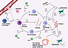
Immune cell–stromal interactions in solid organs drive fibrotic remodelingFigure 2

Immune cell–stromal interactions in solid organs drive fibrotic remodeling through both direct cell–cell communication and the secretion of soluble mediators, including cytokines and growth factors. Tissue injury or inflammation releases damage-associated molecular patterns (DAMPs) and alarmins that are recognized by tissue-resident macrophages via pattern recognition receptors (PRRs), leading to recruitment of circulating monocytes through the CCL2/CCR2 chemokine axis. Recruited Ly6Chi (mouse) or CD14hi (human) monocytes differentiate into monocyte-derived macrophages that polarize toward M1 or M2 states in response to local cues, including CD4+ Th cell cytokines, such as Th1-derived IFN-γ or Th2-derived IL-4 and IL-13. Under profibrotic conditions, Th17-derived interleukin-17A (IL-17A), granulocyte–macrophage colony-stimulating factor (GM-CSF), and megakaryocyte/platelet-derived CXC chemokine ligand 4 (CXCL4), together with transforming growth factor-β (TGF-β), drive differentiation of monocytes/macrophages into secreted phosphoprotein–positive (SPP1+) macrophages. These profibrotic SPP1+ macrophages, along with platelets, secrete mediators such as SPP1, connective tissue growth factor (CTGF), PDGF, and TGF-β, which activate fibroblasts and perivascular stromal cells, including glioma-associated oncogene homolog 1–positive (Gli1+) pericytes, leptin receptor–positive (LepR+) pericytes, PDGF receptor-β–positive (PDGFRβ+) progenitors, and hepatic stellate cells, depending on the tissue context. These cells differentiate into ECM-producing myofibroblasts that drive fibrotic remodeling. In addition, monocyte-derived fibrocytes also contribute to ECM deposition through IL-4–, IL-13–, and TGF-β–dependent signaling pathways.

Macrophage activation states: beyond the M1/M2 dichotomy. Infiltrating monocytes were traditionally framed as M1 (pro-inflammatory) or M2 (repair/regulatory). In fibrosis, macrophage activation is highly plastic and context dependent. For example, subsets of M2-like macrophages (M2a/b/c/d) arise in response to distinct cues and can exert a variety of functions, including tissue repair, regulatory, antiinflammatory/profibrotic, or tumor-promoting activities (13–15). Thus, it has become clear that the simple M1/M2 paradigm is insufficient to capture the diversity of macrophage populations in fibrotic disease.

Scar-associated macrophages. Single-cell and spatial profiling have identified a conserved population of scar-associated macrophages (SAMs) across organs that are enriched for ECM-remodeling genes and high SPP1 (osteopontin) expression (16). In the liver, analogous TREM2+CD9+SPP1+ macrophages activate stellate cells, and similar populations appear in lung, heart, and kidney fibrosis (17–19). IL-17A, GM-CSF, and platelet-derived CXCL4 promote differentiation toward the SPP1+ state (17). Functionally, SPP1+ macrophages secrete osteopontin, TGF-β, and PDGFs, thereby fostering fibroblast proliferation and collagen deposition via SPP1-, FN1-, and SEMA3-mediated macrophage–stromal crosstalk (17). Two transcriptional SAM states have been described so far. SPP1+ macrophages that do not express marker-associated macrophage (MAM) markers appear to be enriched with age, while SPP1+MAM+ macrophages associate with matrix remodeling and predominate during active fibrosis (16).
Adaptive immune modulation of fibrogenic macrophages

Th2 cytokines (IL-4/IL-13) drive alternative macrophage activation via IL-4Rα/STAT6-mediated induction of TGF-β1, which promotes fibrosis and contributes to later remodeling (20–22). Th17 cells sustain neutrophilic inflammation and expand SPP1+ macrophages through IL-17A/GM-CSF (18, 23). In contrast, Th1-derived IFN-γ induces SMAD7, suppresses TGF-β signaling, and is antifibrotic. In lung injury models, Th1 depletion exacerbates fibrosis, while Th17 loss mitigates fibrosis (24, 25). Tregs further complicate fibrosis outcomes, as Treg-derived amphiregulin stimulates fibroblasts, and Treg-specific amphiregulin loss attenuates liver fibrosis in metabolic dysfunction–associated steatohepatitis models (26).

Stromal cell contributions and organ-specific context

Across organs, stromal cells, such as perivascular mesenchymal progenitors, fibroblasts, and pericyte-derived populations, are described to be the dominant source of myofibroblasts (27, 28). Lineage tracing studies have shown that Gli1+, FOXD1-lineage, ADAM12+, and Lepr+ stromal cells proliferate and differentiate into contractile, ECM-producing myofibroblasts after injury in kidney, liver, lung, heart, skin, and BM (27, 29–35). Myofibroblasts are generated in all organs; however, the source of these cells can be organ dependent. In the liver, α-SMA+COL1A1+ myofibroblasts are derived from quiescent hepatic stellate cells (36, 37). Kidney FOXD1+PDGFRβ+ stromal cells undergo STAT3-dependent migration and myofibroblast conversion (38). In the skin and muscle, ADAM12+ perivascular progenitors acquire α-SMA expression and secrete ECM following injury (35). In the BM, heterogeneous Lepr+ MSC subsets (ALPL+CXCL12+ adipogenic stromal cells and SPP1+WIF1+IBSP+SP7+BGLAP+ osteogenic stromal cells) expand and drive fibrosis. Gli1+ stromal progenitors fall within the BM compartment, with an osteolineage focus, and respond to alarmin and platelet-derived CXCL4/PF4 signaling (9, 34, 39, 40). Additionally, hematopoietically derived CD45+COL1A1+ fibrocytes, which arise from monocytes in response to IL-4/IL-13/TGF-β signaling, deposit collagen, and secrete TGF-β, are enriched in IPF lungs and JAK2V617F MF (41, 42).

Beyond fibroblast activation, macrophages also interact extensively with epithelial and endothelial cells, shaping tissue remodeling and fibrosis progression (7). In epithelial tissues such as the lung, liver, and kidney, injured epithelial cells release alarmins and cytokines that recruit and polarize macrophages, while macrophage-derived mediators — including TGF-β, IL-1β, and PDGF — promote epithelial dysfunction, epithelial–mesenchymal signaling, and barrier disruption. In parallel, endothelial cells contribute to the inflammatory–fibrotic niche by producing chemokines that facilitate monocyte recruitment and by undergoing endothelial-to-mesenchymal transition (EndMT), thereby generating additional ECM-producing cells (43, 44). Macrophage-derived cytokines and growth factors can directly promote EndMT and vascular remodeling, linking immune activation to structural alterations of the vascular niche. These interactions illustrate that fibrosis arises not only from fibroblast activation but also from coordinated communication among immune, stromal, epithelial, and vascular compartments.

Innate sensing and inflammatory axes that prime fibrosis

DAMPs and alarmins, including S100A8/A9 HMGB1, engage TLRs to drive cytokine output, often through CCR2–CCL2–dependent immune cell recruitment. In BM fibrosis, S100A8/A9 is upregulated in stromal progenitors, marks disease progression, and when inhibited ameliorates murine MF (9). In MF, circulating HMGB1 and S100A8/A9 correlate with inflammatory activity (45). Across organs, TLR4 activation amplifies fibrosis, while TLR4 deficiency can attenuate fibrotic responses (46, 47). TLRs prime the NLRP3 inflammasome, subsequently activating caspase-1 and IL-1β to reinforce inflammatory–fibrotic loops, and pharmacologic NLRP3 inhibition reduces experimental liver fibrosis in mice.

Cytokine effector landscape acting on macrophages and stroma

While the previous section describes how adaptive immune signals shape macrophage states, the following section focuses on the downstream cytokine networks that directly activate stromal cells and sustain ECM production. A complex cytokine network orchestrates the reciprocal activation of macrophages and stromal cells during fibrosis. Among the most potent mediators are IL-1α and IL-1β, which act as central amplifiers of inflammation and drivers of tissue remodeling. In experimental lung injury, IL-1β cooperates with bleomycin to induce pulmonary fibrosis in an IL-17A–dependent manner (23). In the BM, selective deletion of IL-1β in JAK2V617F mutant hematopoietic cells markedly ameliorates fibrosis, highlighting a pathogenic role for IL-1β in inflammatory niche remodeling (48). IL-6, particularly through its trans-signaling pathway, activates STAT3 in fibroblasts, thereby promoting the expression of ECM–associated genes and enhancing fibroblast proliferation (49). These results establish IL-6 as a critical link between chronic inflammation and sustained matrix production. Similarly, TNF-α bridges immune activation and fibrogenesis by upregulating TGF-β1 expression in fibroblasts, thereby integrating inflammatory and canonical profibrotic pathways. Type 2 cytokines, notably IL-4 and IL-13, further shape both macrophage polarization and fibroblast activation. Acting through the IL-13R–IL-13α2–IL-4Rα receptor complex, IL-13 induces TGF-β1 expression and stimulates myofibroblast differentiation. Myeloid IL-4Rα signaling supports activation of alternative macrophages that promote tissue repair and fibrosis progression but also participate in later phases of resolution (20, 21). Together, these cytokine-mediated pathways establish a tightly interwoven network that sustains inflammation, drives stromal activation, and reinforces the chronicity of fibrotic disease.

HMGB1 is a prototypical DAMP that is elevated across hematologic malignancies and fibrotic disorders (50–52). The redox state of HMGB1 dictates its function, with reduced HMGB1 being chemotactic and disulfide HMGB1 acting as a pro-inflammatory cytokine (53). Fibroblasts migrate directionally toward secreted HMGB1 (54), and HMGB1 can induce expression of inflammatory cytokines (IL-1β, TNF-α, IL-6, IL-18) (55). HMGB1 expression, at both the mRNA and protein level, is significantly elevated in BM fibrosis and also associated with systemic sclerosis, cystic fibrosis, and liver fibrosis (55–58).

Fibrosis emerges from a feed-forward loop in which DAMP-driven monocyte recruitment and context-specific macrophage states (notably SPP1+ SAMs) instruct and amplify stromal myofibroblast programs via a TLR/inflammasome/cytokine axis, with organ-specific stromal progenitors and fibrocytes executing sustained ECM deposition. Together, these observations illustrate that macrophage–stromal interactions form a central regulatory node of fibrogenesis. Consequently, many emerging antifibrotic strategies aim to interrupt these inflammatory–stromal circuits, by targeting macrophage activation states, neutralizing DAMP signaling, or eliminating fibrosis-driving stromal populations.

Therapeutic modulation of fibrosis: from cytokines to matrix remodeling

As outlined above, a wide array of mediators orchestrates activation and persistence of myofibroblasts during fibrosis. Therapeutic efforts have therefore focused on interfering with key cytokine and growth factor pathways that sustain chronic inflammation and excessive ECM deposition. Among these, the IL-1 and IL-6 axes have received considerable attention as central inflammatory drivers linking immune activation to fibrotic remodeling.

IL-1–directed therapies have shown promise in both experimental and clinical settings. The IL-1 receptor antagonist anakinra has demonstrated antifibrotic potential in preclinical models of pulmonary, renal, and arthrofibrosis, primarily by suppressing IL-1–driven inflammatory signaling (59–61). Similarly, the IL-1β–neutralizing mAb canakinumab attenuates systemic inflammation and hematopoietic skewing (62). In the CANTOS trial, which enrolled more than 10,000 patients with cardiovascular risk, canakinumab treatment suppressed hepcidin, myeloid activation, and inflammatory cytokines (63). Strikingly, the strongest responses were observed in patients harboring clonal hematopoiesis mutations, such as TET2 or DNMT3A, highlighting the role of IL-1β in driving inflammation-resistant clonal expansion and ineffective erythropoiesis (64).

IL-6 is implicated in fibrotic remodeling across organs, including the lung, liver, heart, and BM (65–68). Elevated IL-6 levels correlate with disease severity, anemia, and splenomegaly in myeloproliferative neoplasms (MPNs) (69–72). In vitro studies indicate that IL-6 stimulation of MSCs induces myofibroblast-like features, including α-SMA upregulation (73, 74). mAbs that target IL-6 (siltuximab) or its receptor (tocilizumab) have therefore emerged as potential immunotherapies in chronic inflammatory and neoplastic diseases with fibrotic components (75–77).

Beyond individual cytokines, combined blockade of the IL-1 family signaling complex represents a broader antiinflammatory approach to treating fibrosis. The IL-1 receptor accessory protein (IL1RAP) serves as a coreceptor for IL-1R, IL-33R, and IL-36R and is highly upregulated in systemic sclerosis. Targeting IL1RAP with a humanized LALA-mutated IgG1κ antibody (CAN10) inhibited IL-1α/β, IL-33, and IL-36 signaling and reduced collagen deposition and myofibroblast activation in dermal and lung fibrosis models. Moreover, CAN10 may hold promise for hematologic fibrotic disorders such as MF (75, 78). A first-in-human clinical trial using this antibody is currently ongoing in plaque psoriasis (79). While these cytokine-targeted strategies demonstrate tangible benefits, systemic immune modulation carries inherent risks, such as infection and/or off-target suppression, emphasizing the need for organ-specific or locally delivered interventions.

As discussed above, DAMP-driven inflammation reinforces fibrosis by perpetuating macrophage activation and stromal priming; therefore, therapeutic strategies to neutralize innate danger signals are of interest. The S100A8/A9 alarmin complex is an attractive target, as it is markedly upregulated in murine and human MPN–associated BM fibrosis. Moreover, S100A8/A9 alarmin is typically absent in stromal cells, with expression shifting from mutant hematopoietic clones to fibrosis-driving mesenchymal cells during disease progression. Pharmacological inhibition of S100A8/A9 binding with tasquinimod ameliorated cytopenia and markedly reduced BM fibrosis in JAK2V617F models (9, 39, 80). HMGB1-neutralizing Abs have demonstrated efficacy in models of diabetic cardiomyopathy, cystic fibrosis, and arthritis, while the orally available HMGB1 inhibitor SB17170, converted to its active form SB1703, is under phase II evaluation for IPF (81). While the context-dependent function of the HMGB1 redox state complicates pharmacologic targeting, accumulating evidence positions this prototypical DAMP as a promising target that connects inflammation, stromal activation, and clonal hematopoiesis.

TGF-β remains the central profibrotic master regulator. It is produced by multiple cell types, including megakaryocytes, macrophages, platelets, endothelial cells, and stromal cells, and orchestrates myofibroblast differentiation and ECM deposition. TGF-β inhibition has therefore been explored at several mechanistic levels and as potential therapeutic targets. Activation of latent TGF-β, which is secreted in complex with latency-associated peptide, can be prevented by targeting its activators. Inhibition of the matrix protein thrombospondin-1 or αvβ3/αvβ5 integrins suppresses TGF-β1/SMAD3 signaling, collagen production, and fibrosis in peritoneal, renal, and cardiac injury models (82–86). Direct neutralization strategies have included the pan–TGF-β antibody fresolimumab (GC1008), which reduced both myofibroblast markers and TGF-β–inducible genes in patients with systemic sclerosis (87). However, systemic TGF-β blockade causes adverse effects, including cardiovascular damage and impaired wound healing in preclinical models (88). More selective approaches, such as the ligand trap AVID200 targeting TGF-β1 and TGF-β3, showed encouraging results, including improved BM morphology and hematologic parameters without cardiac or dermatologic toxicity, in a phase Ib clinical trial in patients with MF (NCT03895112) (89). Downstream inhibition of TGF-β signaling via ALK5/TGF-βR1 kinase blockade (galunisertib) has been primarily assessed in oncology, but preclinical models of liver, kidney, and lung fibrosis demonstrate substantial antifibrotic potential (90–95). Similarly, neutralization of CTGF with pamrevlumab has proven effective in preclinical cardiac and peritoneal fibrosis and has shown safety and disease-modifying activity in patients with IPF (96–98).

Despite progress in approaches to modulate inflammatory and growth factor pathways, these strategies often fail to fully reverse established fibrosis because they do not eliminate the effector cells responsible for ECM deposition. Consequently, attention has shifted toward direct targeting of fibrosis-driving cells. Elegant preclinical work has demonstrated the feasibility of selective fibroblast depletion using chimeric antigen receptor (CAR) T cell therapy. In cardiac injury models, fibroblast activation protein–specific (FAP-specific) CAR T cells removed activated fibroblasts, reduced interstitial collagen, and improved cardiac function (99). A lipid nanoparticle mRNA delivery platform then enabled transient in vivo generation of FAP-CAR T cells that achieved similar antifibrotic effects without ex vivo manipulation (100). More recently, CAR T cells targeting PDGFRβ+ fibroblasts, which are abundant in chronic kidney disease, effectively reduced fibrosis in mouse models and human kidney organoids, outperforming FAP-CAR T cells (101). Approaches that eradicate or reprogram ECM-producing fibroblasts may enable sustained tissue recovery where cytokine inhibition alone fails.

Finally, the fibrotic matrix itself has emerged as a therapeutic target. Enzymes of the lysyl-oxidase (LOX) and LOX-like (LOXL1–4) families catalyze covalent cross-linking of collagens and elastins, stabilizing the stiff ECM that is characteristic of chronic fibrosis. In preclinical models, Ab-mediated inhibition of LOXL2 with AB0023 attenuated fibrosis across organs (102–104); however, the humanized analog simtuzumab failed in phase II trials for patients with liver fibrosis and MF (105). In contrast, small-molecule LOXL2 inhibitors, such as GB2064, have shown favorable safety, target engagement, and preliminary improvements in BM fibrosis, while the pan-LOX inhibitor PXS-5505 demonstrated good tolerability and early evidence of reduced marrow collagen in patients with MF (106). Broader inhibition of the LOX family, particularly LOXL4, appears more effective than selective LOXL2 blockade, producing stronger suppression of collagen cross-linking and fibrosis resolution in preclinical lung and liver models (107, 108). Together, these results underscore ECM remodeling enzymes as tractable targets for restoring tissue elasticity and interrupting the mechanical feed-forward loop that sustains fibrosis.

Across all therapeutic classes, systemic toxicity and limited tissue penetration remain major challenges. Emerging strategies are increasingly focused on tissue-selective delivery, including Ab drug conjugates, nanoparticles, or prodrugs that preferentially accumulate in fibrotic niches. Together, these multifaceted approaches to attenuate inflammatory cytokines, block DAMP-driven amplification, modulate TGF-β and CTGF pathways, deplete activated fibroblasts, and reduce ECM cross-linking converge on a paradigm for true fibrosis reversal that combines immune modulation, stromal targeting, and matrix remodeling into an integrated therapeutic framework.

BM fibrosis and MF as a paradigm for fibrosis reversal

Among all fibrotic diseases, BM fibrosis in MF provides a rare clinical example where complete structural reversal of fibrosis can occur. In MF, allo-HSCT promotes regression, even with advanced reticulin and collagen fibrosis, that includes restoration of normal marrow architecture and reappearance of adipocytes and hematopoietic niches. Thus, the BM provides a unique and powerful model for true fibrosis regression in humans. Mechanistically, reversal likely reflects a combination of effects: elimination of the inflammatory malignant clone, repopulation by donor-derived hematopoietic and immune cells that suppress profibrotic signaling, and gradual reprogramming or replacement of fibrosis-driving stromal cells. Despite this curative potential, allo-HSCT remains a high-risk and aggressive therapy that is associated with considerable morbidity and mortality. Only a subset of younger and fit patients qualifies for transplantation. A better understanding of the cellular and molecular mechanisms that enable fibrosis reversal in this context will be crucial to replicate this regenerative process through targeted and less toxic interventions in the future.

MF represents the most aggressive form of the Philadelphia chromosome–negative MPN, which also encompasses polycythemia vera and essential thrombocythemia (31, 109). These clonal stem cell disorders arise from somatic mutations in JAK2, CALR, or MPL, all converging on constitutive activation of the JAK/STAT pathway. This signaling promotes myeloproliferation and excessive cytokine release, fueling a chronic inflammatory milieu rich in IL-6, TNF-α, IL-1β, and TGF-β. In the BM, these cytokines activate MSCs and fibroblasts, leading to pathological ECM deposition and reticulin fibrosis. Over time, marrow failure develops, accompanied by cytopenia and compensatory extramedullary hematopoiesis in the spleen. Thus, inflammation and fibrosis are intertwined hallmarks of disease progression, representing both cause and consequence of the disrupted hematopoietic niche.

Profibrotic stromal and hematopoietic crosstalk

In MF, fibrosis primarily originates from mesenchymal stromal progenitors, including Lepr+, Gli1+, and nestin+ perivascular cells (9, 34, 110), which transdifferentiate into α-SMA+ myofibroblasts under cytokine stimulation or promote a neuropathy-like state in the BM. α-SMA+ myofibroblasts deposit collagens, fibronectin, and fibrillar ECM proteins that alter marrow stiffness and impede normal hematopoietic stem cell function. Megakaryocytes serve as central amplifiers of this process by secreting TGF-β, PDGF, and CXCL4/PF4, which drive stromal activation and recruit profibrotic macrophages (39, 111). During disease progression, the marrow microenvironment undergoes a shift from inflammatory to fibrotic signaling, dominated by TGF-β/SMAD, SPP1, and S100A8/A9 pathways, which consolidate a self-sustaining profibrotic feedback loop between the hematopoietic clone and the stromal niche.

Beyond the canonical stromal progenitors, other niche compartments also have been described to contribute to fibrosis. EndMT enables endothelial cells to lose vascular characteristics and acquire mesenchymal, ECM-producing phenotypes, disrupting vascular integrity and amplifying fibrosis (112). Dysregulation of differentiation of cells in the osteolineage and osteosclerosis accompany advanced stages of MF, reflecting abnormal coupling between bone formation and fibrosis. Importantly, alarmins such as S100A8/A9 act as key molecular mediators linking the malignant clone to its fibrotic niche. These proteins are upregulated in mutant hematopoietic cells and are transferred to stromal progenitors, where they activate TLR4 and RAGE signaling, sustaining inflammation, ECM production, and myofibroblast persistence.

Therapeutic landscape and niche-directed interventions

The JAK1/2 inhibitor ruxolitinib remains the standard of care for MF, effectively reducing splenomegaly and inflammatory cytokine burden while improving constitutional symptoms. However, ruxolitinib exerts minimal effect on fibrosis regression, clonal allele burden, or overall survival, and its use is limited by cytopenia and resistance (113, 114). Consequently, current therapeutic strategies increasingly aim to target the fibrotic BM niche and its pathogenic signaling loops (Table 1).

Table 1

Targets and ongoing trials to specifically affect the BM niche in MF

One of the most advanced antifibrotic candidates is PRM-151 (zinpentraxin alfa), a recombinant form of human pentraxin-2 that modulates macrophage differentiation and inhibits myofibroblast activation. In phase II trials, patients with MF were treated with PRM-151 alone or in combination with ruxolitinib, and clinical benefits included a 37% mean spleen size reduction, 54% improvement in symptom score, and measurable regression of MF fibrosis in one-third of patients (115). While PRM-151 is no longer in development for MF, it remains under evaluation for other organ fibroses and underscores the potential of macrophage-modulating antifibrotic therapies. TGF-β signaling is another promising therapeutic target. The selective TGF-β1/3 ligand trap AVID200 inhibits ligand–receptor interaction without affecting the homeostatic TGF-β2 isoform. In an investigator-led phase Ib study in patients with advanced MF, AVID200 treatment reduced plasma TGF-β levels, decreased SMAD2 phosphorylation and stromal proliferation, and resulted in more than 50% symptom reduction in nearly half of patients (89). Clinical improvement was observed particularly after extended treatment cycles, suggesting a time-dependent remodeling effect on the fibrotic niche (116). Additional strategies under exploration include mAbs that directly target TGF-β (e.g., fresolimumab) or small molecules, such as galunisertib, that inhibit the TGF-β receptor I kinase (ALK5), which has shown antifibrotic efficacy in preclinical models.

In parallel, luspatercept, a recombinant fusion protein comprising the activin receptor IIB domain linked to human IgG1 Fc, acts as a ligand trap for activins and GDF11, thereby inhibiting downstream SMAD2/3 signaling and promoting erythroid differentiation. In clinical studies, luspatercept improved anemia in MF, achieving transfusion independence in up to one-third of transfusion-dependent patients receiving concurrent ruxolitinib. It is currently being evaluated in a phase III placebo-controlled trial (117).

Pharmacologic inhibition of Gli1, a key effector of the hedgehog signaling pathway that is expressed by myofibroblast progenitors, represents another avenue for targeting stromal activation. In preclinical MPN models, the small-molecule Gli1 inhibitor GANT61 prevented myofibroblast differentiation and ameliorated BM fibrosis, highlighting a potential strategy to block the stromal activation cascade at its origin (27, 29, 118).

Finally, the identification of S100A8/A9 as a central hematopoietic/stromal signaling axis has led to translational efforts targeting this alarmin complex. The small molecule tasquinimod, which prevents S100A8/A9 binding to its receptors TLR4 and RAGE, markedly reduced fibrosis and improved hematopoiesis in preclinical MPN models (9, 80). Based on these findings, two clinical trials are underway to test tasquinimod as monotherapy or in combination with ruxolitinib, aiming to directly disrupt the inflammatory cross-talk that drives fibrosis progression (119, 120).

From transplantation to regeneration

Allo-HSCT in BM fibrosis provides one of the most compelling clinical proofs of principle that even advanced fibrotic architecture can be reversed in humans. In patients with MF, successful transplantation leads to the elimination of the malignant hematopoietic clone and initiates a cascade of microenvironmental changes that progressively restore tissue homeostasis. Early after transplantation, inflammatory cytokine levels decline and macrophage activation states normalize, reflecting the rapid disruption of the chronic inflammatory circuits that sustain fibrosis. This normalization is followed by gradual remodeling of the ECM, accompanied by the disappearance of reticulin and collagen fibers that previously dominated the marrow architecture. Over time, donor-derived hematopoietic and immune cells repopulate the marrow niche and reestablish balanced immune signaling. As these regulatory networks are restored, fibrosis-driving stromal populations lose their pathological activation state.

These clinical observations fundamentally challenge the long-standing view of fibrosis as a permanent scar and instead support the concept that fibrosis is a dynamic and actively maintained tissue state that depends on continuous signaling between inflammatory, immune, and stromal compartments. In this context, the reversal of BM fibrosis following transplantation can be understood as a so-called “natural experiment” in human regenerative biology: once the initiating driver is removed and inflammatory circuits are reset, the tissue retains a remarkable capacity to remodel itself and regain functional architecture. This regenerative remodeling involves coordinated changes across multiple cellular layers of the niche, including hematopoietic cells, immune regulators, stromal progenitors, and ECM-producing fibroblasts.

The success of allo-HSCT therefore demonstrates that organ fibrosis can normalize when the pathogenic drivers that sustain it are extinguished and stromal homeostasis is reestablished (Figure 3). The critical challenge now is to translate the biological lessons learned from this clinical scenario into therapeutic strategies that reproduce key aspects of the regenerative reset without requiring ablative transplantation. Achieving this goal requires a detailed understanding of how donor hematopoiesis restores immune equilibrium, dampens DAMP signaling, and interrupts the self-reinforcing inflammatory/stromal circuits that maintain fibrosis. Equally important is deciphering how activated stromal populations, including fibrosis-driving fibroblasts and mesenchymal progenitors, either revert to quiescence or are replaced during tissue remodeling. Future studies combining lineage tracing, spatial transcriptomics, and longitudinal marrow biopsies will be critical to determine whether fibrosis-driving stromal cells revert to quiescence, undergo apoptosis, or are replaced by newly generated niche cells during this regenerative process.
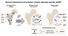
The regenerative reset of BM fibrosis following allo-HSCT.Figure 3

The regenerative reset of BM fibrosis following allo-HSCT. The pathologically altered bone marrow (BM) niche in BM fibrosis is induced by injury in the form of a malignant hematopoietic clone causing high concentrations of inflammatory cytokines that activate macrophages (and megakaryocytes) into a profibrotic state. This induces the fibrotic transformation of stromal cells and ECM deposition over time. Following allogeneic hematopoietic stem cell transplantation (allo-HSCT), a sequential transition occurs: the elimination of the malignant clone (the injury stimulus) leads to a rapid decline in inflammatory signaling, which triggers the normalization of macrophages toward a homeostatic state and facilitates the enzymatic remodeling of the fibrotic matrix. This “regenerative reset” culminates in a restored niche where donor-derived hematopoietic and immune cells maintain balanced signaling, allowing stromal cells to revert to a quiescent phenotype or be replaced by healthy niche cells. This natural experiment demonstrates that removing the initiating driver and reprogramming the immune microenvironment can drive the transition from advanced fibrosis back toward tissue homeostasis.

Importantly, the mechanistic insights gained from BM fibrosis are likely to extend beyond hematologic disease. Across multiple organs — including liver, lung, kidney, and heart — fibrosis emerges from similar feedback loops linking immune activation, macrophage polarization, stromal progenitor activation, and ECM deposition. The BM therefore provides a uniquely informative system in which the full cycle of fibrosis initiation, maintenance, and reversal can be studied in humans. Unlike most solid organs, the BM is readily accessible for longitudinal sampling and high-resolution histologic analysis, allowing researchers to track dynamic changes in cellular composition, inflammatory signaling, and matrix remodeling over time. As such, BM represents a powerful in vivo model system, a living laboratory, for understanding how fibrotic tissues transition back toward homeostasis.

By dissecting the cellular and molecular programs that enable fibrosis regression after allo-HSCT, the field may uncover broadly applicable strategies to therapeutically reprogram fibrotic tissues. Such strategies could include targeted modulation of inflammatory drivers, manipulation of macrophage states, elimination or reprogramming of fibrosis-driving stromal cells, and stimulation of regenerative niche formation. Ultimately, insights derived from this uniquely accessible regenerative process may help guide the development of molecularly targeted therapies capable of inducing true fibrosis reversal across organs.
Conclusions and outlook

Across organs, fibrosis is increasingly recognized not as a terminal scarring endpoint but as a dynamic and potentially reversible process. The BM provides a particularly powerful paradigm for this concept. In MF, mutant hematopoietic and progenitor cells orchestrate a chronic inflammatory environment that activates Gli1+ and Lepr+ stromal progenitors to transdifferentiate into ECM-producing myofibroblasts. When these profibrotic and inflammatory signals are dampened — whether through JAK inhibition, TGF-β blockade, alarmin targeting, or hematopoietic replacement after allo-HSCT — stromal cells can gradually revert toward a homeostatic, hematopoiesis-supporting phenotype. These observations demonstrate that BM fibrosis is not fixed but plastic and that its resolution can be therapeutically induced. Because of its well-characterized cellular composition, accessibility, and regenerative capacity, the BM represents an exceptional model system for studying the principles of fibrosis reversal. Lessons from this tissue illuminate a broader truth: across organs, the interplay between chronic inflammation, immune–stromal crosstalk, and mechanical remodeling drives fibrogenesis, and interventions that restore this balance can re-enable endogenous repair.

Looking ahead, the integration of single-cell, spatial, and temporal omics technologies will fundamentally reshape our understanding of how inflammatory, stromal, and vascular compartments interact to sustain or resolve fibrosis. In MF and beyond, these approaches will allow precise mapping of lineage trajectories, signaling networks, and spatial cell–cell interactions that determine the fate of fibrotic lesions. Such insights will guide the development of microenvironment-directed therapies that can be combined with oncogene- or mutation-targeted treatments, tackling both the clonal driver and its supportive niche. The next generation of antifibrotic therapies will likely move beyond symptom control to achieve true disease modification — restoring organ architecture, function, and regenerative capacity. MF thus serves not only as a clinical challenge but also as an experimental blueprint for how reversing the inflammatory and stromal circuits that sustain fibrosis could ultimately enable organ regeneration across systems.

Conflict of interest

RKS is a cofounder and shareholder of Sequantrix GmbH and has received a research grant from Active Biotech.

Funding support
  • HG by a Gilead Research Scholar Award in Oncology/Hematology, a ZonMW VENI grant, an Erasmus Medical Center Fellowship, a Dutch Cancer Society (KWF) Exploration grant, and an NWO ENW M1 grant.
  • RKS by European Research Council (ERC) grants (Rewind-MF ERC-CoG 101124542; deFIBER ERC-StG 757339 and PoC DeAlarmin) and a ZonMW VIDI grant.
  • Grants of the Deutsche Forschungsgemeinschaft (DFG) (German Research Foundation) to HG (417911533) and RKS (504777725; 417911533; 514007497).
  • RKS as member of the E:MED Consortia Fibromap and the consortium CureFib by the German Ministry of Education and Science (BMBF).
  • LG as part of the Research Training Group (RTG 2375) by the DFG.
Acknowledgments

RKS is an Oncode Institute investigator.

Address correspondence to: Rebekka K. Schneider, Pauwelsstrasse 30, 52074 Aachen, Germany. Email: reschneider@ukaachen.de.

Footnotes

Copyright: © 2026, Greven et al. This is an open access article published under the terms of the Creative Commons Attribution 4.0 International License.

Reference information: JCI Insight. 2026;11(13):e202062. https://doi.org/10.1172/jci.insight.202062.

References
  1. Wynn TA, Ramalingam TR. Mechanisms of fibrosis: therapeutic translation for fibrotic disease. Nat Med. 2012;18(7):1028–1040.
    View this article via: CrossRef PubMed Google Scholar
  2. Henderson NC, et al. Fibrosis: from mechanisms to medicines. Nature. 2020;587(7835):555–566.
    View this article via: CrossRef PubMed Google Scholar
  3. Distler JHW, et al. Shared and distinct mechanisms of fibrosis. Nat Rev Rheumatol. 2019;15(12):705–730.
    View this article via: CrossRef PubMed Google Scholar
  4. Kheirollahi V, et al. Metformin induces lipogenic differentiation in myofibroblasts to reverse lung fibrosis. Nat Commun. 2019;10(1):2987.
    View this article via: CrossRef PubMed Google Scholar
  5. Mescher AL. Macrophages and fibroblasts during inflammation and tissue repair in models of organ regeneration. Regeneration (Oxf). 2017;4(2):39–53.
    View this article via: CrossRef PubMed Google Scholar
  6. Wynn TA. Cellular and molecular mechanisms of fibrosis. J Pathol. 2008;214(2):199–210.
    View this article via: CrossRef PubMed Google Scholar
  7. Wynn TA, Vannella KM. Macrophages in tissue repair, regeneration, and fibrosis. Immunity. 2016;44(3):450–462.
    View this article via: CrossRef PubMed Google Scholar
  8. Gleitz HFE, et al. Still a burning question: the interplay between inflammation and fibrosis in myeloproliferative neoplasms. Curr Opin Hematol. 2021;28(5):364–371.
    View this article via: CrossRef PubMed Google Scholar
  9. Leimkühler NB, et al. Heterogeneous bone-marrow stromal progenitors drive myelofibrosis via a druggable alarmin axis. Cell Stem Cell. 2021;28(4):637–652.
    View this article via: CrossRef PubMed Google Scholar
  10. Li R, et al. A proinflammatory stem cell niche drives myelofibrosis through a targetable galectin-1 axis. Sci Transl Med. 2024;16(768):eadj7552.
    View this article via: CrossRef PubMed Google Scholar
  11. Gibbons MA, et al. Ly6Chi monocytes direct alternatively activated profibrotic macrophage regulation of lung fibrosis. Am J Respir Crit Care Med. 2011;184(5):569–581.
    View this article via: CrossRef PubMed Google Scholar
  12. Misharin AV, et al. Monocyte-derived alveolar macrophages drive lung fibrosis and persist in the lung over the life span. J Exp Med. 2017;214(8):2387–2404.
    View this article via: CrossRef PubMed Google Scholar
  13. Zizzo G, et al. Efficient clearance of early apoptotic cells by human macrophages requires M2c polarization and MerTK induction. J Immunol. 2012;189(7):3508–3520.
    View this article via: CrossRef PubMed Google Scholar
  14. Wang L-X, et al. M2b macrophage polarization and its roles in diseases. J Leukoc Biol. 2019;106(2):345–358.
    View this article via: CrossRef PubMed Google Scholar
  15. Murray PJ, Wynn TA. Protective and pathogenic functions of macrophage subsets. Nat Rev Immunol. 2011;11(11):723–737.
    View this article via: CrossRef PubMed Google Scholar
  16. Ouyang JF, et al. Systems level identification of a matrisome-associated macrophage polarisation state in multi-organ fibrosis. Elife. 2023;12:e85530.
    View this article via: CrossRef PubMed Google Scholar
  17. Hoeft K, et al. Platelet-instructed SPP1+ macrophages drive myofibroblast activation in fibrosis in a CXCL4-dependent manner. Cell Rep. 2023;42(2):112131.
    View this article via: CrossRef PubMed Google Scholar
  18. Fabre T, et al. Identification of a broadly fibrogenic macrophage subset induced by type 3 inflammation. Sci Immunol. 2023;8(82):eadd8945.
    View this article via: CrossRef PubMed Google Scholar
  19. Ramachandran P, et al. Resolving the fibrotic niche of human liver cirrhosis at single-cell level. Nature. 2019;575(7783):512–518.
    View this article via: CrossRef PubMed Google Scholar
  20. Fichtner-Feigl S, et al. IL-13 signaling through the IL-13alpha2 receptor is involved in induction of TGF-beta1 production and fibrosis. Nat Med. 2006;12(1):99–106.
    View this article via: CrossRef PubMed Google Scholar
  21. Weng S-Y, et al. IL-4 Receptor alpha signaling through macrophages differentially regulates liver fibrosis progression and reversal. EBioMedicine. 2018;29:92–103.
    View this article via: CrossRef PubMed Google Scholar
  22. Melo-Cardenas J, et al. IL-13/IL-4 signaling contributes to fibrotic progression of the myeloproliferative neoplasms. Blood. 2022;140(26):2805–2817.
    View this article via: CrossRef PubMed Google Scholar
  23. Wilson MS, et al. Bleomycin and IL-1beta-mediated pulmonary fibrosis is IL-17A dependent. J Exp Med. 2010;207(3):535–552.
    View this article via: CrossRef PubMed Google Scholar
  24. Wen F-Q, et al. Interferon-gamma inhibits transforming growth factor-beta production in human airway epithelial cells by targeting Smads. Am J Respir Cell Mol Biol. 2004;30(6):816–822.
    View this article via: CrossRef PubMed Google Scholar
  25. Šarc I, et al. Elevated Th1 and terminally differentiated cytotoxic T cells with suppressed Tc17 lymphocytes in lung tissue of advanced COPD and IPF patients undergoing lung transplantation. Front Immunol. 2025;16:1646711.
    View this article via: CrossRef PubMed Google Scholar
  26. Savage TM, et al. Amphiregulin from regulatory T cells promotes liver fibrosis and insulin resistance in non-alcoholic steatohepatitis. Immunity. 2024;57(2):303–318.
    View this article via: CrossRef PubMed Google Scholar
  27. Kramann R, et al. Perivascular Gli1+ progenitors are key contributors to injury-induced organ fibrosis. Cell Stem Cell. 2015;16(1):51–66.
    View this article via: CrossRef PubMed Google Scholar
  28. Lin S-L, et al. Pericytes and perivascular fibroblasts are the primary source of collagen-producing cells in obstructive fibrosis of the kidney. Am J Pathol. 2008;173(6):1617–1627.
    View this article via: CrossRef PubMed Google Scholar
  29. Schneider RK, et al. Gli1+ mesenchymal stromal cells are a key driver of bone marrow fibrosis and an important cellular therapeutic target. Cell Stem Cell. 2018;23(2):308–309.
    View this article via: CrossRef PubMed Google Scholar
  30. Kuppe C, et al. Decoding myofibroblast origins in human kidney fibrosis. Nature. 2021;589(7841):281–286.
    View this article via: CrossRef PubMed Google Scholar
  31. Kramann R, Schneider RK. The identification of fibrosis-driving myofibroblast precursors reveals new therapeutic avenues in myelofibrosis. Blood. 2018;131(19):2111–2119.
    View this article via: CrossRef PubMed Google Scholar
  32. Agha EE, et al. Mesenchymal stem cells in fibrotic disease. Cell Stem Cell. 2017;21(2):166–177.
    View this article via: CrossRef PubMed Google Scholar
  33. Humphreys BD, et al. Fate tracing reveals the pericyte and not epithelial origin of myofibroblasts in kidney fibrosis. Am J Pathol. 2010;176(1):85–97.
    View this article via: CrossRef PubMed Google Scholar
  34. Decker M, et al. Leptin-receptor-expressing bone marrow stromal cells are myofibroblasts in primary myelofibrosis. Nat Cell Biol. 2017;19(6):677–688.
    View this article via: CrossRef PubMed Google Scholar
  35. Dulauroy S, et al. Lineage tracing and genetic ablation of ADAM12(+) perivascular cells identify a major source of profibrotic cells during acute tissue injury. Nat Med. 2012;18(8):1262–1270.
    View this article via: CrossRef PubMed Google Scholar
  36. Mederacke I, et al. Fate tracing reveals hepatic stellate cells as dominant contributors to liver fibrosis independent of its aetiology. Nat Commun. 2013;4:2823.
    View this article via: CrossRef PubMed Google Scholar
  37. Tsuchida T, Friedman SL. Mechanisms of hepatic stellate cell activation. Nat Rev Gastroenterol Hepatol. 2017;14(7):397–411.
    View this article via: CrossRef PubMed Google Scholar
  38. Ajay AK, et al. Deletion of STAT3 from Foxd1 cell population protects mice from kidney fibrosis by inhibiting pericytes trans-differentiation and migration. Cell Rep. 2022;38(10):110473.
    View this article via: CrossRef PubMed Google Scholar
  39. Gleitz HFE, et al. Increased CXCL4 expression in hematopoietic cells links inflammation and progression of bone marrow fibrosis in MPN. Blood. 2020;136(18):2051–2064.
    View this article via: CrossRef PubMed Google Scholar
  40. Prasad P, Cancelas JA. From marrow to bone and fat: exploring the multifaceted roles of leptin receptor positive bone marrow mesenchymal stromal cells. Cells. 2024;13(11):910.
    View this article via: CrossRef PubMed Google Scholar
  41. Ozono Y, et al. Neoplastic fibrocytes play an essential role in bone marrow fibrosis in Jak2V617F-induced primary myelofibrosis mice. Leukemia. 2021;35(2):454–467.
    View this article via: CrossRef PubMed Google Scholar
  42. Heukels P, et al. Fibrocytes are increased in lung and peripheral blood of patients with idiopathic pulmonary fibrosis. Respir Res. 2018;19(1):90.
    View this article via: CrossRef PubMed Google Scholar
  43. Piera-Velazquez S, Jimenez SA. Endothelial to mesenchymal transition: role in physiology and in the pathogenesis of human diseases. Physiol Rev. 2019;99(2):1281–1324.
    View this article via: CrossRef PubMed Google Scholar
  44. Alonso-Herranz L, et al. Macrophages promote endothelial-to-mesenchymal transition via MT1-MMP/TGFβ1 after myocardial infarction. Elife. 2020;9:e57920.
    View this article via: CrossRef PubMed Google Scholar
  45. De Luca G, et al. Elevated levels of damage-associated molecular patterns HMGB1 and S100A8/A9 coupled with toll-like receptor-triggered monocyte activation are associated with inflammation in patients with myelofibrosis. Front Immunol. 2024;15:1365015.
    View this article via: CrossRef PubMed Google Scholar
  46. Bhattacharyya S, et al. TLR4-dependent fibroblast activation drives persistent organ fibrosis in skin and lung. JCI Insight. 2018;3(13):e98850.
    View this article via: JCI Insight CrossRef PubMed Google Scholar
  47. Jun YK, et al. Toll-like receptor 4 regulates intestinal fibrosis via cytokine expression and epithelial-mesenchymal transition. Sci Rep. 2020;10(1):19867.
    View this article via: CrossRef PubMed Google Scholar
  48. Rai S, et al. IL-1β promotes MPN disease initiation by favoring early clonal expansion of JAK2-mutant hematopoietic stem cells. Blood Adv. 2024;8(5):1234–1249.
    View this article via: CrossRef PubMed Google Scholar
  49. Chen W, et al. Blocking interleukin-6 trans-signaling protects against renal fibrosis by suppressing STAT3 activation. Theranostics. 2019;9(14):3980–3991.
    View this article via: CrossRef PubMed Google Scholar
  50. Yasinska IM, et al. High mobility group box 1 (HMGB1) acts as an “alarmin” to promote acute myeloid leukaemia progression. Oncoimmunology. 2018;7(6):e1438109.
    View this article via: CrossRef PubMed Google Scholar
  51. Apodaca-Chávez E, et al. Circulating HMGB1 is increased in myelodysplastic syndrome but not in other bone marrow failure syndromes: proof-of-concept cross-sectional study. Ther Adv Hematol. 2022;13:20406207221125990.
    View this article via: CrossRef PubMed Google Scholar
  52. Wu L, et al. HMGB1 as a biomarker for myeloproliferative neoplasm complicated with atherosclerosis. Eur J Med Res. 2025;30(1):392.
    View this article via: CrossRef PubMed Google Scholar
  53. Venereau E, et al. Mutually exclusive redox forms of HMGB1 promote cell recruitment or proinflammatory cytokine release. J Exp Med. 2012;209(9):1519–1528.
    View this article via: CrossRef PubMed Google Scholar
  54. Palumbo R, Bianchi ME. High mobility group box 1 protein, a cue for stem cell recruitment. Biochem Pharmacol. 2004;68(6):1165–1170.
    View this article via: CrossRef PubMed Google Scholar
  55. Tripathi A, et al. HMGB1 protein as a novel target for cancer. Toxicol Rep. 2019;6:253–261.
    View this article via: CrossRef PubMed Google Scholar
  56. Rowe SM, et al. Potential role of high-mobility group box 1 in cystic fibrosis airway disease. Am J Respir Crit Care Med. 2008;178(8):822–831.
    View this article via: CrossRef PubMed Google Scholar
  57. Yoshizaki A, et al. Clinical significance of serum HMGB-1 and sRAGE levels in systemic sclerosis: association with disease severity. J Clin Immunol. 2009;29(2):180–189.
    View this article via: CrossRef PubMed Google Scholar
  58. Kao Y-H, et al. Involvement of the nuclear high mobility group B1 peptides released from injured hepatocytes in murine hepatic fibrogenesis. Biochim Biophys Acta. 2014;1842(9):1720–1732.
    View this article via: CrossRef PubMed Google Scholar
  59. Ling YH, et al. Anakinra reduces blood pressure and renal fibrosis in one kidney/DOCA/salt-induced hypertension. Pharmacol Res. 2017;116:77–86.
    View this article via: CrossRef PubMed Google Scholar
  60. Gasse P, et al. IL-1R1/MyD88 signaling and the inflammasome are essential in pulmonary inflammation and fibrosis in mice. J Clin Invest. 2007;117(12):3786–3799.
    View this article via: JCI PubMed CrossRef Google Scholar
  61. Kallianos SA, et al. Interleukin-1 receptor antagonist inhibits arthrofibrosis in a post-traumatic knee immobilization model. Knee. 2021;33:210–215.
    View this article via: CrossRef PubMed Google Scholar
  62. Woo J, et al. Effects of IL-1β inhibition on anemia and clonal hematopoiesis in the randomized CANTOS trial. Blood Adv. 2023;7(24):7471–7484.
    View this article via: CrossRef PubMed Google Scholar
  63. Ridker PM, et al. Antiinflammatory therapy with canakinumab for atherosclerotic disease. N Engl J Med. 2017;377(12):1119–1131.
    View this article via: CrossRef PubMed Google Scholar
  64. Svensson EC, et al. TET2-driven clonal hematopoiesis and response to canakinumab: an exploratory analysis of the CANTOS randomized clinical trial. JAMA Cardiol. 2022;7(5):521–528.
    View this article via: CrossRef PubMed Google Scholar
  65. Kishimoto T. IL-6: from its discovery to clinical applications. Int Immunol. 2010;22(5):347–352.
    View this article via: CrossRef PubMed Google Scholar
  66. Tsujimura Y, et al. Basophils play a pivotal role in immunoglobulin-G-mediated but not immunoglobulin-E-mediated systemic anaphylaxis. Immunity. 2008;28(4):581–589.
    View this article via: CrossRef PubMed Google Scholar
  67. Wheeler DS, et al. Interleukin 6 trans-signaling is a critical driver of lung allograft fibrosis. Am J Transplant. 2021;21(7):2360–2371.
    View this article via: CrossRef PubMed Google Scholar
  68. O’Reilly S, et al. Interleukin-6: a new therapeutic target in systemic sclerosis? Clin Transl Immunology. 2013;2(4):e4.
    View this article via: CrossRef PubMed Google Scholar
  69. Wang Y, Zuo X. Cytokines frequently implicated in myeloproliferative neoplasms. Cytokine X. 2019;1(1):100005.
    View this article via: CrossRef PubMed Google Scholar
  70. Hasselbalch HC. The role of cytokines in the initiation and progression of myelofibrosis. Cytokine Growth Factor Rev. 2013;24(2):133–145.
    View this article via: CrossRef PubMed Google Scholar
  71. Vaidya R, et al. Plasma cytokines in polycythemia vera: phenotypic correlates, prognostic relevance, and comparison with myelofibrosis. Am J Hematol. 2012;87(11):1003–1005.
    View this article via: CrossRef PubMed Google Scholar
  72. Fleischman AG, et al. TNFα facilitates clonal expansion of JAK2V617F positive cells in myeloproliferative neoplasms. Blood. 2011;118(24):6392–6398.
    View this article via: CrossRef PubMed Google Scholar
  73. Gallucci RM, et al. IL-6 modulates alpha-smooth muscle actin expression in dermal fibroblasts from IL-6-deficient mice. J Invest Dermatol. 2006;126(3):561–568.
    View this article via: CrossRef PubMed Google Scholar
  74. Alter C, et al. IL-6 in the infarcted heart is preferentially formed by fibroblasts and modulated by purinergic signaling. J Clin Invest. 2023;133(11):e163799.
    View this article via: JCI CrossRef PubMed Google Scholar
  75. van Rhee F, et al. Siltuximab for multicentric Castleman’s disease: a randomised, double-blind, placebo-controlled trial. Lancet Oncol. 2014;15(9):966–974.
    View this article via: CrossRef PubMed Google Scholar
  76. Rose-John S. Interleukin-6 family cytokines. Cold Spring Harb Perspect Biol. 2018;10(2):a028415.
    View this article via: CrossRef PubMed Google Scholar
  77. Ogata A, Tanaka T. Tocilizumab for the treatment of rheumatoid arthritis and other systemic autoimmune diseases: current perspectives and future directions. Int J Rheumatol. 2012;2012:946048.
    View this article via: CrossRef PubMed Google Scholar
  78. Fields JK, et al. Antibodies targeting the shared cytokine receptor IL-1 receptor accessory protein invoke distinct mechanisms to block all cytokine signaling. Cell Rep. 2024;43(5):114099.
    View this article via: CrossRef PubMed Google Scholar
  79. Otsuka Pharmaceutical Development & Commercialization, Inc. A study to investigate the safety and tolerability of CAN10 antibody in healthy subjects and in subjects with plaque psoriasis. ClinicalTrials.gov identifier: NCT06143371. Updated February 5, 2026. Accessed April 24, 2026. https://clinicaltrials.gov/study/NCT06143371.
  80. Gleitz HFE, et al. Inhibiting the alarmin-driven hematopoiesis-stromal cell crosstalk in primary myelofibrosis ameliorates bone marrow fibrosis. Hemasphere. 2025;9(8):e70179.
    View this article via: CrossRef PubMed Google Scholar
  81. SPARK Biopharma. SB17170 Phase 2 Trial in IPF Patients. ClinicalTrials.gov identifier: NCT06747923. Updated November 19, 2025. Accessed April 24, 2026. https://clinicaltrials.gov/study/NCT06747923.
  82. Bagnato GL, et al. Dual αvβ3 and αvβ5 blockade attenuates fibrotic and vascular alterations in a murine model of systemic sclerosis. Clin Sci (Lond). 2018;132(2):231–242.
    View this article via: CrossRef PubMed Google Scholar
  83. Li C, et al. Increased activation of latent TGF-β1 by αVβ3 in human Crohn’s disease and fibrosis in TNBS colitis can be prevented by cilengitide. Inflamm Bowel Dis. 2013;19(13):2829–2839.
    View this article via: CrossRef PubMed Google Scholar
  84. Sui S, Hou Y. Dual integrin αvβ3 and αvβ5 blockade attenuates cardiac dysfunction by reducing fibrosis in a rat model of doxorubicin-induced cardiomyopathy. Scand Cardiovasc J. 2021;55(5):287–296.
    View this article via: CrossRef PubMed Google Scholar
  85. Jiang N, et al. Blockade of thrombospondin-1 ameliorates high glucose-induced peritoneal fibrosis through downregulation of TGF-β1/Smad3 signaling pathway. J Cell Physiol. 2020;235(1):364–379.
    View this article via: CrossRef PubMed Google Scholar
  86. Zhou Y, et al. microRNA-221 inhibits latent TGF-β1 activation through targeting thrombospondin-1 to attenuate kidney failure-induced cardiac fibrosis. Mol Ther Nucleic Acids. 2020;22:803–814.
    View this article via: CrossRef PubMed Google Scholar
  87. Rice LM, et al. Fresolimumab treatment decreases biomarkers and improves clinical symptoms in systemic sclerosis patients. J Clin Invest. 2015;125(7):2795–2807.
    View this article via: JCI CrossRef PubMed Google Scholar
  88. Mitra MS, et al. A potent pan-TGFβ neutralizing monoclonal antibody elicits cardiovascular toxicity in mice and cynomolgus monkeys. Toxicol Sci. 2020;175(1):24–34.
    View this article via: CrossRef PubMed Google Scholar
  89. Mascarenhas J, et al. A Phase Ib trial of AVID200, a TGFβ 1/3 trap, in patients with myelofibrosis. Clin Cancer Res. 2023;29(18):3622–3632.
    View this article via: CrossRef PubMed Google Scholar
  90. Kelley RK, et al. A phase 2 study of galunisertib (TGF-β1 Receptor Type I Inhibitor) and sorafenib in patients with advanced hepatocellular carcinoma. Clin Transl Gastroenterol. 2019;10(7):e00056.
    View this article via: CrossRef PubMed Google Scholar
  91. Nadal E, et al. A phase Ib/II study of galunisertib in combination with nivolumab in solid tumors and non-small cell lung cancer. BMC Cancer. 2023;23(1):708.
    View this article via: CrossRef PubMed Google Scholar
  92. Yamazaki T, et al. Galunisertib plus neoadjuvant chemoradiotherapy in patients with locally advanced rectal cancer: a single-arm, phase 2 trial. Lancet Oncol. 2022;23(9):1189–1200.
    View this article via: CrossRef PubMed Google Scholar
  93. Santini V, et al. Phase II study of the ALK5 inhibitor galunisertib in very low-, low-, and intermediate-risk myelodysplastic syndromes. Clin Cancer Res. 2019;25(23):6976–6985.
    View this article via: CrossRef PubMed Google Scholar
  94. Masuda A, et al. Promotion of liver regeneration and antifibrotic effects of the TGF-β receptor kinase inhibitor galunisertib in CCl4-treated mice. Int J Mol Med. 2020;46(1):427–438.
    View this article via: CrossRef PubMed Google Scholar
  95. Dadrich M, et al. Combined inhibition of TGFβ and PDGF signaling attenuates radiation-induced pulmonary fibrosis. Oncoimmunology. 2016;5(5):e1123366.
    View this article via: CrossRef PubMed Google Scholar
  96. Vainio LE, et al. Connective tissue growth factor inhibition enhances cardiac repair and limits fibrosis after myocardial infarction. JACC Basic Transl Sci. 2019;4(1):83–94.
    View this article via: CrossRef PubMed Google Scholar
  97. Sakai N, et al. Inhibition of CTGF ameliorates peritoneal fibrosis through suppression of fibroblast and myofibroblast accumulation and angiogenesis. Sci Rep. 2017;7(1):5392.
    View this article via: CrossRef PubMed Google Scholar
  98. Richeldi L, et al. Pamrevlumab, an anti-connective tissue growth factor therapy, for idiopathic pulmonary fibrosis (PRAISE): a phase 2, randomised, double-blind, placebo-controlled trial. Lancet Respir Med. 2020;8(1):25–33.
    View this article via: CrossRef PubMed Google Scholar
  99. Aghajanian H, et al. Targeting cardiac fibrosis with engineered T cells. Nature. 2019;573(7774):430–433.
    View this article via: CrossRef PubMed Google Scholar
  100. Rurik JG, et al. CAR T cells produced in vivo to treat cardiac injury. Science. 2022;375(6576):91–96.
    View this article via: CrossRef PubMed Google Scholar
  101. Zhao S, et al. Targeting ECM-producing cells with CAR-T therapy alleviates fibrosis in chronic kidney disease. Cell Stem Cell. 2025;32(9):1390–1402.
    View this article via: CrossRef PubMed Google Scholar
  102. Barry-Hamilton V, et al. Allosteric inhibition of lysyl oxidase-like-2 impedes the development of a pathologic microenvironment. Nat Med. 2010;16(9):1009–1017.
    View this article via: CrossRef PubMed Google Scholar
  103. Ikenaga N, et al. Selective targeting of lysyl oxidase-like 2 (LOXL2) suppresses hepatic fibrosis progression and accelerates its reversal. Gut. 2017;66(9):1697–1708.
    View this article via: CrossRef PubMed Google Scholar
  104. Yang J, et al. Targeting LOXL2 for cardiac interstitial fibrosis and heart failure treatment. Nat Commun. 2016;7:13710.
    View this article via: CrossRef PubMed Google Scholar
  105. Raghu G, et al. Efficacy of simtuzumab versus placebo in patients with idiopathic pulmonary fibrosis: a randomised, double-blind, controlled, phase 2 trial. Lancet Respir Med. 2017;5(1):22–32.
    View this article via: CrossRef PubMed Google Scholar
  106. Vachhani P, et al. A phase I/IIa trial of PXS-5505, a novel pan-lysyl oxidase inhibitor, in advanced myelofibrosis. Haematologica. 2025;110(10):2376–2387.
    View this article via: PubMed CrossRef Google Scholar
  107. Ma H-Y, et al. LOXL4, but not LOXL2, is the critical determinant of pathological collagen cross-linking and fibrosis in the lung. Sci Adv. 2023;9(21):eadf0133.
    View this article via: CrossRef PubMed Google Scholar
  108. Yao Y, et al. Pan-lysyl oxidase inhibitor PXS-5505 ameliorates multiple-organ fibrosis by inhibiting collagen crosslinks in rodent models of systemic sclerosis. Int J Mol Sci. 2022;23(10):5533.
    View this article via: CrossRef PubMed Google Scholar
  109. Schieber M, et al. Myelofibrosis in 2019: moving beyond JAK2 inhibition. Blood Cancer J. 2019;9(9):74.
    View this article via: CrossRef PubMed Google Scholar
  110. Arranz L, et al. Neuropathy of haematopoietic stem cell niche is essential for myeloproliferative neoplasms. Nature. 2014;512(7512):78–81.
    View this article via: CrossRef PubMed Google Scholar
  111. Gleitz HF, et al. Understanding deregulated cellular and molecular dynamics in the haematopoietic stem cell niche to develop novel therapeutics for bone marrow fibrosis. J Pathol. 2018;245(2):138–146.
    View this article via: CrossRef PubMed Google Scholar
  112. Erba BG, et al. Endothelial-to-mesenchymal transition in bone marrow and spleen of primary myelofibrosis. Am J Pathol. 2017;187(8):1879–1892.
    View this article via: CrossRef PubMed Google Scholar
  113. Cervantes F, Pereira A. Does ruxolitinib prolong the survival of patients with myelofibrosis? Blood. 2017;129(7):832–837.
    View this article via: CrossRef PubMed Google Scholar
  114. Cervantes F, et al. Efficacy and safety of a novel dosing strategy for ruxolitinib in the treatment of patients with myelofibrosis and anemia: the REALISE phase 2 study. Leukemia. 2021;35(12):3455–3465.
    View this article via: CrossRef PubMed Google Scholar
  115. Verstovsek S, et al. PRM-151 in Myelofibrosis: Efficacy and Safety in an Open Label Extension Study. Blood. 2018;132(suppl 1):686.
    View this article via: CrossRef Google Scholar
  116. Varricchio L, et al. TGF-β1 protein trap AVID200 beneficially affects hematopoiesis and bone marrow fibrosis in myelofibrosis. JCI Insight. 2021;6(18):e145651.
    View this article via: JCI Insight CrossRef PubMed Google Scholar
  117. Gerds AT, et al. Safety and efficacy of luspatercept for the treatment of anemia in patients with myelofibrosis. Blood Adv. 2024;8(17):4511–4522.
    View this article via: CrossRef PubMed Google Scholar
  118. Kramann R, et al. Pharmacological GLI2 inhibition prevents myofibroblast cell-cycle progression and reduces kidney fibrosis. J Clin Invest. 2015;125(8):2935–2951.
    View this article via: JCI CrossRef PubMed Google Scholar
  119. Stichting Hemato-Oncologie voor Volwassenen Nederland. Tasquinimod in Patients with Myelofibrosis Refractory to or Intolerant for JAK2 Inhibition (HOVON 172 MF). ClinicalTrials.gov indentifier: NCT06605586. Updated March 7, 2025. Accessed April 24, 2026. https://clinicaltrials.gov/study/NCT06605586.
  120. M.D. Anderson Cancer Center. Open Label Phase 1/​2 Study of Tasquinimod in Patients With Primary Myelofibrosis (PMF), Post-Polycythemia Vera Myelofibrosis (Post-PV MF), or Post-Essential Thrombocytosis Myelofibrosis (Post-ET MF). ClinicalTrials.gov identifier: NCT06327100. Udpated. Accessed April 24, 2026. https://clinicaltrials.gov/study/NCT06327100.; supplemental material available online with this article; https://doi.org/10.1172/jci.insight.202062DS1.
  121. Mascarenhas J, et al. Anti-transforming growth factor-β therapy in patients with myelofibrosis. Leuk Lymphoma. 2013;55(2):450–452.
    View this article via: CrossRef PubMed Google Scholar
  122. Herbertz S, et al. Clinical development of galunisertib (LY2157299 monohydrate), a small molecule inhibitor of transforming growth factor-beta signaling pathway. Drug Des Devel Ther. 2015;9:4479–4499.
    View this article via: PubMed CrossRef Google Scholar
  123. Yue L, et al. Preclinical efficacy of TGF-Beta receptor I kinase inhibitor, galunisertib, in myelofibrosis. Blood. 2015;126(23):603.
    View this article via: CrossRef Google Scholar
  124. Jutzi JS, et al. LSD1 inhibition prolongs survival in mouse models of MPN by selectively targeting the disease clone. Hemasphere. 2018;2(3):e54.
    View this article via: CrossRef PubMed Google Scholar
  125. Drexler B, et al. The sympathomimetic agonist mirabegron did not lower JAK2-V617F allele burden, but restored nestin-positive cells and reduced reticulin fibrosis in patients with myeloproliferative neoplasms: results of phase II study SAKK 33/14. Haematologica. 2019;104(4):710–716.
    View this article via: CrossRef PubMed Google Scholar
  126. Verstovsek S, et al. A phase 2 study of simtuzumab in patients with primary, post-polycythaemia vera or post-essential thrombocythaemia myelofibrosis. Br J Haematol. 2017;176(6):939–949.
    View this article via: CrossRef PubMed Google Scholar
  127. Bose P, et al. Phase-2 study of sotatercept (ACE-011) in myeloproliferative neoplasm-associated myelofibrosis and anemia. Blood. 2016;128(22):478.
    View this article via: CrossRef Google Scholar
  128. Gerds AT, et al. A phase 2 study of luspatercept in patients with myelofibrosis-associated anemia. Blood. 2019;134(suppl 1):557.
    View this article via: CrossRef Google Scholar
Version history
  • Version 1 (July 8, 2026): Electronic publication

Article tools

  • View PDF
  • Download citation information
  • Send a comment
  • Terms of use
  • Standard abbreviations
  • Need help? Email the journal

Metrics

  • Article usage
  • Citations to this article

Go to

  • Top
  • Abstract
  • The common language of fibrosis across organs
  • Macrophage–stromal crosstalk as a driver of fibrosis
  • Adaptive immune modulation of fibrogenic macrophages
  • Stromal cell contributions and organ-specific context
  • Innate sensing and inflammatory axes that prime fibrosis
  • Cytokine effector landscape acting on macrophages and stroma
  • Therapeutic modulation of fibrosis: from cytokines to matrix remodeling
  • BM fibrosis and MF as a paradigm for fibrosis reversal
  • Profibrotic stromal and hematopoietic crosstalk
  • Therapeutic landscape and niche-directed interventions
  • From transplantation to regeneration
  • Conclusions and outlook
  • Conflict of interest
  • Funding support
  • Acknowledgments
  • Footnotes
  • References
  • Version history
Advertisement
Advertisement

Copyright © 2026 American Society for Clinical Investigation
ISSN 2379-3708

Sign up for email alerts