
Go to The Journal of Clinical Investigation
  • About
  • Editors
  • Consulting Editors
  • For authors
  • Publication ethics
  • Publication alerts by email
  • Transfers
  • Advertising
  • Job board
  • Contact
  • Physician-Scientist Development
  • Current issue
  • Past issues
  • By specialty
    • COVID-19
    • Cardiology
    • Immunology
    • Metabolism
    • Nephrology
    • Oncology
    • Pulmonology
    • All ...
  • Videos
  • Collections
    • In-Press Preview
    • Resource and Technical Advances
    • Clinical Research and Public Health
    • Research Letters
    • Editorials
    • Perspectives
    • Physician-Scientist Development
    • Reviews
    • Top read articles

  • Current issue
  • Past issues
  • Specialties
  • In-Press Preview
  • Resource and Technical Advances
  • Clinical Research and Public Health
  • Research Letters
  • Editorials
  • Perspectives
  • Physician-Scientist Development
  • Reviews
  • Top read articles
  • About
  • Editors
  • Consulting Editors
  • For authors
  • Publication ethics
  • Publication alerts by email
  • Transfers
  • Advertising
  • Job board
  • Contact
Top
  • View PDF
  • Download citation information
  • Send a comment
  • Terms of use
  • Standard abbreviations
  • Need help? Email the journal
  • Top
  • Abstract
  • Introduction
  • Mechanisms of fibrosis
  • The evolving landscape of omics studies in fibrosis
  • Targeting profibrotic mechanisms: past, present, and future
  • Conclusion
  • Conflict of interest
  • Acknowledgments
  • Footnotes
  • References
  • Version history
  • Article usage
  • Citations to this article
Advertisement

Review Open Access | 10.1172/jci.insight.200155

Unleashing innovative cross-organ fibrosis therapies by harnessing the omics revolution

Cynthia Lebeaupin, Katelyn L. Donahue, Ken Dower, Thomas A. Wynn, Kevin M. Hart, and Thomas Fabre

Pfizer Inc., Cambridge, Massachusetts, USA.

Address correspondence to: Thomas Fabre, Pfizer Inc., 1 Portland Street, Cambridge, Massachusetts 02139, USA. Email: Thomas.Fabre@pfizer.com.

Find articles by Lebeaupin, C. in: PubMed | Google Scholar

Pfizer Inc., Cambridge, Massachusetts, USA.

Address correspondence to: Thomas Fabre, Pfizer Inc., 1 Portland Street, Cambridge, Massachusetts 02139, USA. Email: Thomas.Fabre@pfizer.com.

Find articles by Donahue, K. in: PubMed | Google Scholar

Pfizer Inc., Cambridge, Massachusetts, USA.

Address correspondence to: Thomas Fabre, Pfizer Inc., 1 Portland Street, Cambridge, Massachusetts 02139, USA. Email: Thomas.Fabre@pfizer.com.

Find articles by Dower, K. in: PubMed | Google Scholar

Pfizer Inc., Cambridge, Massachusetts, USA.

Address correspondence to: Thomas Fabre, Pfizer Inc., 1 Portland Street, Cambridge, Massachusetts 02139, USA. Email: Thomas.Fabre@pfizer.com.

Find articles by Wynn, T. in: PubMed | Google Scholar

Pfizer Inc., Cambridge, Massachusetts, USA.

Address correspondence to: Thomas Fabre, Pfizer Inc., 1 Portland Street, Cambridge, Massachusetts 02139, USA. Email: Thomas.Fabre@pfizer.com.

Find articles by Hart, K. in: PubMed | Google Scholar

Pfizer Inc., Cambridge, Massachusetts, USA.

Address correspondence to: Thomas Fabre, Pfizer Inc., 1 Portland Street, Cambridge, Massachusetts 02139, USA. Email: Thomas.Fabre@pfizer.com.

Find articles by Fabre, T. in: PubMed | Google Scholar

Published July 8, 2026 - More info

Published in Volume 11, Issue 13 on July 8, 2026
JCI Insight. 2026;11(13):e200155. https://doi.org/10.1172/jci.insight.200155.
© 2026 Lebeaupin et al. This work is licensed under the Creative Commons Attribution 4.0 International License. To view a copy of this license, visit http://creativecommons.org/licenses/by/4.0/.
Published July 8, 2026 - Version history
View PDF
Abstract

Fibrosis is a major cause of mortality and morbidity worldwide with limited therapeutic options. Our understanding of fibrosis has significantly improved and led to the identification of “core” fibrogenic mechanisms that fuel a self-sustaining vicious cycle following the initial insult. The fibrotic niche is the result of complex cellular and molecular interactions that need to be disrupted to achieve transformational therapies. In this Review, we describe the current understanding of fibrogenic mechanisms, the progress and limitations of omics approaches in the identification of novel fibrotic pathways, and advances in therapeutic modalities that all together have the potential to unleash innovative cross-organ antifibrotic therapies.

Introduction

Progressive tissue fibrosis is a hallmark of many chronic human diseases. Despite fibrosis being a major contributor to morbidity and mortality, highly effective antifibrotic therapies are lacking. The reasons underlying this paucity include an incomplete understanding of pathogenic versus protective mechanisms, challenges in identifying tractable therapeutic targets that have acceptable safety margins, and a focus on addressing factors that initiate disease and upstream inducers of epithelial cell damage rather than understanding the core mechanisms that cause normal tissue remodeling following injury to transition to pathological scar formation.

Scar tissue, which characterizes all progressive fibrotic diseases, triggers additional pathogenic changes in the tissue that over time affect nutrient and waste diffusion (e.g., gas exchange, bile); destroy blood vessels, leading to more severe hypertension (e.g., coronary artery disease, esophageal varices, pulmonary hypertension); and impair normal cell function. These effects together disrupt normal tissue architecture, leading to progressive organ stiffness and dysfunction, as seen in chronic kidney disease, interstitial lung disease (ILD), idiopathic pulmonary fibrosis (IPF), myocardial fibrosis, and liver cirrhosis (1, 2). These tissue changes progressively worsen disease outcomes by exacerbating cell death in an already-stressed cellular environment, driving additional inflammation and promoting further expansion of scarring in a self-sustaining feedback loop. These processes appear to be augmented with age-associated disease in liver (3, 4), lung (5), kidney (6), and heart (7), with aging causing changes in fibroblasts that decrease their support of homeostatic niches, increase activation after injury, and reduce contraction during resolution (8). Indeed, fibrosis is a clear independent correlate for patient outcomes and survival in cirrhosis and many other chronic inflammatory diseases (9, 10).

However, the belief that extensive scarring in diseased tissue is progressive and often irreversible has been challenged by the advent of curative hepatitis C agents, which have convincingly demonstrated regression of fibrosis in some patients, despite confirmatory follow-up studies largely relying on noninvasive measurements (11). Similarly, data from patients undergoing gastric bypass (12) or those in trials of metabolic dysfunction–associated steatohepatitis (MASH) therapies (13) suggest that even highly efficacious mechanisms of reducing epithelial death and disease activity score (NASH activity score [NAS]/MASH activity score) may be insufficient to induce robust fibrosis resolution in a large proportion of patients. Multiple treatments have shown improved fibrosis in only a small percentage of patients — resmetirom: ~12% placebo adjusted (14), semaglutide: 14.4% placebo adjusted (15), obeticholic acid: 11% placebo adjusted at high dose (16), pegozafermin (modified FGF21): 20% at high dose (17), efruxifermin: 10% and 16% placebo adjusted at top two doses in cirrhosis and 21% placebo adjusted in patients with F2/3 MASH (18, 19). Indeed, complete reversal of disease likely occurs in only a small fraction of treated patients, with those with more severe fibrosis prior to treatment showing little to no reversal and many displaying an ongoing worsening of disease (20). Despite treatment of the underlying cause, many patients with fibrosis remain at increased risk for mortality and tumor development (reviewed in refs. 21–24). Collectively, it is increasingly clear that patients will require bona fide antifibrotic therapies as standalone or in combination with epithelium-centric treatments to achieve robust resolution of their pathological fibrosis. Reversing the function of or eliminating senescent cells and other pathogenic cell players that respond to tissue damage and identifying critical central mechanisms that induce progressive fibrogenesis are enabled by advances in biotechnologies, which, if leveraged correctly, may drive a wave of innovative target identification and transformative pan-organ therapies.

Mechanisms of fibrosis

Our current understanding of fibrosis biology stems from foundational mechanistic discoveries and experimental frameworks, which remain the bedrock of omics-driven discovery. Omics technologies expand our ability to map molecular landscapes, while serving to reinforce and contextualize discoveries first made through classical hypothesis-driven studies. Many of these concepts are introduced and synthesized in this Review as the launching point for cross-organ therapeutic innovation.

Insult to injury. Fibrosis is a maladaptive wound-healing response driven by a self-sustaining, four-stage cycle (Figure 1). Fibrosis often begins with epithelial (or, in some cases, endothelial) injury triggered by a variety of insults, including environmental toxins, pathogens, immune reactions, lifestyle factors, or genetic predispositions (as reviewed in refs. 25–27). Genomic studies in individuals with organ-specific fibrosis have identified susceptibility loci (28), such as PNPLA3 and HSD17B13 in liver (29, 30), TERT (31), MUC5B (32), in lung, and SLC2A12 and HFE in the heart (33). The resulting injury triggers cell stress and often death of vulnerable cell types, such as hepatocytes in the liver or proximal tubular epithelial cells in the kidney, due to metabolic or hypoxic insults. Early injury across organs involves epithelial disruption, microvascular damage, and in some cases microbiome imbalances that together trigger inflammation (34, 35).

The feed-forward cycle of injury, inflammation, differentiation, and scarriFigure 1

The feed-forward cycle of injury, inflammation, differentiation, and scarring in chronic fibrotic disease. Chronic fibrotic diseases across organs are driven by a conserved, self-perpetuating cycle of injury, inflammation, cell differentiation, and scarring. An insult (e.g., environmental toxins, pathogens, genetic predisposition) begins the cycle. Insult leads to injury via cellular stress that activates the endoplasmic reticulum unfolded protein response (UPR), releases alarmins and mitochondrial reactive oxygen species, and culminates in epithelial cell death. These events initiate the inflammatory phase due to platelet infiltration, recruitment of innate immune cells (e.g., monocytes/macrophages, neutrophils, dendritic cells, DCs), and activation of adaptive immune responses (Th2 cells, Th17 cells, Tregs, B cells), orchestrated primarily by a milieu of type 2 and type 3 cytokines. This immune activation induces the loss of tissue homeostasis and primes the fibrotic niche. During the differentiation phase, endothelial cells undergo defenestration with loss of endothelium integrity and a switch to a pro-inflammatory/recruitment phenotype, while resident macrophages are depleted and replaced by monocyte-derived profibrotic/pathogenic macrophages. Fibroblasts differentiate into myofibroblasts, acquiring contractile and extracellular matrix–producing (ECM-producing) phenotypes aimed first at injury resolution and tissue repair. If resolution and repair are unsuccessful, the scarring phase is perpetuated by excessive ECM deposition, reduced matrix degradation due to MMP/tissue inhibitor of metalloproteinases (TIMP) imbalance, and increased tissue stiffness that contribute to scar blockade. These changes impair epithelial regeneration, restrict perfusion, and reinforce the fibrotic microenvironment, thereby alimenting the cycle. Failure to interrupt this cycle leads to progressive fibrosis and organ dysfunction. The right panel shows currently approved antifibrotic therapies (in bold and larger font: Esbriet [pirfedinone] for IPF, Ofev [nintedanib] and Jascayd [nerandomilast] for IPF/progressive pulmonary fibrosis, Actemra [tocilizumab] for systemic sclerosis–ILD, Rezdiffra [resmetirom] and Wegovy [semaglutide] for MASH; in strikethrough: active or stopped phase II/III assets and their relative impact on the “core” fibrotic mechanisms of injury, inflammation, differentiation, and scarring).

Cellular endoplasmic reticulum stress (36) activates the UPR and integrated stress response (37), whereas mitochondrial dysfunction leads to ROS production (38). Both pathways can initiate NLRP3 inflammasome activation (39, 40) and drive sterile inflammation, releasing damage- or pathogen-associated molecular patterns and alarmins, such as IL-33 (41) and HMGB1 (42, 43). Cell death pathways (as defined in ref. 44), namely apoptosis and pyroptosis, engage in crosstalk with innate immunity and compromise epithelial integrity (45). These conserved features across different diseases are actively being investigated as therapeutic targets: e.g., pan-caspase inhibitor emricasan (46), necroptosis-related RIPK3 (47), and pyroptosis-related gasdermin D (48). As the nature of the initiating insult or predisposition may remain organ specific, and sometimes unknown (idiopathic), omics approaches may connect disparate biological threads into coherent, targetable pathways across organ systems (49).

The immune cascade from inflammation to scarring. Following injury, inflammation is rapidly initiated by platelet degranulation, consisting of the release of bioactive molecules, including growth factors (e.g., TGF-β, PDGF), adhesion molecules, cytokines/chemokines, and other mediators that prime endothelial cells and fibroblasts, enhance vascular permeability, and promote leukocyte recruitment (50). Concomitantly, activated platelets can upregulate surface CD62P (P-selectin) and CD40L (CD154), release α-granule cargo, and expose phosphatidylserine, collectively creating a procoagulant scaffold that amplifies tissue factor-driven thrombin generation. A platelet-dependent activation of the coagulation cascade promotes fibrin deposition and stabilizes the provisional wound matrix, while facilitating platelet-leukocyte interactions that shape early inflammatory and stromal responses (51, 52). Activated platelets (51) and fibroblasts secrete cytokines and chemokines that attract neutrophils and monocytes, which polarize and differentiate into pro-inflammatory macrophages. Crosstalk between macrophages and stromal cells is a core mechanism in fibrogenesis. An inflammatory niche is established, particularly skewing toward type 2 (53, 54) and type 3 inflammation (55, 56), also reviewed (57). Type 2 inflammation, characterized by IL-4, IL-5, and IL-13 cytokines, promotes fibroblast activation and macrophage polarization (58). In parallel, type 3 inflammation, characterized by IL-17 and IL-22/IL-23, enhances fibroblast TGF-β1 receptor expression and neutrophil recruitment (55, 59, 60). Neutrophils defend against insults through phagocytosis, degranulation, and the formation of neutrophil extracellular traps, which are linked to organ fibrosis (61–63). Adaptive immune cells are also recruited, though their role as central mediators (as opposed to bystanders) across fibrotic indications is less clear, as the antigens that drive their activation are not well characterized. Calculating the neutrophil-to-lymphocyte ratio was presented as an accessible marker of immune imbalance, linked to disease progression, hospitalization risk, and mortality in pulmonary fibrosis (64). Nonetheless, lymphocytes can reinforce certain inflammatory niches (65), as through the autoaggressive T cell phenotype (66). Autoantibodies from B cells — long seen as diagnostic markers — contribute to chronic damage in kidney nephropathies, rheumatoid arthritis, and systemic sclerosis (67), which can lead to interstitial fibrosis. In contrast, Tregs and NKT cells tend to suppress fibrogenic pathways, but their impact as either protective or pathogenic remains complex and context dependent (68–70). Rebound inflammation can occur in response to some antifibrotic treatments and may further exacerbate the mechanisms that promote fibrosis. Thus, a deeper understanding of the cellular mediators and pathways that promote, terminate, and reverse fibrosis is needed to develop the most effective antifibrotic treatments (53, 71–73).

The chronic presence of pro-inflammatory cytokines, such as IL-1β, IL-6, and TNF-α, sustains inflammation and primes fibroblasts for differentiation (74). Fibroblasts lose their quiescent, homeostatic identity and acquire a contractile, ECM-producing phenotype as myofibroblasts (25) in a transition further orchestrated by TGF-β1 (75), IL-11 (76), and PDGF (77). Moreover, defects in macrophage efferocytosis can perpetuate disease, as homeostatic clearance of apoptotic cells is critical in inflammation and fibrosis resolution (78–83). With sustained fibrosis, myofibroblasts express α–smooth muscle actin (α-SMA) and secrete large amounts of collagen and fibronectin, contributing to scar formation that is reinforced by mechanical cues (84). Excessive ECM deposition, primarily collagens I and III, disrupts normal tissue architecture and function. This accumulation is exacerbated by underlysis or reduced ECM degradation due to an imbalance between MMPs and their inhibitors (TIMPs) (85–87). The resulting ECM stiffness not only prolongs fibroblast activation, as driven by YAP/TAZ (88–90) with PIEZO activation (91) or αvβ integrins (92) and NOTCH (93, 94), but also impairs epithelial regeneration (95, 96), immune cell mobility, and perfusion/nutrient delivery via defects in glucose, lipid, or amino acid metabolism (97). More than a consequence of injury, fibrosis nurtures a dynamic immunological and cellular bottleneck that traps tissues in a cycle of chronic dysfunction and impaired repair.

Myeloid/stromal dynamics in the fibrotic niche. Shared and divergent features between fibrosis across different organ systems, as reviewed (25), reveal a common interplay between fibroblasts and macrophages in both tissue homeostasis and fibrosis (98). Tissue-resident macrophages, which can arise from yolk sac or bone marrow, typically self-renew and contribute to tissue homeostasis (99–104). In response to injury, the resulting inflammation and fibrosis often lead to a depletion of resident macrophages (105), which are replaced by monocyte-derived macrophages that either rescue tissue sentinel function (106) or perpetuate inflammation and fibrosis (107, 108). Infiltrating monocytes differentiate into a pathogenic population of SPP1+ macrophages (109–112), also reported as scar-associated macrophages and lipid-associated macrophages, that appear to be largely conserved across fibrotic diseases. Indeed, an integrated human single-cell RNA-seq (scRNA-seq) atlas curated from liver and lung fibrosis patient data identified a subset of macrophages coexpressing five markers (SPP1, GPNMB, FABP5, CD63, and CD9) that is enriched at the edges of fibrotic scars and closely associated with activated mesenchymal cells and neutrophils producing IL-17A, GM-CSF, and MMP9 (109). The monocyte to pathogenic macrophage transition during liver disease was recently linked to NOTCH/RBPJ signaling (113). It is increasingly clear that macrophages are heterogeneous and require more elaborate nomenclature than previously used (114). Macrophage identity is, and continues to be, shaped by developmental origin, tissue-specific cues, and environmental context (115). Understanding the spatiotemporal dynamics of monocyte-to-macrophage transitions after injury is critical for designing strategies to target pathogenic macrophages in fibrosis.

Aside from the well-established role of α-SMA+ myofibroblasts in pathological ECM deposition, a human fibroblast atlas revealed surprisingly diverse subtypes of fibroblasts where distinct pathological conditions reshaped fibroblast transcriptomes, leading to the description of myofibroblast subsets, like LRRC15+ or MMP1+ fibroblasts, that form potent immunosuppressive niches (116). Certain resident fibroblasts are important sentinel cells in all tissues that produce leukocyte-recruiting chemokines following injury or infection (117). Similar to macrophages, fibroblasts are dynamic in their identity, spatial distribution, and function, as explored by transcriptomic characterization of lineage tracing models and pseudotime analyses in various fibrotic organs (118). Spatial transcriptomics identified injury-specific microenvironments defined by localized epithelial-fibroblast-immune interactions, such as Clcf1/Crlf1 (IL-6 family member) (116) or Jag1/Notch3 (119) signaling and Snai1 (120) in the kidney, that orchestrate inflammation and fibrosis following acute injury. Fibroblasts may become locked in a profibrotic state because of epigenetic reprogramming of epithelial-mesenchymal transition, driven by regulators that inhibit CDH1 (encoding key adherens junction protein E-cadherin) or enhance BRD4-associated (121) and MEOX1-associated (122) pathways, making reversal challenging (123). Fibrosis is simplified as a linear progression of injury, inflammation, and fibrotic remodeling. In reality, these occur cyclically and asynchronously across spatially distinct niches within the organ.

The evolving landscape of omics studies in fibrosis

The fibrosis research community is rapidly adopting high-resolution and high-parameter omics technologies in applications, such as scRNA-seq, single-nuclei RNA sequencing (snRNA-seq), spatial transcriptomics, Epi Multiome assay for transposase-accessible chromatin with sequencing (ATAC-seq), ChIP-seq, and more, resulting in robust, publicly available datasets that are revealing novel targets and core profibrotic pathways, with innovative artificial intelligence (AI)/machine learning (ML) technologies likely to rapidly advance our understanding and treatment paradigms for fibrotic disease in the very near future. The resulting effects and current pitfalls of omics integration to fibrosis studies need to be evaluated. The implementation of omics technologies in fibrosis research has evolved with new technical and computational methodologies (as reviewed in ref. 124).

Wrangling cells of interest. The study of fibrosis with single-cell transcriptomics continues to be optimized because of challenges in capturing intact-cell scRNA-seq for some tricky populations (e.g., hepatic mesenchymal cells). To circumvent this issue, investigators have implemented snRNA-seq to yield datasets with increased stromal coverage (125). In human livers, tumors, or fibrotic kidneys, snRNA-seq showed increased recovery of several cell types, including mesenchymal cells, mesangial cells, podocytes, and endothelial cells, that were poorly detected by scRNA-seq (126–128). Although snRNA-seq data displayed reduced technical noise compared with scRNA-seq data, snRNA-seq can be combined with gradient centrifugation and cell- or nucleus-based sorting to optimize representation of mesenchymal cells and other challenging populations (129). Furthermore, snRNA-seq is compatible with fixed and frozen samples, thus enabling greater flexibility with tissue banking approaches. While snRNA-seq improves sensitivity of epithelial and mesenchymal cell coverage, scRNA-seq has superior profiling of immune cells, making a combination of both technologies ideal to compare the healthy and diseased tissue landscape, as reviewed (125).

Mapping fibrosis with transcriptomic atlases. Transcriptomic atlases, which combine multiple datasets (109, 118, 130–133), have the fundamental benefit of including a great number of cells, organs, and patients, therefore capturing rare and heterogeneous cell populations. Increased power from transcriptomic atlases and trajectory inference more readily identifies relationships between phenotypes and transitional populations.

However, transcriptomic profiling is complicated by varying patient sample source and procurement, sample preparation (e.g., tissue digestion and batching), sequencing technologies, sequencing depth, and genome alignment. This hinders uniform transcriptomic profiling due to missing data, collinearity, and dimensionality variabilities (49, 134). Bearing in mind these covariates, multiple approaches have been taken to perform accurate dataset integration (132, 134, 135). The resulting atlases highlight similarities and differences in the annotation of key pathology-associated cell subsets (such as SPP1+ macrophages) (109, 135) or dive deeper into broadly annotated cell types (e.g., DCs divided into DC1/DC2) (136). Combining multiple fibrotic compartments (liver, lung, heart, kidney) has revealed potential interventions for cross-organ therapies (109, 137).

Multiomic approaches unlock understanding. Although transcriptomics offers a wealth of information, it has limitations, such as cell population recovery, low-expressed gene detection, and innate differences in RNA/protein abundance (138). Many advanced applications extract predictions without conclusively demonstrating cellular activity from transcriptomic data, namely pathway activation, transcription factor activity (139, 140), and cell-to-cell communication (141). Better predictions can be made by combining multiple omics modalities. For example, cellular indexing of transcriptomes and epitopes with sequencing (CITE-seq) combines surface protein abundance measurements via antibody binding with gene expression to profile both transcriptome and proteome (137, 139, 142). Furthermore, populations of interest can be identified by unbiased RNA-seq and localized with spatial transcriptomics of disease area versus healthy parenchyma (143). Combining sc/snRNA-seq data with spatial transcriptomics to maintain the depth and breadth of transcriptomic profiling with the distribution of ligands and receptors improves cellular communication predictions (144, 145). Multiome/scATAC-seq are other powerful techniques that complement transcriptomic data (102, 137, 139). These technologies detect transcription factor activity and inform on cell ontogeny by profiling the epigenome (146, 147), which is beneficial to translate conclusions from mouse lineage tracing studies to human samples. The combination of epigenome and transcriptome informs on a cell’s past experiences and origin as well as its present role in disease as snapshots into dynamic disease (102). Realistically, longitudinal patient samples are necessary but challenging to obtain, particularly from patients with fibrosis compared with those with cancer, due to the invasive nature of sample collection needed to accurately characterize disease progression.

Omics in the driver’s seat of functional biology. The current application of omics technologies provides observations that are phenotypic rather than functional in nature. Molecular pathway signatures established in vitro are inherently biased by cell type and the lack of complex cell-to-cell interactions, thus requiring contextualization for broader biological relevance. New systems are emerging for omics (e.g., Perturb-seq, ref. 148, and Drug-seq, ref. 149) to accurately characterize the dynamic disease environment. For example, spatial transcriptomic profiling of various CRISPR-edited lung cancer cells identified Tgfbr2 as a key modulator of tumor growth in mice (150). A combination of in vitro human CRISPR screen with transcriptomic atlas revealed gene perturbation relationships, with ZEB2 as a master switch controlling tumor-associated macrophages (151). Transcription factor perturbations ex vivo by CRISPR activation revealed KLF2, KLF4, and PLAGL1 as key regulators of pro-inflammatory fibroblasts (140). Implementing omics approaches in high-throughput perturbation screens to map out healthy versus diseased states will continue to uncover essential mechanisms of disease, and consequentially, potential therapeutic opportunities.

Targeting profibrotic mechanisms: past, present, and future

Despite significant progress in our biological understanding of fibrosis, the number of approved antifibrotic therapies can be counted on two hands (Figure 1). Most importantly, none of the approved therapies have demonstrated pan-tissue activity in the clinic, because they target a mechanism that is disease specific, or expansion to other indications may be limited by tolerability considerations. This underscores the challenge for developing antifibrotic medicines with multi-indication potential. Approved therapies largely slow fibrosis progression rather than causing substantial reversal of fibrosis. However, at this current inflection point in multiomics technologies, new modalities, and AI/ML, the tide may slowly shift.

Elementary steps for antifibrotic therapies. Drug modalities for the treatment of fibrosis have principally relied on small and large molecules. Antifibrotic therapies are developed around antagonism of the key pathological features of disease (Figure 1) or agonism of proresolution/quiescent pathways. Development includes building confidence in rationale around a predefined therapeutic hypothesis using preclinical models. These models span 2D (primary cells) and 3D (organoids) in vitro, in vivo, and ex vivo models, such as tissue slices (152). Drug development also involves establishing a sufficient therapeutic index (benefit versus risk), clinical biomarkers, and robust clinical trial design. These steps are critical to ensure clinical success in fibrosis trials, which are often long, difficult, and costly. Early intervention in fibrotic disease remains a major challenge, as most current imaging modalities lack the sensitivity to detect early-stage fibrogenesis, and patients are frequently asymptomatic during this window. Consequently, most clinical trials enroll patients with more advanced fibrosis in order to reduce trial duration and maximize the likelihood of detecting a measurable therapeutic delta, particularly given the detection limits of current endpoints and the narrow therapeutic index of many core fibrotic pathways. The development of sensitive, mechanism-informed biomarkers, potentially enabled by multiomic approaches, represents a holy grail for enabling earlier diagnosis, rational patient stratification, and credible evaluation of therapeutic impact.

The currently approved therapies for IPF/ILD, nintedanib, pirfenidone, and nerandomilast (153), are complex kinase inhibitors and PDE4B blockers that target growth factor signaling (nintedanib target PDGF, for example, is considered a core fibrogenic pathway) (154). While these drugs meet all the initial criteria, they fail to demonstrate safe and significant efficacy outside of the lungs and therefore cannot be considered pan-antifibrotic drugs. This limitation stems from an unclear mechanism of action due to the polypharmacology and low tolerability of such molecules (155). In contrast, despite their robust efficacy in preclinical models across tissues, both small (higher polypharmacology) and large molecule (more specific) TGF-β inhibitors have failed in the clinic due to toxicity and lack of tolerability. This led to development of new modalities around TGF-β biology to improve safety while maintaining efficacy, which includes antibodies against active TGF-β1, inhibition of specific TGF-β isoforms (1, 2, or 3), and the various pathways and mechanisms that contribute to TGF-β activation (e.g., αv integrins) (92, 156, 157). Integrin inhibitors carried both significant efficacy and improved tolerability in preclinical models but recently failed in the clinic due to worsening of disease (158). Finally, several mechanisms that were safe and well tolerated failed to meet their clinical endpoints despite showing promising positive trends in trials (e.g., anti–IL-13, CTGF), speaking to the importance of well-designed and statistically powered trials with acceptable endpoints (159–161). Thus, combining safe and well-tolerated mechanisms may be necessary to achieve significant outcome in patients.

Connecting data with AI/ML. Recent technological innovations, like gene editing, molecular glues, antibody-conjugates (162), multifunctional antibodies, lipid nanoparticles (LNPs), and AI/ML are enabling and accelerating the development of new therapies, in part due to the transformative impact of the 50-year discovery of mAb technology (163). In the era of multiomics datasets, it is attractive to quickly query data to identify and develop safe and innovative therapeutics. The number of data packages and models developed to mine genetic, transcriptomic, and proteomic datasets for target identification, structure-activity relationships, peptide molecular design, epitope mapping, and protein-protein interactions continues to expand at a breathtaking pace (164–166). In pulmonary fibrosis, using an AI/ML pipeline, a small molecule kinase inhibitor targeting TRAF2 and NCK interacting kinase (TNIK) was identified, preclinically validated, synthetized, and nominated for clinical use within 18 months (167). TNIK was selected based on a so-called druggability filter, which is designed to assess whether a target has therapeutic potential. While clearly advantageous for speed, few targets are considered undruggable in this era of ever evolving therapeutic modalities that include degraders (i.e., PROTAC, LYTAC), molecular glues, proton pump inhibitors, macrocyclic peptides, siRNAs, ADCs, LNPs, modified mRNAs, splicing modulators, and multifunctional antibodies, which together have ushered in a major paradigm shift in the pharmaceutical industry that used to rely heavily on traditional small molecules (168). Indeed, the TNIK-targeting molecule showed early signs of efficacy 12 months later in a phase IIa trial in pulmonary fibrosis, highlighting the potential of these innovative approaches to drug development (167, 169). Time will tell whether targeting TNIK will be transformational for patients, as the real hurdle for antifibrotic drug development has often been the transition from the smaller phase II studies to much larger phase III clinical trials (170, 171). Moreover, a virtual lab of human-created AI research scientists recently developed a novel computational pipeline for drug discovery that designed nanobodies with promising binding profiles to SARS-CoV-2 variants (172). As such, it is important to consider that current AI/ML models are as good or accurate as the data feeding them. The next wave of datasets from perturbation (gene-editing or drug based) screening in vitro and ex vivo from clinical trials will improve the foundation of knowledge needed to build better predictions (148, 149, 173–176). As an example, gene signatures generated from human precision-cut lung slices treated with antifibrotic compounds were compared with an existing comprehensive IPF atlas for clinical relevance (177). A new frontier of perturbation sequencing will come from in vivo screens that better recapitulate the complex interactions between cells in the fibrotic niche. This iterative loop of data from perturbation and drug screening will more efficiently expand fundamental knowledge and therapy development. Ultimately, the successes and failures of such approaches in the clinic will refine models to develop the next generation of therapeutics.

Adding strength through combination therapies. Combination therapies have significantly improved patient outcomes in oncology and are being applied to dermatology, rheumatology, gastroenterology, and respiratory diseases (178–180). In contrast, fibrosis has been slower to evaluate this strategy beyond MASLD/MASH. Metabolic drugs and antisteatotics were combined with antiinflammatory/antifibrotic molecules to treat patients with late-stage disease (181, 182), but no beneficial effects were observed over monotherapies (183). Recently, a combination of an FGF21 analog and GLP-1 agonists demonstrated additional antifibrotic efficacy while maintaining significant improvement of weight loss and NAS (184). Reanalysis of preclinical, clinical, and safety data with AI/ML could help design combination therapies targeting nonoverlapping and orthogonal fibrotic pathways. Progress made on protein engineering (such as bi- or multispecific antibodies) (185) opens a door to therapies affecting multiple pathways while maintaining higher specific and lower off-target effects compared with small molecules (Figure 2). This is a clear trend for biologics across therapeutic areas where transformational efficacy may be achieved with more complex biologics. This approach will not be restricted to large molecules, as high-horsepower small molecules that target core fibrogenic mechanisms can be combined with other molecules that target different disease parameters.
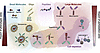
Representative overview of existing and novel therapeutic modalities.Figure 2

Representative overview of existing and novel therapeutic modalities. Therapeutic modalities are organized from small (left) to large (right) molecules. Untargeted therapeutics are located on the top side of the figure while the bottom part represents cell-specific modalities. Each light color shading represents a specific modality: small molecules (blue), oligos (yellow), peptides (pink), large molecules (red), and cell depletion strategies (purple). Watercolor overlap highlights therapeutics combining different modalities, such as antibody drug-conjugate (ADC) or antibody oligo-conjugate (AOC). Nanoparticles deliver both small molecules and oligos can be further directed to relevant cell types when combined with a targeting arm (green). The left and right gradients show the relative impact (blue low, red high) of phenotypic omics and perturbation screen on the discovery of novel therapeutic targets (top) versus cell-targeting strategies (bottom).

Creative targeting of master mechanisms. Central pathways in fibrosis, such as TGF-β, YAP/TAZ, and NOTCH, also participate in homeostasis, normal wound healing, and systemic processes, thereby limiting therapeutic index (186, 187). This limitation may be overcome using tissue- or cell-specific delivery systems. For instance, reformulation of nintedanib into an aerosol is being explored to enhance lung exposure and limit off-target effects (188, 189). A secondary approach is to engineer inactive compounds that require proteolytic activation by proteases expressed specifically in disease areas, known as protease “unmasking” (190). Unmasking is not new; however, significant advances in chemistry have increased its potential beyond oncology to fibrosis because of the similar proteolytic nature of the fibrotic niche. Cell-targeted strategies (Figure 2) are expanding to selectively deliver therapeutic payloads (e.g., small molecules, oligonucleotides: siRNA, mRNA) with nanoparticles or antibodies. For nanoparticles, cell specificity can be achieved by manipulating sugar and lipid content or adding targeted antibodies against a cell type of interest (191). Antibody drug- or oligo-conjugates (ADCs and AOCs, respectively) are gaining traction as viable modalities to treat chronic diseases but require proper identification of cell surface targets that are specific and have the right molecular properties (192, 193). Despite these challenges, several interesting approaches have demonstrated efficacy in preclinical models. For example, a LOXi-ADC targeted to profibrotic macrophages, an anti–SPP1-AOC targeted to TREM2+ scar-associated macrophages, and an LNP siRNA anti–TGF-β receptor 1 targeted to hepatic stellate cells had preclinical efficacy in models of muscle, heart, and liver fibrosis, respectively (194–196). Multiomics datasets may provide additional means to identify robust cell-targeting arms. As RNA levels may not always correlate with protein expression, combining single-cell transcriptomics with proteomics is essential to validate cell surface protein expression (e.g., CITE-seq; ref. 142) and subcellular protein localization (197, 198) to yield selectivity.

Depletion strategies of pathogenic players. A promising concept is to “reset” the fibrotic niche by depleting established pathogenic cells, such as B cells, myofibroblasts, or profibrotic macrophages (199). Depletion strategies include antibody-dependent cell-mediated cytotoxicity, ADCs with cytotoxic payloads (typically microtubule or DNA topoisomerase inhibitors), T cell engagers (TCEs), or chimeric antigen receptors (CARs), which can be expressed in a variety of immune cells with clearance function (e.g., T, NK, and myeloid cells) (200, 201). While many of these strategies focus on treating tumors, the translation of B cell–depleting therapies such as B cell maturation antigen and CD19 TCEs and/or CAR T from oncology to inflammatory diseases is opening the door for such approaches in fibrotic diseases (202, 203). B cell depletion (e.g., rituximab) was as efficacious as the standard of care (i.e., cyclophosphamide) in diffuse systemic sclerosis, including in lung endpoints, to improve symptoms and lung function (204, 205). Preclinically, myofibroblasts’ depletion using ADCs or CAR T cells successfully achieved antifibrotic activity in hepatic and cardiac fibrosis, respectively (206, 207). Furthermore, there is growing evidence of epigenetic imprinting of injury in multiple cell types, including mesenchymal and myeloid cells. Cell depletion will remove the associated pathogenic functions and reduce epigenetic memory that contributes to accelerated progression following rechallenge (102, 208). Repopulation of cells educated in the homeostatic niche may support long-term remission.
Conclusion

After a long pause following approval of the first two antifibrotic drugs, new waves of drugs covering a range of novel mechanisms are close to or exceeding clinical benefit expectations across a number of fibrotic diseases. This includes the first approvals for MASH, in which important therapeutic endpoints were finally achieved in some patients, leading to improvements in patients’ lives (14, 15), and the recent approval of nerandomilast (153) for IPF. The omics revolution is providing a deeper understanding of key mechanisms at the cellular and molecular level and revealing mechanistic hallmarks of fibrosis that span multiple tissues and fibrotic diseases. Omics readouts help clarify tolerability concerns with antifibrotic drugs by revealing essential overlapping homeostatic functions of individual targets. Omics data also reveal cell-selective targeting options and additional insights that drive new modality selection to improve the treatment of fibrotic diseases. Whether the enhanced biologic sensitivity afforded by omics and AI/ML can improve early detection of fibrogenesis, increase ability to detect therapeutic efficacy in trials, or open new windows into early intervention remains to be seen. Thus, the call to arms for omics technology applications will be to go beyond high-resolution phenotypic understanding of disease. Functional, kinetic, and dynamic insights on pathobiology will enable novel therapeutics and combinations to achieve rapid and robust resolution of fibrosis, amelioration of disease, and regeneration of homeostatic organ architecture and function.

Conflict of interest

CL, KLD, KD, TAW, KMH, and TF are employees of Pfizer Inc.

Acknowledgments

The studies referenced in this Review represent only a subset of the extensive literature on fibrosis. Readers are encouraged to consult the cited review articles and their referenced works for a more comprehensive understanding of additional and complementary aspects of the biology not covered here.

Address correspondence to: Thomas Fabre, Pfizer Inc., 1 Portland Street, Cambridge, Massachusetts 02139, USA. Email: Thomas.Fabre@pfizer.com.

Footnotes

Copyright: © 2026, Lebeaupin et al. This is an open access article published under the terms of the Creative Commons Attribution 4.0 International License.

Reference information: JCI Insight. 2026;11(13):e200155. https://doi.org/10.1172/jci.insight.200155.

References
  1. Angulo P, et al. Liver fibrosis, but no other histologic features, is associated with long-term outcomes of patients with nonalcoholic fatty liver disease. Gastroenterology. 2015;149(2):389–397.
    View this article via: CrossRef PubMed Google Scholar
  2. Ng CH, et al. Mortality outcomes by fibrosis stage in nonalcoholic fatty liver disease: a systematic review and meta-analysis. Clin Gastroenterol Hepatol. 2023;21(4):931–939.
    View this article via: CrossRef PubMed Google Scholar
  3. Poynard T, et al. Natural history of liver fibrosis progression in patients with chronic hepatitis C. The OBSVIRC, METAVIR, CLINIVIR, and DOSVIRC groups. Lancet. 1997;349(9055):825–832.
    View this article via: CrossRef PubMed Google Scholar
  4. Angulo P, et al. Independent predictors of liver fibrosis in patients with nonalcoholic steatohepatitis. Hepatology. 1999;30(6):1356–1362.
    View this article via: CrossRef PubMed Google Scholar
  5. Mortimer KM, et al. Characterizing health outcomes in idiopathic pulmonary fibrosis using US health claims data. Respiration. 2020;99(2):108–118.
    View this article via: CrossRef PubMed Google Scholar
  6. Sato Y, et al. Heterogeneous fibroblasts underlie age-dependent tertiary lymphoid tissues in the kidney. JCI Insight. 2016;1(11):e87680.
    View this article via: JCI Insight CrossRef PubMed Google Scholar
  7. Burkauskiene A, et al. Age-related changes in myocardial nerve and collagen networks of the auricle of the right atrium. Acta Cardiol. 2006;61(5):513–518.
    View this article via: CrossRef PubMed Google Scholar
  8. Lingampally A, et al. Unraveling alveolar fibroblast and activated myofibroblast heterogeneity and differentiation trajectories during lung fibrosis development and resolution in young and old mice. Aging Cell. 2025;24(5):e14503.
    View this article via: CrossRef PubMed Google Scholar
  9. Sanyal AJ, et al. Cirrhosis regression is associated with improved clinical outcomes in patients with nonalcoholic steatohepatitis. Hepatology. 2022;75(5):1235–1246.
    View this article via: CrossRef PubMed Google Scholar
  10. Sanyal AJ, et al. Prospective study of outcomes in adults with nonalcoholic fatty liver disease. N Engl J Med. 2021;385(17):1559–1569.
    View this article via: CrossRef PubMed Google Scholar
  11. Rockey DC, Friedman SL. Fibrosis regression after eradication of hepatitis C virus: from bench to bedside. Gastroenterology. 2021;160(5):1502–1520.
    View this article via: CrossRef PubMed Google Scholar
  12. Pais R, et al. Persistence of severe liver fibrosis despite substantial weight loss with bariatric surgery. Hepatology. 2022;76(2):456–468.
    View this article via: CrossRef PubMed Google Scholar
  13. Targher G, et al. Metabolic dysfunction-associated steatotic liver disease. N Engl J Med. 2025;393(7):683–698.
    View this article via: CrossRef PubMed Google Scholar
  14. Harrison SA, et al. A phase 3, randomized, controlled trial of resmetirom in NASH with liver fibrosis. N Engl J Med. 2024;390(6):497–509.
    View this article via: CrossRef PubMed Google Scholar
  15. Sanyal AJ, et al. Phase 3 trial of semaglutide in metabolic dysfunction-associated steatohepatitis. N Engl J Med. 2025;392(21):2089–2099.
    View this article via: CrossRef PubMed Google Scholar
  16. Younossi ZM, et al. Obeticholic acid for the treatment of non-alcoholic steatohepatitis: interim analysis from a multicentre, randomised, placebo-controlled phase 3 trial. Lancet. 2019;394(10215):2184–2196.
    View this article via: CrossRef PubMed Google Scholar
  17. Loomba R, et al. Randomized, controlled trial of the FGF21 analogue pegozafermin in NASH. N Engl J Med. 2023;389(11):998–1008.
    View this article via: CrossRef PubMed Google Scholar
  18. Harrison SA, et al. Safety and efficacy of once-weekly efruxifermin versus placebo in non-alcoholic steatohepatitis (HARMONY): a multicentre, randomised, double-blind, placebo-controlled, phase 2b trial. Lancet Gastroenterol Hepatol. 2023;8(12):1080–1093.
    View this article via: CrossRef PubMed Google Scholar
  19. Noureddin M, et al. Efruxifermin in compensated liver cirrhosis caused by MASH. N Engl J Med. 2025;392(24):2413–2424.
    View this article via: CrossRef PubMed Google Scholar
  20. Elsharkawy A, et al. Fibrosis regression following hepatitis C antiviral therapy. World J Hepatol. 2022;14(6):1120–1130.
    View this article via: CrossRef PubMed Google Scholar
  21. Yang C, et al. Surveillance and management of hepatocellular carcinoma after treatment of hepatitis C with direct-acting antiviral drugs. Ann Hepatol. 2024;30(2):101582.
    View this article via: CrossRef PubMed Google Scholar
  22. Carrieri P, et al. Severe liver fibrosis in the HCV cure era: major effects of social vulnerability, diabetes, and unhealthy behaviors. JHEP Rep. 2022;4(6):100481.
    View this article via: CrossRef PubMed Google Scholar
  23. Ponzetto A, Figura N. Risk of Hepatocellular carcinoma after cure of hepatitis C virus. Gastroenterology. 2018;154(5):1549.
    View this article via: CrossRef PubMed Google Scholar
  24. Hamdane N, et al. HCV-induced epigenetic changes associated with liver cancer risk persist after sustained virologic response. Gastroenterology. 2019;156(8):2313–2329.
    View this article via: CrossRef PubMed Google Scholar
  25. Rieder F, et al. Fibrosis: cross-organ biology and pathways to development of innovative drugs. Nat Rev Drug Discov. 2025;24(7):543–569.
    View this article via: CrossRef PubMed Google Scholar
  26. Barron AMS, et al. Distinct fibroblast functions associated with fibrotic and immune-mediated inflammatory diseases and their implications for therapeutic development. F1000Res. 2024;13:54.
    View this article via: CrossRef PubMed Google Scholar
  27. Kisseleva T, Brenner D. Molecular and cellular mechanisms of liver fibrosis and its regression. Nat Rev Gastroenterol Hepatol. 2021;18(3):151–166.
    View this article via: CrossRef PubMed Google Scholar
  28. Bhattacharya M, Ramachandran P. Immunology of human fibrosis. Nat Immunol. 2023;24(9):1423–1433.
    View this article via: CrossRef PubMed Google Scholar
  29. Schwantes-An TH, et al. Genome-wide association study and meta-analysis on alcohol-associated liver cirrhosis identifies genetic risk factors. Hepatology. 2021;73(5):1920–1931.
    View this article via: CrossRef PubMed Google Scholar
  30. Anstee QM, et al. Genome-wide association study of non-alcoholic fatty liver and steatohepatitis in a histologically characterised cohort٭. J Hepatol. 2020;73(3):505–515.
    View this article via: CrossRef PubMed Google Scholar
  31. Tsakiri KD, et al. Adult-onset pulmonary fibrosis caused by mutations in telomerase. Proc Natl Acad Sci U S A. 2007;104(18):7552–7557.
    View this article via: CrossRef PubMed Google Scholar
  32. Seibold MA, et al. A common MUC5B promoter polymorphism and pulmonary fibrosis. N Engl J Med. 2011;364(16):1503–1512.
    View this article via: CrossRef PubMed Google Scholar
  33. Nauffal V, et al. Genetics of myocardial interstitial fibrosis in the human heart and association with disease. Nat Genet. 2023;55(5):777–786.
    View this article via: CrossRef PubMed Google Scholar
  34. Kennedy MS, Chang EB. Emerging concepts and shifting paradigms for understanding the microbial basis of inflammatory bowel diseases. J Clin Invest. 2025;135(17):e193969.
    View this article via: JCI CrossRef PubMed Google Scholar
  35. Chioma OS, et al. Gut microbiota modulates lung fibrosis severity following acute lung injury in mice. Commun Biol. 2022;5(1):1401.
    View this article via: CrossRef PubMed Google Scholar
  36. Kropski JA, Blackwell TS. Endoplasmic reticulum stress in the pathogenesis of fibrotic disease. J Clin Invest. 2018;128(1):64–73.
    View this article via: JCI CrossRef PubMed Google Scholar
  37. Grootjans J, et al. The unfolded protein response in immunity and inflammation. Nat Rev Immunol. 2016;16(8):469–484.
    View this article via: CrossRef PubMed Google Scholar
  38. Mansouri A, et al. Mitochondrial dysfunction and signaling in chronic liver diseases. Gastroenterology. 2018;155(3):629–647.
    View this article via: CrossRef PubMed Google Scholar
  39. Billingham LK, et al. Mitochondrial electron transport chain is necessary for NLRP3 inflammasome activation. Nat Immunol. 2022;23(5):692–704.
    View this article via: CrossRef PubMed Google Scholar
  40. Lebeaupin C, et al. ER stress induces NLRP3 inflammasome activation and hepatocyte death. Cell Death Dis. 2015;6(9):e1879.
    View this article via: CrossRef PubMed Google Scholar
  41. Faas M, et al. IL-33-induced metabolic reprogramming controls the differentiation of alternatively activated macrophages and the resolution of inflammation. Immunity. 2021;54(11):2531–2546.
    View this article via: CrossRef PubMed Google Scholar
  42. Tian J, et al. Toll-like receptor 9-dependent activation by DNA-containing immune complexes is mediated by HMGB1 and RAGE. Nat Immunol. 2007;8(5):487–496.
    View this article via: CrossRef PubMed Google Scholar
  43. Deng M, et al. The endotoxin delivery protein HMGB1 mediates caspase-11-dependent lethality in sepsis. Immunity. 2018;49(4):740–753.
    View this article via: CrossRef PubMed Google Scholar
  44. Galluzzi L, et al. Molecular mechanisms of cell death: recommendations of the nomenclature committee on cell death 2018. Cell Death Differ. 2018;25(3):486–541.
    View this article via: CrossRef PubMed Google Scholar
  45. Blander JM. Death in the intestinal epithelium-basic biology and implications for inflammatory bowel disease. FEBS J. 2016;283(14):2720–2730.
    View this article via: CrossRef PubMed Google Scholar
  46. Frenette CT, et al. Emricasan improves liver function in patients with cirrhosis and high model for end-stage liver disease scores compared with placebo. Clin Gastroenterol Hepatol. 2019;17(4):774–783.
    View this article via: CrossRef PubMed Google Scholar
  47. Shi Y, et al. RIPK3 blockade attenuates tubulointerstitial fibrosis in a mouse model of diabetic nephropathy. Sci Rep. 2020;10(1):10458.
    View this article via: CrossRef PubMed Google Scholar
  48. Song M, et al. Inhibition of gasdermin D-dependent pyroptosis attenuates the progression of silica-induced pulmonary inflammation and fibrosis. Acta Pharm Sin B. 2022;12(3):1213–1224.
    View this article via: CrossRef PubMed Google Scholar
  49. Morabito A, et al. Algorithms and tools for data-driven omics integration to achieve multilayer biological insights: a narrative review. J Transl Med. 2025;23(1):425.
    View this article via: CrossRef PubMed Google Scholar
  50. Gros A, et al. Platelets in inflammation: regulation of leukocyte activities and vascular repair. Front Immunol. 2014;5:678.
    View this article via: PubMed CrossRef Google Scholar
  51. Malehmir M, et al. Platelet GPIbα is a mediator and potential interventional target for NASH and subsequent liver cancer. Nat Med. 2019;25(4):641–655.
    View this article via: CrossRef PubMed Google Scholar
  52. Kommoss KS, et al. Single cell mapping identifies a distinct platelet-phenotype in psoriatic type III inflammation. Nat Commun. 2025;16(1):10881.
    View this article via: CrossRef PubMed Google Scholar
  53. Hart KM, et al. Type-2 immunity is protective in metabolic disease but exacerbates NAFLD collaboratively with TGF-β. Sci Transl Med. 2017;9(396):eaal3694.
    View this article via: CrossRef PubMed Google Scholar
  54. Gieseck RL, et al. Interleukin-13 activates distinct cellular pathways leading to ductular reaction, steatosis, and fibrosis. Immunity. 2016;45(1):145–158.
    View this article via: CrossRef PubMed Google Scholar
  55. Fabre T, et al. Type 3 cytokines IL-17A and IL-22 drive TGF-β-dependent liver fibrosis. Sci Immunol. 2018;3(28):eaar7754.
    View this article via: CrossRef PubMed Google Scholar
  56. Meng F, et al. Interleukin-17 signaling in inflammatory, Kupffer cells, and hepatic stellate cells exacerbates liver fibrosis in mice. Gastroenterology. 2012;143(3):765–776.
    View this article via: CrossRef PubMed Google Scholar
  57. Wynn TA. Fibrotic disease and the T(H)1/T(H)2 paradigm. Nat Rev Immunol. 2004;4(8):583–594.
    View this article via: CrossRef PubMed Google Scholar
  58. Singh B, et al. Repetitive intradermal bleomycin injections evoke T-helper cell 2 cytokine-driven pulmonary fibrosis. Am J Physiol Lung Cell Mol Physiol. 2017;313(5):L796–L806.
    View this article via: CrossRef PubMed Google Scholar
  59. Fabre T, et al. IL-17A enhances the expression of profibrotic genes through upregulation of the TGF-β receptor on hepatic stellate cells in a JNK-dependent manner. J Immunol. 2014;193(8):3925–3933.
    View this article via: CrossRef PubMed Google Scholar
  60. Wilson MS, et al. Bleomycin and IL-1beta-mediated pulmonary fibrosis is IL-17A dependent. J Exp Med. 2010;207(3):535–552.
    View this article via: CrossRef PubMed Google Scholar
  61. Wu D, et al. Common signatures of neutrophils in diverse disease conditions. Cell Discov. 2025;11(1):66.
    View this article via: CrossRef PubMed Google Scholar
  62. Li Y, et al. Current perspectives and trends of neutrophil extracellular traps in organ fibrosis: a bibliometric and visualization study. Front Immunol. 2025;16:1508909.
    View this article via: CrossRef PubMed Google Scholar
  63. Xia Y, et al. Neutrophil extracellular traps promote MASH fibrosis by metabolic reprogramming of HSC. Hepatology. 2025;81(3):947–961.
    View this article via: CrossRef PubMed Google Scholar
  64. Monteleone G, et al. A simple ratio in a complex disease: exploring the neutrophil-to-lymphocyte ratio in idiopathic pulmonary fibrosis. J Clin Med. 2025;14(14):5100.
    View this article via: CrossRef PubMed Google Scholar
  65. Hammerich L, Tacke F. Hepatic inflammatory responses in liver fibrosis. Nat Rev Gastroenterol Hepatol. 2023;20(10):633–646.
    View this article via: CrossRef PubMed Google Scholar
  66. Dudek M, et al. Auto-aggressive CXCR6+ CD8 T cells cause liver immune pathology in NASH. Nature. 2021;592(7854):444–449.
    View this article via: CrossRef PubMed Google Scholar
  67. Volkov M, et al. Autoantibodies and B cells: the ABC of rheumatoid arthritis pathophysiology. Immunol Rev. 2020;294(1):148–163.
    View this article via: CrossRef PubMed Google Scholar
  68. Park O, et al. Diverse roles of invariant natural killer T cells in liver injury and fibrosis induced by carbon tetrachloride. Hepatology. 2009;49(5):1683–1694.
    View this article via: CrossRef PubMed Google Scholar
  69. Birjandi SZ, et al. CD4(+)CD25(hi)Foxp3(+) cells exacerbate bleomycin-induced pulmonary fibrosis. Am J Pathol. 2016;186(8):2008–2020.
    View this article via: CrossRef PubMed Google Scholar
  70. Claassen MA, et al. Abundant numbers of regulatory T cells localize to the liver of chronic hepatitis C infected patients and limit the extent of fibrosis. J Hepatol. 2010;52(3):315–321.
    View this article via: CrossRef PubMed Google Scholar
  71. Wynn TA, et al. Immunopathogenesis of schistosomiasis. Immunol Rev. 2004;201:156–167.
    View this article via: CrossRef PubMed Google Scholar
  72. Noster R, et al. IL-17 and GM-CSF expression are antagonistically regulated by human T helper cells. Sci Transl Med. 2014;6(241):241ra80.
    View this article via: CrossRef PubMed Google Scholar
  73. Ramalingam TR, et al. Enhanced protection from fibrosis and inflammation in the combined absence of IL-13 and IFN-γ. J Pathol. 2016;239(3):344–354.
    View this article via: CrossRef PubMed Google Scholar
  74. Zou AE, et al. Fibroblasts in immune responses, inflammatory diseases and therapeutic implications. Nat Rev Rheumatol. 2025;21(6):336–354.
    View this article via: CrossRef PubMed Google Scholar
  75. Kim KK, et al. TGF-β1 signaling and tissue fibrosis. Cold Spring Harb Perspect Biol. 2018;10(4):a022293.
    View this article via: CrossRef PubMed Google Scholar
  76. Ng B, et al. Interleukin-11 is a therapeutic target in idiopathic pulmonary fibrosis. Sci Transl Med. 2019;11(511):eaaw1237.
    View this article via: CrossRef PubMed Google Scholar
  77. Yao L, et al. Temporal control of PDGFRα regulates the fibroblast-to-myofibroblast transition in wound healing. Cell Rep. 2022;40(7):111192.
    View this article via: CrossRef PubMed Google Scholar
  78. Scolaro B, et al. Caloric restriction promotes resolution of atherosclerosis in obese mice, while weight regain accelerates its progression. J Clin Invest. 2025;135(18):e172198.
    View this article via: JCI CrossRef PubMed Google Scholar
  79. Doran AC, et al. Efferocytosis in health and disease. Nat Rev Immunol. 2020;20(4):254–267.
    View this article via: CrossRef PubMed Google Scholar
  80. Liebold I, et al. Apoptotic cell identity induces distinct functional responses to IL-4 in efferocytic macrophages. Science. 2024;384(6691):eabo7027.
    View this article via: CrossRef PubMed Google Scholar
  81. Wang Y, et al. Stiffness sensing via Piezo1 enhances macrophage efferocytosis and promotes the resolution of liver fibrosis. Sci Adv. 2024;10(23):eadj3289.
    View this article via: CrossRef PubMed Google Scholar
  82. Chen S, et al. Myeloid MAS-driven macrophage efferocytosis promotes resolution in ischemia-stressed mouse and human livers. Sci Transl Med. 2025;17(806):eadr2725.
    View this article via: CrossRef PubMed Google Scholar
  83. Cai B, et al. Macrophage MerTK promotes liver fibrosis in nonalcoholic steatohepatitis. Cell Metab. 2020;31(2):406–421.
    View this article via: CrossRef PubMed Google Scholar
  84. Hadzipasic M, et al. ROCK-dependent mechanotransduction of macroscale forces drives fibrosis in degenerative spinal disease. Nat Biomed Eng. 2025;9(10):1677–1690.
    View this article via: CrossRef PubMed Google Scholar
  85. Robert S, et al. Characterization of the MMP/TIMP imbalance and collagen production induced by IL-1β or TNF-α release from human hepatic stellate cells. PLoS One. 2016;11(4):e0153118.
    View this article via: CrossRef PubMed Google Scholar
  86. Todd JL, et al. Circulating matrix metalloproteinases and tissue metalloproteinase inhibitors in patients with idiopathic pulmonary fibrosis in the multicenter IPF-PRO registry cohort. BMC Pulm Med. 2020;20(1):64.
    View this article via: CrossRef PubMed Google Scholar
  87. Hemmann S, et al. Expression of MMPs and TIMPs in liver fibrosis - a systematic review with special emphasis on anti-fibrotic strategies. J Hepatol. 2007;46(5):955–975.
    View this article via: CrossRef PubMed Google Scholar
  88. He X, et al. Myofibroblast YAP/TAZ activation is a key step in organ fibrogenesis. JCI Insight. 2022;7(4):e146243.
    View this article via: JCI Insight CrossRef PubMed Google Scholar
  89. Wagner DE, et al. Inhibition of epithelial cell YAP-TEAD/LOX signaling attenuates pulmonary fibrosis in preclinical models. Nat Commun. 2025;16(1):7099.
    View this article via: CrossRef PubMed Google Scholar
  90. Segel M, et al. Niche stiffness underlies the ageing of central nervous system progenitor cells. Nature. 2019;573(7772):130–134.
    View this article via: CrossRef PubMed Google Scholar
  91. Xu L, et al. PIEZO1 mediates periostin+ myofibroblast activation and pulmonary fibrosis in mice. J Clin Invest. 2025;135(11):e184158.
    View this article via: JCI CrossRef PubMed Google Scholar
  92. Henderson NC, et al. Targeting of αv integrin identifies a core molecular pathway that regulates fibrosis in several organs. Nat Med. 2013;19(12):1617–1624.
    View this article via: CrossRef PubMed Google Scholar
  93. Kretschmer M, et al. Matrix stiffness regulates Notch signaling activity in endothelial cells. J Cell Sci. 2023;136(2):jcs260442.
    View this article via: CrossRef PubMed Google Scholar
  94. Gori JL, et al. Vascular niche promotes hematopoietic multipotent progenitor formation from pluripotent stem cells. J Clin Invest. 2015;125(3):1243–1254.
    View this article via: JCI CrossRef PubMed Google Scholar
  95. Griffin MF, et al. Growth arrest specific-6 and angiotoxin receptor-like signaling drive oral regenerative wound repair. Sci Transl Med. 2025;17(805):eadk2101.
    View this article via: CrossRef PubMed Google Scholar
  96. Mascharak S, et al. Preventing Engrailed-1 activation in fibroblasts yields wound regeneration without scarring. Science. 2021;372(6540):eaba2374.
    View this article via: CrossRef PubMed Google Scholar
  97. Romani P, et al. Crosstalk between mechanotransduction and metabolism. Nat Rev Mol Cell Biol. 2021;22(1):22–38.
    View this article via: CrossRef PubMed Google Scholar
  98. Wynn TA, Vannella KM. Macrophages in tissue repair, regeneration, and fibrosis. Immunity. 2016;44(3):450–462.
    View this article via: CrossRef PubMed Google Scholar
  99. Guilliams M, Scott CL. Does niche competition determine the origin of tissue-resident macrophages? Nat Rev Immunol. 2017;17(7):451–460.
    View this article via: CrossRef PubMed Google Scholar
  100. Ginhoux F, Guilliams M. Tissue-resident macrophage ontogeny and homeostasis. Immunity. 2016;44(3):439–449.
    View this article via: CrossRef PubMed Google Scholar
  101. Sakai M, et al. Liver-derived signals sequentially reprogram myeloid enhancers to initiate and maintain kupffer cell identity. Immunity. 2019;51(4):655–670.
    View this article via: CrossRef PubMed Google Scholar
  102. Huang H, et al. Kupffer cell programming by maternal obesity triggers fatty liver disease. Nature. 2025;644(8077):790–798.
    View this article via: CrossRef PubMed Google Scholar
  103. van de Laar L, et al. Yolk Sac macrophages, fetal liver, and adult monocytes can colonize an empty niche and develop into functional tissue-resident macrophages. Immunity. 2016;44(4):755–768.
    View this article via: CrossRef PubMed Google Scholar
  104. Mass E, et al. Tissue-specific macrophages: how they develop and choreograph tissue biology. Nat Rev Immunol. 2023;23(9):563–579.
    View this article via: CrossRef PubMed Google Scholar
  105. Fima R, et al. Loss of embryonically-derived Kupffer cells during hypercholesterolemia accelerates atherosclerosis development. Nat Commun. 2024;15(1):8341.
    View this article via: CrossRef PubMed Google Scholar
  106. Peiseler M, et al. Kupffer cell-like syncytia replenish resident macrophage function in the fibrotic liver. Science. 2023;381(6662):eabq5202.
    View this article via: CrossRef PubMed Google Scholar
  107. Jiang Y, et al. Macrophages in organ fibrosis: from pathogenesis to therapeutic targets. Cell Death Discov. 2024;10(1):487.
    View this article via: CrossRef PubMed Google Scholar
  108. Parthasarathy G, et al. Macrophage RAGE activation is proinflammatory in NASH. JCI Insight. 2024;9(3):e169138.
    View this article via: JCI Insight CrossRef PubMed Google Scholar
  109. Fabre T, et al. Identification of a broadly fibrogenic macrophage subset induced by type 3 inflammation. Sci Immunol. 2023;8(82):eadd8945.
    View this article via: CrossRef PubMed Google Scholar
  110. Guilliams M, et al. Spatial proteogenomics reveals distinct and evolutionarily conserved hepatic macrophage niches. Cell. 2022;185(2):379–396.
    View this article via: CrossRef PubMed Google Scholar
  111. Ramachandran P, et al. Resolving the fibrotic niche of human liver cirrhosis at single-cell level. Nature. 2019;575(7783):512–518.
    View this article via: CrossRef PubMed Google Scholar
  112. Hoeft K, et al. Platelet-instructed SPP1+ macrophages drive myofibroblast activation in fibrosis in a CXCL4-dependent manner. Cell Rep. 2023;42(2):112131.
    View this article via: CrossRef PubMed Google Scholar
  113. Guo W, et al. Notch signaling regulates macrophage-mediated inflammation in metabolic dysfunction-associated steatotic liver disease. Immunity. 2024;57(10):2310–2327.
    View this article via: CrossRef PubMed Google Scholar
  114. Murray PJ, et al. Macrophage activation and polarization: nomenclature and experimental guidelines. Immunity. 2014;41(1):14–20.
    View this article via: CrossRef PubMed Google Scholar
  115. Bleriot C, et al. Determinants of resident tissue macrophage identity and function. Immunity. 2020;52(6):957–970.
    View this article via: CrossRef PubMed Google Scholar
  116. Polonsky M, et al. Spatial transcriptomics defines injury specific microenvironments and cellular interactions in kidney regeneration and disease. Nat Commun. 2024;15(1):7010.
    View this article via: CrossRef PubMed Google Scholar
  117. Cavagnero KJ, et al. CXCL12+ dermal fibroblasts promote neutrophil recruitment and host defense by recognition of IL-17. J Exp Med. 2024;221(4):e20231425.
    View this article via: CrossRef PubMed Google Scholar
  118. Gao Y, et al. Cross-tissue human fibroblast atlas reveals myofibroblast subtypes with distinct roles in immune modulation. Cancer Cell. 2024;42(10):1764–1783.
    View this article via: CrossRef PubMed Google Scholar
  119. Yu J, et al. Hepatocyte TLR4 triggers inter-hepatocyte Jagged1/Notch signaling to determine NASH-induced fibrosis. Sci Transl Med. 2021;13(599):eabe1692.
    View this article via: CrossRef PubMed Google Scholar
  120. Grande MT, et al. Snail1-induced partial epithelial-to-mesenchymal transition drives renal fibrosis in mice and can be targeted to reverse established disease. Nat Med. 2015;21(9):989–997.
    View this article via: CrossRef PubMed Google Scholar
  121. Stratton MS, et al. Dynamic chromatin targeting of BRD4 stimulates cardiac fibroblast activation. Circ Res. 2019;125(7):662–677.
    View this article via: CrossRef PubMed Google Scholar
  122. Alexanian M, et al. A transcriptional switch governs fibroblast activation in heart disease. Nature. 2021;595(7867):438–443.
    View this article via: CrossRef PubMed Google Scholar
  123. Talbott HE, et al. Wound healing, fibroblast heterogeneity, and fibrosis. Cell Stem Cell. 2022;29(8):1161–1180.
    View this article via: CrossRef PubMed Google Scholar
  124. Heumos L, et al. Best practices for single-cell analysis across modalities. Nat Rev Genet. 2023;24(8):550–572.
    View this article via: CrossRef PubMed Google Scholar
  125. Kim N, et al. Perspectives on single-nucleus RNA sequencing in different cell types and tissues. J Pathol Transl Med. 2023;57(1):52–59.
    View this article via: CrossRef PubMed Google Scholar
  126. Brazovskaja A, et al. Cell atlas of the regenerating human liver after portal vein embolization. Nat Commun. 2024;15(1):5827.
    View this article via: CrossRef PubMed Google Scholar
  127. Wu H, et al. Advantages of single-nucleus over single-cell RNA sequencing of adult kidney: rare cell types and novel cell states revealed in fibrosis. J Am Soc Nephrol. 2019;30(1):23–32.
    View this article via: CrossRef PubMed Google Scholar
  128. Slyper M, et al. A single-cell and single-nucleus RNA-seq toolbox for fresh and frozen human tumors. Nat Med. 2020;26(5):792–802.
    View this article via: CrossRef PubMed Google Scholar
  129. Richter ML, et al. Single-nucleus RNA-seq2 reveals functional crosstalk between liver zonation and ploidy. Nat Commun. 2021;12(1):4264.
    View this article via: CrossRef PubMed Google Scholar
  130. Tsukui T, et al. Alveolar fibroblast lineage orchestrates lung inflammation and fibrosis. Nature. 2024;631(8021):627–634.
    View this article via: CrossRef PubMed Google Scholar
  131. Adams TS, et al. Single-cell RNA-seq reveals ectopic and aberrant lung-resident cell populations in idiopathic pulmonary fibrosis. Sci Adv. 2020;6(28):eaba1983.
    View this article via: CrossRef PubMed Google Scholar
  132. Patrick R, et al. Integration mapping of cardiac fibroblast single-cell transcriptomes elucidates cellular principles of fibrosis in diverse pathologies. Sci Adv. 2024;10(25):eadk8501.
    View this article via: CrossRef PubMed Google Scholar
  133. Habermann AC, et al. Single-cell RNA sequencing reveals profibrotic roles of distinct epithelial and mesenchymal lineages in pulmonary fibrosis. Sci Adv. 2020;6(28):eaba1972.
    View this article via: CrossRef PubMed Google Scholar
  134. Hrovatin K, et al. Considerations for building and using integrated single-cell atlases. Nat Methods. 2025;22(1):41–57.
    View this article via: CrossRef PubMed Google Scholar
  135. Sikkema L, et al. An integrated cell atlas of the lung in health and disease. Nat Med. 2023;29(6):1563–1577.
    View this article via: CrossRef PubMed Google Scholar
  136. Dutertre CA, et al. Single-cell analysis of human mononuclear phagocytes reveals subset-defining markers and identifies circulating inflammatory dendritic cells. Immunity. 2019;51(3):573–589.
    View this article via: CrossRef PubMed Google Scholar
  137. Amrute JM, et al. Targeting immune-fibroblast cell communication in heart failure. Nature. 2024;635(8038):423–433.
    View this article via: CrossRef PubMed Google Scholar
  138. Qiu P. Embracing the dropouts in single-cell RNA-seq analysis. Nat Commun. 2020;11(1):1169.
    View this article via: CrossRef PubMed Google Scholar
  139. Wlodarczyk T, et al. Epiregulon: single-cell transcription factor activity inference to predict drug response and drivers of cell states. Nat Commun. 2025;16(1):7118.
    View this article via: CrossRef PubMed Google Scholar
  140. Southard KM, et al. Comprehensive transcription factor perturbations recapitulate fibroblast transcriptional states. Nat Genet. 2025;57(9):2323–2334.
    View this article via: CrossRef PubMed Google Scholar
  141. Browaeys R, et al. NicheNet: modeling intercellular communication by linking ligands to target genes. Nat Methods. 2020;17(2):159–162.
    View this article via: CrossRef PubMed Google Scholar
  142. Stoeckius M, et al. Simultaneous epitope and transcriptome measurement in single cells. Nat Methods. 2017;14(9):865–868.
    View this article via: CrossRef PubMed Google Scholar
  143. Vannan A, et al. Spatial transcriptomics identifies molecular niche dysregulation associated with distal lung remodeling in pulmonary fibrosis. Nat Genet. 2025;57(3):647–658.
    View this article via: CrossRef PubMed Google Scholar
  144. Chen Z, et al. Single cell multi-omics of fibrotic kidney reveal epigenetic regulation of antioxidation and apoptosis within proximal tubule. Cell Mol Life Sci. 2024;81(1):56.
    View this article via: CrossRef PubMed Google Scholar
  145. Matchett KP, et al. Multimodal decoding of human liver regeneration. Nature. 2024;630(8015):158–165.
    View this article via: CrossRef PubMed Google Scholar
  146. Espinosa-Martinez M, et al. The molecular basis of cell memory in mammals: the epigenetic cycle. Sci Adv. 2024;10(9):eadl3188.
    View this article via: CrossRef PubMed Google Scholar
  147. Aoyama K, et al. Epigenetic memories in hematopoietic stem and progenitor cells. Cells. 2022;11(14):2187.
    View this article via: CrossRef PubMed Google Scholar
  148. Dixit A, et al. Perturb-seq: dissecting molecular circuits with scalable single-cell RNA profiling of pooled genetic screens. Cell. 2016;167(7):1853–1866.
    View this article via: CrossRef PubMed Google Scholar
  149. Ye C, et al. DRUG-seq for miniaturized high-throughput transcriptome profiling in drug discovery. Nat Commun. 2018;9(1):4307.
    View this article via: CrossRef PubMed Google Scholar
  150. Dhainaut M, et al. Spatial CRISPR genomics identifies regulators of the tumor microenvironment. Cell. 2022;185(7):1223–1239.
    View this article via: CrossRef PubMed Google Scholar
  151. Sheban F, et al. ZEB2 is a master switch controlling the tumor-associated macrophage program. Cancer Cell. 2025;43(7):1227–1241.
    View this article via: CrossRef PubMed Google Scholar
  152. Oakley F, et al. Implementation of pre-clinical methodologies to study fibrosis and test anti-fibrotic therapy. Curr Opin Pharmacol. 2019;49:95–101.
    View this article via: CrossRef PubMed Google Scholar
  153. Richeldi L, et al. Nerandomilast in patients with idiopathic pulmonary fibrosis. N Engl J Med. 2025;392(22):2193–2202.
    View this article via: CrossRef PubMed Google Scholar
  154. Wollin L, et al. Antifibrotic and anti-inflammatory activity of the tyrosine kinase inhibitor nintedanib in experimental models of lung fibrosis. J Pharmacol Exp Ther. 2014;349(2):209–220.
    View this article via: CrossRef PubMed Google Scholar
  155. Fletcher SV, et al. Safety and tolerability of nintedanib for the treatment of idiopathic pulmonary fibrosis in routine UK clinical practice. ERJ Open Res. 2018;4(4):00049–2018.
    View this article via: CrossRef PubMed Google Scholar
  156. Sciurba JC, et al. Fibroblast-specific integrin-alpha V differentially regulates type 17 and type 2 driven inflammation and fibrosis. J Pathol. 2019;248(1):16–29.
    View this article via: CrossRef PubMed Google Scholar
  157. Sun T, et al. TGFβ2 and TGFβ3 isoforms drive fibrotic disease pathogenesis. Sci Transl Med. 2021;13(605):eabe0407.
    View this article via: CrossRef PubMed Google Scholar
  158. Decaris ML, et al. Dual inhibition of αvβ6 and αvβ1 reduces fibrogenesis in lung tissue explants from patients with IPF. Respir Res. 2021;22(1):265.
    View this article via: CrossRef PubMed Google Scholar
  159. Maher TM, et al. Phase 2 trial to assess lebrikizumab in patients with idiopathic pulmonary fibrosis. Eur Respir J. 2021;57(2):1902442.
    View this article via: CrossRef PubMed Google Scholar
  160. Parker JM, et al. A phase 2 randomized controlled study of tralokinumab in subjects with idiopathic pulmonary fibrosis. Am J Respir Crit Care Med. 2018;197(1):94–103.
    View this article via: CrossRef PubMed Google Scholar
  161. Richeldi L, et al. Pamrevlumab, an anti-connective tissue growth factor therapy, for idiopathic pulmonary fibrosis (PRAISE): a phase 2, randomised, double-blind, placebo-controlled trial. Lancet Respir Med. 2020;8(1):25–33.
    View this article via: CrossRef PubMed Google Scholar
  162. Mullard A. Dual-payload ADCs move into first oncology clinical trials. Nat Rev Drug Discov. 2025;24(8):573–576.
    View this article via: CrossRef PubMed Google Scholar
  163. Chan AC, et al. Fifty years of monoclonals: the past, present and future of antibody therapeutics. Nat Rev Immunol. 2025;25(10):745–765.
    View this article via: CrossRef PubMed Google Scholar
  164. Jarallah SJ, et al. Artificial intelligence revolution in drug discovery: a paradigm shift in pharmaceutical innovation. Int J Pharm. 2025;680:125789.
    View this article via: CrossRef PubMed Google Scholar
  165. Pun FW, et al. Identification of therapeutic targets for amyotrophic lateral sclerosis using PandaOmics - an AI-enabled biological target discovery platform. Front Aging Neurosci. 2022;14:914017.
    View this article via: CrossRef PubMed Google Scholar
  166. Ochoa D, et al. Open targets platform: supporting systematic drug-target identification and prioritisation. Nucleic Acids Res. 2021;49(d1):D1302–D1310.
    View this article via: CrossRef PubMed Google Scholar
  167. Ren F, et al. A small-molecule TNIK inhibitor targets fibrosis in preclinical and clinical models. Nat Biotechnol. 2025;43(1):63–75.
    View this article via: CrossRef PubMed Google Scholar
  168. Deshaies RJ, Potts PR. Load and lock: an emerging class of therapeutics that influence macromolecular dissociation. Science. 2025;389(6762):eadx3595.
    View this article via: CrossRef PubMed Google Scholar
  169. Xu Z, et al. A generative AI-discovered TNIK inhibitor for idiopathic pulmonary fibrosis: a randomized phase 2a trial. Nat Med. 2025;31(8):2602–2610.
    View this article via: CrossRef PubMed Google Scholar
  170. Zamora AC, et al. When the third time is not the charm-trial outcomes in idiopathic pulmonary fibrosis. JAMA. 2024;332(5):374–376.
    View this article via: CrossRef PubMed Google Scholar
  171. Raghu G, Fleming TR. Moving forward in IPF: lessons learned from clinical trials. Lancet Respir Med. 2024;12(8):583–585.
    View this article via: CrossRef PubMed Google Scholar
  172. Swanson K, et al. The virtual lab of AI agents designs new SARS-CoV-2 nanobodies. Nature. 2025;646(8085):716–723.
    View this article via: CrossRef PubMed Google Scholar
  173. Thomas T, et al. A longitudinal single-cell atlas of anti-tumour necrosis factor treatment in inflammatory bowel disease. Nat Immunol. 2024;25(11):2152–2165.
    View this article via: CrossRef PubMed Google Scholar
  174. Metzner E, et al. Multiome Perturb-seq unlocks scalable discovery of integrated perturbation effects on the transcriptome and epigenome. Cell Syst. 2025;16(1):101161.
    View this article via: CrossRef PubMed Google Scholar
  175. Replogle JM, et al. Combinatorial single-cell CRISPR screens by direct guide RNA capture and targeted sequencing. Nat Biotechnol. 2020;38(8):954–961.
    View this article via: CrossRef PubMed Google Scholar
  176. Yao D, et al. Scalable genetic screening for regulatory circuits using compressed Perturb-seq. Nat Biotechnol. 2024;42(8):1282–1295.
    View this article via: CrossRef PubMed Google Scholar
  177. Lang NJ, et al. Ex vivo tissue perturbations coupled to single-cell RNA-seq reveal multilineage cell circuit dynamics in human lung fibrogenesis. Sci Transl Med. 2023;15(725):eadh0908.
    View this article via: CrossRef PubMed Google Scholar
  178. Yang J, et al. A target map of clinical combination therapies in oncology: an analysis of clinicaltrials.gov. Discov Oncol. 2023;14(1):151.
    View this article via: CrossRef PubMed Google Scholar
  179. Felten R, et al. Novel therapeutic strategies for autoimmune and inflammatory rheumatic diseases. Drug Discov Today. 2023;28(7):103612.
    View this article via: CrossRef PubMed Google Scholar
  180. Bayat Mokhtari R, et al. Combination therapy in combating cancer. Oncotarget. 2017;8(23):38022–38043.
    View this article via: CrossRef PubMed Google Scholar
  181. Ratziu V, Charlton M. Rational combination therapy for NASH: insights from clinical trials and error. J Hepatol. 2023;78(5):1073–1079.
    View this article via: CrossRef PubMed Google Scholar
  182. Dufour JF, et al. Combination therapy for non-alcoholic steatohepatitis: rationale, opportunities and challenges. Gut. 2020;69(10):1877–1884.
    View this article via: CrossRef PubMed Google Scholar
  183. Anstee QM, et al. Tropifexor plus cenicriviroc combination versus monotherapy in nonalcoholic steatohepatitis: results from the phase 2b TANDEM study. Hepatology. 2023;78(4):1223–1239.
    View this article via: CrossRef PubMed Google Scholar
  184. Xiang L, et al. Safety and efficacy of GLP-1/FGF21 dual agonist HEC88473 in MASLD and T2DM: a randomized, double-blind, placebo-controlled study. J Hepatol. 2025;82(6):967–978.
    View this article via: CrossRef PubMed Google Scholar
  185. Deshaies RJ. How multispecific molecules are transforming pharmacotherapy. Nat Rev Drug Discov. 2025;24(12):945–957.
    View this article via: CrossRef PubMed Google Scholar
  186. Mitra MS, et al. A potent pan-TGFβ neutralizing monoclonal antibody elicits cardiovascular toxicity in mice and cynomolgus monkeys. Toxicol Sci. 2020;175(1):24–34.
    View this article via: CrossRef PubMed Google Scholar
  187. Kovacs RJ, et al. Cardiac safety of TGF-β receptor I kinase inhibitor LY2157299 monohydrate in cancer patients in a first-in-human dose study. Cardiovasc Toxicol. 2015;15(4):309–323.
    View this article via: CrossRef PubMed Google Scholar
  188. Aboud HM, et al. Optimization and appraisal of nintedanib-loaded mixed polymeric micelles as a potential nanovector for non-invasive pulmonary fibrosis mitigation. Pharmaceuticals (Basel). 2024;17(10):1275.
    View this article via: CrossRef PubMed Google Scholar
  189. Andrade da Silva LH, et al. Development of nintedanib nanosuspension for inhaled treatment of experimental silicosis. Bioeng Transl Med. 2023;8(2):e10401.
    View this article via: CrossRef PubMed Google Scholar
  190. Poreba M. Protease-activated prodrugs: strategies, challenges, and future directions. FEBS J. 2020;287(10):1936–1969.
    View this article via: CrossRef PubMed Google Scholar
  191. Han X, et al. Ligand-tethered lipid nanoparticles for targeted RNA delivery to treat liver fibrosis. Nat Commun. 2023;14(1):75.
    View this article via: CrossRef PubMed Google Scholar
  192. Fu Z, et al. Antibody drug conjugate: the “biological missile” for targeted cancer therapy. Signal Transduct Target Ther. 2022;7(1):93.
    View this article via: CrossRef PubMed Google Scholar
  193. Dugal-Tessier J, et al. Antibody-oligonucleotide conjugates: a twist to antibody-drug conjugates. J Clin Med. 2021;10(4):838.
    View this article via: CrossRef PubMed Google Scholar
  194. Chen YC, et al. Development of a lipid-encapsulated TGFβRI-siRNA drug for liver fibrosis induced by schistosoma mansoni. PLoS Negl Trop Dis. 2024;18(9):e0012502.
    View this article via: CrossRef PubMed Google Scholar
  195. Odeh A, et al. Anti-fibrotic, muscle-promoting antibody-drug conjugates for the improvement and treatment of DMD. iScience. 2025;28(5):112335.
    View this article via: CrossRef PubMed Google Scholar
  196. Momin N, et al. Therapeutic Spp1 silencing in TREM2+ cardiac macrophages suppresses atrial fibrillation [preprint]. https://doi.org/10.1101/2024.08.10.607461 Posted on bioRxiv August 10, 2024.
  197. Remmel A. Where do proteins go in cells? Next-generation methods map the molecules’ hidden lives. Nature. 2025;640(8058):556–560.
    View this article via: CrossRef PubMed Google Scholar
  198. Thul PJ, et al. A subcellular map of the human proteome. Science. 2017;356(6340):eaal3321.
    View this article via: CrossRef PubMed Google Scholar
  199. Adler M, et al. Principles of cell circuits for tissue repair and fibrosis. iScience. 2020;23(2):100841.
    View this article via: CrossRef PubMed Google Scholar
  200. Look T, et al. CAR T cells, CAR NK cells, and CAR macrophages exhibit distinct traits in glioma models but are similarly enhanced when combined with cytokines. Cell Rep Med. 2025;6(2):101931.
    View this article via: CrossRef PubMed Google Scholar
  201. Tsuchikama K, et al. Exploring the next generation of antibody-drug conjugates. Nat Rev Clin Oncol. 2024;21(3):203–223.
    View this article via: CrossRef PubMed Google Scholar
  202. Michaelson JS, Baeuerle PA. CD19-directed T cell-engaging antibodies for the treatment of autoimmune disease. J Exp Med. 2024;221(5):e20240499.
    View this article via: CrossRef PubMed Google Scholar
  203. Baeuerle PA, Wesche H. T-cell-engaging antibodies for the treatment of solid tumors: challenges and opportunities. Curr Opin Oncol. 2022;34(5):552–558.
    View this article via: CrossRef PubMed Google Scholar
  204. Maher TM, et al. Rituximab versus intravenous cyclophosphamide in patients with connective tissue disease-associated interstitial lung disease in the UK (RECITAL): a double-blind, double-dummy, randomised, controlled, phase 2b trial. Lancet Respir Med. 2023;11(1):45–54.
    View this article via: CrossRef PubMed Google Scholar
  205. Schett G, et al. CAR T-cell therapy in autoimmune diseases. Lancet. 2023;402(10416):2034–2044.
    View this article via: CrossRef PubMed Google Scholar
  206. Liu S, et al. Antibody-drug conjugates targeting CD248 inhibits liver fibrosis through specific killing on myofibroblasts. Mol Med. 2022;28(1):37.
    View this article via: CrossRef PubMed Google Scholar
  207. Rurik JG, et al. CAR T cells produced in vivo to treat cardiac injury. Science. 2022;375(6576):91–96.
    View this article via: CrossRef PubMed Google Scholar
  208. Kisseleva T, et al. Myofibroblasts revert to an inactive phenotype during regression of liver fibrosis. Proc Natl Acad Sci U S A. 2012;109(24):9448–9453.
    View this article via: CrossRef PubMed Google Scholar
Version history
  • Version 1 (July 8, 2026): Electronic publication

Article tools

  • View PDF
  • Download citation information
  • Send a comment
  • Terms of use
  • Standard abbreviations
  • Need help? Email the journal

Metrics

  • Article usage
  • Citations to this article

Go to

  • Top
  • Abstract
  • Introduction
  • Mechanisms of fibrosis
  • The evolving landscape of omics studies in fibrosis
  • Targeting profibrotic mechanisms: past, present, and future
  • Conclusion
  • Conflict of interest
  • Acknowledgments
  • Footnotes
  • References
  • Version history
Advertisement
Advertisement

Copyright © 2026 American Society for Clinical Investigation
ISSN 2379-3708

Sign up for email alerts