
Research ArticleCardiologyCell biology
Open Access |  10.1172/jci.insight.195805
Slick K+ channels contribute to cardiac remodeling, fibrosis, and dysfunction in postinfarction hearts
Jiaqi Yang,1 Lin Zhu,1 David Spähn,1 Melanie Cruz Santos,1 Sophia Schanz,1 Selina Maier,1 Lena Birkenfeld,1 Helmut Bischof,1 Anna Roslan,1 Nina Wettschureck,2 Oliver Borst,3 Lucas Matt,1 and Robert Lukowski1
1Department of Pharmacology, Toxicology and Clinical Pharmacy, Institute of Pharmacy, University of Tübingen, Tübingen, Germany.
2Max-Planck-Institute for Heart and Lung Research, Department of Pharmacology, Bad Nauheim, Germany.
3DFG Heisenberg Group Thrombocardiology, Tübingen, Germany, and Department of Cardiology, Angiology and Cardiovascular Medicine, University of Tübingen, Tübingen, Germany.
Address correspondence to: Robert Lukowski, Experimental Pharmacology, Department of Pharmacology, Toxicology and Clinical Pharmacy, Institute of Pharmacy, University of Tübingen, Tübingen, Germany. Phone: 49.7071.29.74550; Email: robert.lukowski@uni-tuebingen.de.
Find articles by Yang, J. in: PubMed | Google Scholar
1Department of Pharmacology, Toxicology and Clinical Pharmacy, Institute of Pharmacy, University of Tübingen, Tübingen, Germany.
2Max-Planck-Institute for Heart and Lung Research, Department of Pharmacology, Bad Nauheim, Germany.
3DFG Heisenberg Group Thrombocardiology, Tübingen, Germany, and Department of Cardiology, Angiology and Cardiovascular Medicine, University of Tübingen, Tübingen, Germany.
Address correspondence to: Robert Lukowski, Experimental Pharmacology, Department of Pharmacology, Toxicology and Clinical Pharmacy, Institute of Pharmacy, University of Tübingen, Tübingen, Germany. Phone: 49.7071.29.74550; Email: robert.lukowski@uni-tuebingen.de.
Find articles by Zhu, L. in: PubMed | Google Scholar
1Department of Pharmacology, Toxicology and Clinical Pharmacy, Institute of Pharmacy, University of Tübingen, Tübingen, Germany.
2Max-Planck-Institute for Heart and Lung Research, Department of Pharmacology, Bad Nauheim, Germany.
3DFG Heisenberg Group Thrombocardiology, Tübingen, Germany, and Department of Cardiology, Angiology and Cardiovascular Medicine, University of Tübingen, Tübingen, Germany.
Address correspondence to: Robert Lukowski, Experimental Pharmacology, Department of Pharmacology, Toxicology and Clinical Pharmacy, Institute of Pharmacy, University of Tübingen, Tübingen, Germany. Phone: 49.7071.29.74550; Email: robert.lukowski@uni-tuebingen.de.
Find articles by Spähn, D. in: PubMed | Google Scholar
1Department of Pharmacology, Toxicology and Clinical Pharmacy, Institute of Pharmacy, University of Tübingen, Tübingen, Germany.
2Max-Planck-Institute for Heart and Lung Research, Department of Pharmacology, Bad Nauheim, Germany.
3DFG Heisenberg Group Thrombocardiology, Tübingen, Germany, and Department of Cardiology, Angiology and Cardiovascular Medicine, University of Tübingen, Tübingen, Germany.
Address correspondence to: Robert Lukowski, Experimental Pharmacology, Department of Pharmacology, Toxicology and Clinical Pharmacy, Institute of Pharmacy, University of Tübingen, Tübingen, Germany. Phone: 49.7071.29.74550; Email: robert.lukowski@uni-tuebingen.de.
Find articles by Cruz Santos, M. in: PubMed | Google Scholar
1Department of Pharmacology, Toxicology and Clinical Pharmacy, Institute of Pharmacy, University of Tübingen, Tübingen, Germany.
2Max-Planck-Institute for Heart and Lung Research, Department of Pharmacology, Bad Nauheim, Germany.
3DFG Heisenberg Group Thrombocardiology, Tübingen, Germany, and Department of Cardiology, Angiology and Cardiovascular Medicine, University of Tübingen, Tübingen, Germany.
Address correspondence to: Robert Lukowski, Experimental Pharmacology, Department of Pharmacology, Toxicology and Clinical Pharmacy, Institute of Pharmacy, University of Tübingen, Tübingen, Germany. Phone: 49.7071.29.74550; Email: robert.lukowski@uni-tuebingen.de.
Find articles by Schanz, S. in: PubMed | Google Scholar
1Department of Pharmacology, Toxicology and Clinical Pharmacy, Institute of Pharmacy, University of Tübingen, Tübingen, Germany.
2Max-Planck-Institute for Heart and Lung Research, Department of Pharmacology, Bad Nauheim, Germany.
3DFG Heisenberg Group Thrombocardiology, Tübingen, Germany, and Department of Cardiology, Angiology and Cardiovascular Medicine, University of Tübingen, Tübingen, Germany.
Address correspondence to: Robert Lukowski, Experimental Pharmacology, Department of Pharmacology, Toxicology and Clinical Pharmacy, Institute of Pharmacy, University of Tübingen, Tübingen, Germany. Phone: 49.7071.29.74550; Email: robert.lukowski@uni-tuebingen.de.
Find articles by Maier, S. in: PubMed | Google Scholar
1Department of Pharmacology, Toxicology and Clinical Pharmacy, Institute of Pharmacy, University of Tübingen, Tübingen, Germany.
2Max-Planck-Institute for Heart and Lung Research, Department of Pharmacology, Bad Nauheim, Germany.
3DFG Heisenberg Group Thrombocardiology, Tübingen, Germany, and Department of Cardiology, Angiology and Cardiovascular Medicine, University of Tübingen, Tübingen, Germany.
Address correspondence to: Robert Lukowski, Experimental Pharmacology, Department of Pharmacology, Toxicology and Clinical Pharmacy, Institute of Pharmacy, University of Tübingen, Tübingen, Germany. Phone: 49.7071.29.74550; Email: robert.lukowski@uni-tuebingen.de.
Find articles by Birkenfeld, L. in: PubMed | Google Scholar
1Department of Pharmacology, Toxicology and Clinical Pharmacy, Institute of Pharmacy, University of Tübingen, Tübingen, Germany.
2Max-Planck-Institute for Heart and Lung Research, Department of Pharmacology, Bad Nauheim, Germany.
3DFG Heisenberg Group Thrombocardiology, Tübingen, Germany, and Department of Cardiology, Angiology and Cardiovascular Medicine, University of Tübingen, Tübingen, Germany.
Address correspondence to: Robert Lukowski, Experimental Pharmacology, Department of Pharmacology, Toxicology and Clinical Pharmacy, Institute of Pharmacy, University of Tübingen, Tübingen, Germany. Phone: 49.7071.29.74550; Email: robert.lukowski@uni-tuebingen.de.
Find articles by Bischof, H. in: PubMed | Google Scholar
1Department of Pharmacology, Toxicology and Clinical Pharmacy, Institute of Pharmacy, University of Tübingen, Tübingen, Germany.
2Max-Planck-Institute for Heart and Lung Research, Department of Pharmacology, Bad Nauheim, Germany.
3DFG Heisenberg Group Thrombocardiology, Tübingen, Germany, and Department of Cardiology, Angiology and Cardiovascular Medicine, University of Tübingen, Tübingen, Germany.
Address correspondence to: Robert Lukowski, Experimental Pharmacology, Department of Pharmacology, Toxicology and Clinical Pharmacy, Institute of Pharmacy, University of Tübingen, Tübingen, Germany. Phone: 49.7071.29.74550; Email: robert.lukowski@uni-tuebingen.de.
Find articles by Roslan, A. in: PubMed | Google Scholar
1Department of Pharmacology, Toxicology and Clinical Pharmacy, Institute of Pharmacy, University of Tübingen, Tübingen, Germany.
2Max-Planck-Institute for Heart and Lung Research, Department of Pharmacology, Bad Nauheim, Germany.
3DFG Heisenberg Group Thrombocardiology, Tübingen, Germany, and Department of Cardiology, Angiology and Cardiovascular Medicine, University of Tübingen, Tübingen, Germany.
Address correspondence to: Robert Lukowski, Experimental Pharmacology, Department of Pharmacology, Toxicology and Clinical Pharmacy, Institute of Pharmacy, University of Tübingen, Tübingen, Germany. Phone: 49.7071.29.74550; Email: robert.lukowski@uni-tuebingen.de.
Find articles by Wettschureck, N. in: PubMed | Google Scholar
1Department of Pharmacology, Toxicology and Clinical Pharmacy, Institute of Pharmacy, University of Tübingen, Tübingen, Germany.
2Max-Planck-Institute for Heart and Lung Research, Department of Pharmacology, Bad Nauheim, Germany.
3DFG Heisenberg Group Thrombocardiology, Tübingen, Germany, and Department of Cardiology, Angiology and Cardiovascular Medicine, University of Tübingen, Tübingen, Germany.
Address correspondence to: Robert Lukowski, Experimental Pharmacology, Department of Pharmacology, Toxicology and Clinical Pharmacy, Institute of Pharmacy, University of Tübingen, Tübingen, Germany. Phone: 49.7071.29.74550; Email: robert.lukowski@uni-tuebingen.de.
Find articles by Borst, O. in: PubMed | Google Scholar
1Department of Pharmacology, Toxicology and Clinical Pharmacy, Institute of Pharmacy, University of Tübingen, Tübingen, Germany.
2Max-Planck-Institute for Heart and Lung Research, Department of Pharmacology, Bad Nauheim, Germany.
3DFG Heisenberg Group Thrombocardiology, Tübingen, Germany, and Department of Cardiology, Angiology and Cardiovascular Medicine, University of Tübingen, Tübingen, Germany.
Address correspondence to: Robert Lukowski, Experimental Pharmacology, Department of Pharmacology, Toxicology and Clinical Pharmacy, Institute of Pharmacy, University of Tübingen, Tübingen, Germany. Phone: 49.7071.29.74550; Email: robert.lukowski@uni-tuebingen.de.
Find articles by Matt, L. in: PubMed | Google Scholar
1Department of Pharmacology, Toxicology and Clinical Pharmacy, Institute of Pharmacy, University of Tübingen, Tübingen, Germany.
2Max-Planck-Institute for Heart and Lung Research, Department of Pharmacology, Bad Nauheim, Germany.
3DFG Heisenberg Group Thrombocardiology, Tübingen, Germany, and Department of Cardiology, Angiology and Cardiovascular Medicine, University of Tübingen, Tübingen, Germany.
Address correspondence to: Robert Lukowski, Experimental Pharmacology, Department of Pharmacology, Toxicology and Clinical Pharmacy, Institute of Pharmacy, University of Tübingen, Tübingen, Germany. Phone: 49.7071.29.74550; Email: robert.lukowski@uni-tuebingen.de.
Find articles by Lukowski, R. in: PubMed | Google Scholar
Published March 17, 2026 - More info
JCI Insight. 2026;11(13):e195805. https://doi.org/10.1172/jci.insight.195805.
© 2026 Yang et al. This work is licensed under the Creative Commons Attribution 4.0 International License. To view a copy of this license, visit http://creativecommons.org/licenses/by/4.0/.
Received: May 21, 2025; Accepted: March 12, 2026

-
Results
Expression and functional characterization of Slick in RCFs and CMFs in vitro. To examine the expression of Slick at the transcript and protein levels, we first performed alkaline phosphatase–based IHC on healthy cardiac tissue sections derived from WT and global Slick KO mice (Figure 1A). After tissue treatment with a specific anti-Slick antibody, WT heart sections developed a strong blue coloration, indicative of Slick expression, while KO heart sections remained unstained.
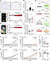 Figure 1
Expression and functional characterization of Slick in RCFs and CMFs. (A) Representative alkaline phosphatase staining for Slick protein (blue) in cryosections from WT and KO hearts. Scale bar: 500 μm. (B) RT-qPCR analysis of Kcnt2 transcripts in isolated primary CMs, RCFs, and cultured CMFs, normalized to Hprt. One-way ANOVA with Tukey’s multiple-comparison test, **P < 0.01 (n = 11 hearts for CMs, n = 12 hearts for RCFs, and n = 5 hearts for CMFs). (C) Representative FRET-based live-cell [K+]cyto images in RCFs after 15 minutes of monensin treatment. Higher FRET/CFP ratios appeared as red-white signals. Scale bar: 50 μm. Right panel: normalized average FRET/CFP ratio over time. (D) Δ FRET/CFP ratios in WT and KO RCFs. Δ was calculated as R20min – R5min, with R0 defined as the mean ratio during the first 5 minutes. Mann-Whitney U test, *P < 0.05 (n = 8 hearts per genotype). (E) Representative FRET images and average ratio over time in CMFs during monensin treatment. Scale bar: 50 μm. (F) Δ FRET/CFP ratios in WT and Slick KO CMFs. Two-tailed unpaired Student’s t test, *P < 0.05 (n = 8 hearts per genotype). (G) Representative whole-cell currents at baseline and after 5 minutes of NFA. (H and I) Current-voltage (I-V) relationships of persistent currents in WT and KO RCFs before and after NFA treatment. Two-way ANOVA with Šidák’s multiple-comparison test, ***P < 0.001. (J) Δ current density at 100 mV. Nested t test, **P < 0.01 (WT: 6 cells from 5 hearts; KO: 11 cells from 4 hearts). (K and L) I-V relationships of persistent currents in CMFs. Two-way ANOVA with Šidák’s multiple-comparison test, ***P < 0.001. (M) Δ current density at 100 mV. Nested t test, **P < 0.01 (WT: 13 cells; KO: 15 cells from 4 hearts).
To further delineate the cell-specific expression profile of Slick channels in the heart, we assessed Kcnt2 transcript levels, encoding murine Slick, in freshly isolated RCFs and in RCF-derived CMFs after 10 days in vitro. The identity and purity of the isolated populations, as well as their successful transition from RCF to CMF, were confirmed by increased expression of CMF markers Acta2 (encoding α-SMA) and Postn (encoding periostin), in comparison to cell-specific markers identified in endothelial and immune cells or CMs (9, 34) (Supplemental Figure 1A; supplemental material available online with this article; https://doi.org/10.1172/jci.insight.195805DS1). Consistent with single-cell RNA-Seq data from the Tabula Muris consortium (31), RT-qPCR analysis revealed that Kcnt2 was highly expressed in both RCFs and cultured CMFs, whereas its expression was markedly lower in primary adult CMs (Figure 1B). These data suggest that Slick channels are primarily expressed in fibroblast populations, both at rest and after activation in vitro.
To investigate potential compensatory mechanisms in the absence of Slick, we further analyzed the expression of several other K+ channel genes previously reported in healthy and diseased cardiac myo-/fibroblasts (35–37). However, no significant compensatory up- or downregulation was observed in Slick-deficient CMFs (Supplemental Figure 1, B–H). Notably, Kcnt2 expression levels (Figure 1B) exceeded those of other tested K+ channels, underscoring the predominant role of Slick in cardiac fibroblast physiology.
Next, we assessed whether the expected Slick-mediated net K+ outflow affects cytosolic K+ ([K+]cyto) in live RCFs/CMFs using the genetically encoded fluorescence resonance energy transfer–based (FRET-based) K+ indicator GEPII 1.0 NES lc-LysM (GEPII 1.0) (38). This indicator incorporates a bacterial K+-binding protein (Kbp) flanked by mseCFP (cyan) and cpV (yellow) fluorescent protein (YFP). Interaction of [K+]cyto with Kbp eventually results in energy transfer from mseCFP to FRET (YFP) signal, which can be visualized under a fluorescent microscope (38). GEPII 1.0 has been demonstrated to function effectively in both primary cells (38–40) and multiple cell lines tested (41, 42). Again, we showed that GEPII 1.0 was functionally expressed in RCFs and CMFs of both genotypes, as evidenced by reciprocal changes of CFP and FRET signals observed in real-time response to increasing extracellular K+ ([K+]ex) concentrations ranging from 1 to 300 mM (Supplemental Figure 1, I–L). Given the [Na+]i sensitivity of the Slick channel, we next applied monensin (Na+-permeable ionophore) to induce Na+ uptake and thereby an increase in intracellular Na+ ([Na+]i) (43). Consequently, a small but significant decline in the normalized average FRET/CFP ratio signal was observed in WT RCFs (Figure 1, C and D) and CMFs (Figure 1, E and F) compared with respective KO cells, suggesting a role of Slick in regulating the Na+-induced K+ efflux, whereby it functionally affects [K+]cyto dynamics in these cells.
To investigate the electrophysiological properties of RCF and CMF in the presence and absence of Slick channels, we performed whole-cell patch-clamp recordings. To this end, niflumic acid (NFA), a previously validated Slick channel opener (44, 45), was applied to evaluate its effect on Slick-mediated outward current components (ISlick). In WT RCFs, NFA treatment resulted in a pronounced increase in outward ISlick current density, whereas NFA did not alter current recordings performed in KO RCFs at voltages ranging from −80 mV to 100 mV (Figure 1, G–J). In WT CMFs, we consistently observed a small increase in outward current density in response to NFA, indicative of ISlick, while in KO CMFs, the application of NFA unexpectedly repressed whole-cell current density (Figure 1, G and K–M). This phenomenon may be attributed to the lower expression of Slick transcripts in cultured CMFs compared with freshly isolated RCFs (Figure 1B). Alternatively, it might also result from inhibition of NFA-sensitive Cl– currents, potentially mediated by TMEM16A (46, 47), a Ca2+-activated Cl– channel known to be highly expressed in myo-/fibroblasts (48, 49). Given that NFA also inhibits TMEM16A, the resulting decrease in outward Cl– current may counteract ISlick. This effect might also explain the rather small increase in current density in WT CMFs. Given that KO CMFs lack Slick and thus outward-directed ISlick, they demonstrated an unexpected decrease in current density. Furthermore, to validate the role of Slick in Na+-induced K+ efflux, as previously demonstrated by FRET-based [K+]cyto measurements (Figure 1, E and F), we applied monensin during patch-clamp recordings. In KO CMFs, monensin-induced Na+ influx led to a marked reduction in outward current amplitude, whereas this decrease was significantly attenuated in WT CMFs, likely due to simultaneous Slick-dependent K+ efflux (Supplemental Figure 1, M–Q).
In summary, these live-cell imaging results and patch-clamp recordings provide strong evidence of functional Slick channels at the plasma membrane of RCFs and CMFs that regulate the K+ movement from intracellular to extracellular spaces.
Loss of Slick in RCFs and CMFs disrupts intracellular Ca2+ ([Ca2+]i) dynamics. K+ channels have been shown to modulate cellular excitability and [Ca2+]i dynamics through multiple mechanisms. These include affecting the influx of extracellular Ca2+ ([Ca2+]ex) and facilitating Ca2+ release from intracellular stores, both of which collectively shape [Ca2+]i signaling (39, 50, 51). Therefore, we investigated whether and how Slick-dependent K+ dynamics affect [Ca2+]i in RCFs and CMFs employing Fura-2 (a Ca2+-binding fluorescent dye). Initially, we examined the basal [Ca2+]i storage content in RCFs and CMFs. We applied ionomycin, a Ca2+-permeable ionophore, combined with a gradient change from 2 mM [Ca2+]ex buffer to a Ca2+-free buffer supplemented by EGTA. Given that this series of experiments was performed with an EGTA-containing Ca2+-free buffer, an increase in [Ca2+]i reflected the release of Ca2+ from intracellular stores rather than entry of Ca2+ from the extracellular space. This enabled us to measure the Ca2+ content of cardiac fibroblasts’ intracellular stores by calculating the reduction in [Ca2+]i following this protocol. Indeed, we observed a transient increase in [Ca2+]i in both RCFs and CMFs, likely reflecting Ca2+ release from intracellular stores such as the ER. The subsequent drop was indicative of an ionomycin-mediated Ca2+ leakage from the cell, reflecting the total [Ca2+]i storage capacity, which was significantly higher in WT compared with KO RCFs (Figure 2, A and B) and CMFs (Figure 2, C and D). This indicates that Slick deletion interferes with ER and total [Ca2+]i storage in RCFs and CMFs in a reproducible manner.
![Loss of Slick in RCFs and CMFs disrupts [Ca2+]i dynamics.](//df6sxcketz7bb.cloudfront.net/manuscripts/195000/195805/small/jci.insight.195805.f2.gif "Loss of Slick in RCFs and CMFs disrupts [Ca2+]i dynamics.") Figure 2
Figure 2Loss of Slick in RCFs and CMFs disrupts [Ca2+]i dynamics. (A) Normalized average 340 nm/380 nm ratios over time in RCFs during [Ca2+]ex depletion. (B) Δ [Ca2+]i (Rmax – Rmin) in WT and KO RCFs. Mann-Whitney U test, *P < 0.05 (n = 4 hearts per genotype). (C) Normalized average 340 nm/380 nm ratios in CMFs during [Ca2+]ex depletion. (D) Δ [Ca2+]i in WT and Slick KO CMFs. Two-tailed unpaired Student’s t test, **P < 0.01 (n = 4 hearts per genotype). (E) Normalized average 340 nm/380 nm ratios in RCFs during niclosamide treatment. (F) Δ [Ca2+]i (R20min – R5min) in WT and KO RCFs. Two-tailed unpaired Student’s t test, *P < 0.05 (n = 10 hearts per genotype). (G) Normalized average 340 nm/380 nm ratios in CMFs during niclosamide treatment. (H) Δ [Ca2+]i in WT and Slick KO CMFs. Mann-Whitney U test, **P < 0.01 (n = 10 hearts per genotype). (I) Normalized average 340 nm/380 nm ratios during SOCE in RCFs. SOCE was induced by ER Ca2+ depletion with BHQ, followed by re-addition of 2 mM [Ca2+]ex. Niclosamide was applied to open Slick channels 5 minutes after baseline recording. (J) Δ SOCE (Rmax – Rmin during Ca2+ re-addition) in WT and Slick KO RCFs. Two-tailed unpaired Student’s t test (n = 8 hearts per genotype). (K) Normalized average 340 nm/380 nm ratios during SOCE in CMFs. (L) Δ SOCE in WT and Slick KO CMFs. Two-tailed unpaired Student’s t test, *P < 0.05 (n = 8 hearts per genotype).
To investigate [Ca2+]i dynamics upon mimicking Slick channel activation, we applied niclosamide, previously described as a selective KNa channel opener (52, 53). Although niclosamide reproducibly activates Slack channels in CMs (40), its effects on Slick channels remain ambiguous. To address this, we employed HEK293 cells cotransfected with pcDNA3.1-Slick and pcDNA3.1-mRuby3. The inclusion of pcDNA3.1-mRuby3 not only served as negative control (NC) but also facilitated the identification of successfully Slick-cotransfected cells via its red fluorescence. Immunofluorescence staining confirmed the expression of Slick protein in red fluorescent protein–positive cells within the cotransfected group, whereas Slick was not detectable in the NC group (Supplemental Figure 2A), confirming our Western blot analysis of protein lysates obtained from these cells (Supplemental Figure 2B). At the basal level, cells overexpressing Slick exhibited a relatively lower membrane potential, likely due to an increased K+ “leakage” resulting from higher Slick expression (Supplemental Figure 2C). After niclosamide treatment, [K+]cyto efflux was significantly increased in Slick-overexpressing cells compared with NC cells, expressing the FRET-based [K+]cyto-indicator GEPII 1.0 (Supplemental Figure 2, D and E). Consistent with these K+ imaging experiments, patch-clamp recordings showed that niclosamide induces a pronounced K+ outward current in HEK293 cells overexpressing Slick, and, importantly, that the current amplitudes were significantly higher compared with NC cells (Supplemental Figure 2, F–J). These findings support a role for niclosamide in modulating the activity of Slick.
Upon validating this effect in Slick-proficient HEK293 cells, we applied niclosamide to investigate [Ca2+]i dynamics in RCFs and CMFs. This approach allowed us to assess the differential effects of Slick channel activation on [Ca2+]i regulation. In WT cells, this treatment resulted in a rather small and transient increment, potentially due to Ca2+ released from ER to the cytosol, followed by a steady increase in [Ca2+]i. The latter response was largely attenuated in KO cells (Figure 2, E–H). This observation prompted us to examine potential origins of the observed Ca2+ signals induced by niclosamide in more detail. [Ca2+]i in nonexcitable cells, particularly myo-/fibroblasts, rises due to the actions of store-operated calcium entry (SOCE) (54–56). SOCE, mediated by a Ca2+ influx through Ca2+-release–activated Ca2+ (CRAC, aka Orai) channels, is initiated by a preceding ER Ca2+ depletion. To determine whether SOCE is the main pathway through which Slick affects Ca2+ signaling in RCFs and CMFs, we used 2-Di-t-butyl-1,4-benzohydroquinone (BHQ), an effective inhibitor of sarco-endoplasmic reticulum Ca2+-ATPase (SERCA), to deplete ER Ca2+ stores (57). Subsequently, cells were superfused with a buffer containing 2 mM [Ca2+]ex, allowing Ca2+ entry via SOCE as observed in different cell types (56, 58, 59). On top of that, we opened Slick with niclosamide to identify the Slick-dependent component of SOCE in both cell types. A schematic illustrating the experimental procedure is shown in Supplemental Figure 3A. Accordingly, Slick-proficient cells exhibited a higher magnitude of Ca2+ signals than KO cells via SOCE, an effect that reached significance in CMFs but not in RCFs (Figure 2, I–L). To further characterize the SOCE pathway in CMFs, we utilized GSK7975A, a pan-inhibitor of all 3 Orai protein family members, although it predominantly inhibits Orai1 and has only minor effects on Orai3 (60). Treatment with GSK7975A reduced SOCE signals in WT cells compared with KO levels (Supplemental Figure 3, B and C). Importantly, we excluded the possibility that the observed difference in SOCE levels between WT and KO CMFs originates from altered ER Ca2+ release capacity upon activation of Slick by niclosamide. This conclusion is supported by a comparable [Ca2+]i increase observed under Ca2+-free buffer perfusion in both genotypes (Supplemental Figure 3D). Moreover, transcript levels of the main ER Ca2+ release channels, ryanodine receptor and inositol 1,4,5-trisphosphate receptor (IP3R) subtypes, did not differ between WT and KO CMFs (Supplemental Figure 3, E–J).
To gain insights into the attenuation of SOCE in KO CMFs, we analyzed the expression profiles of key SOCE regulators, namely stromal interaction molecules (STIM) and Orai channel subtypes. The transcript levels of STIM1 and STIM2 did not differ between genotypes (Supplemental Figure 3, K and L). However, we observed a shift in the expression pattern of Orai subtypes, with an increased Orai3/Orai1 ratio in KO CMFs at both transcript and protein levels (Supplemental Figure 3, M–O). These data imply that the formation of heteromultimeric Orai1-Orai3 is favored in the absence of Slick and that the altered Orai stoichiometry could, in turn, ensure a graded attenuation of SOCE regulation and/or activity in KO CMFs (61).
Lastly, since niclosamide is a known mitochondrial uncoupling agent capable of inducing mitochondrial Ca2+ release (62), it remained unclear whether niclosamide-induced mitochondrial dynamics might confound our SOCE results. To address this, we performed oxygen consumption rate (OCR) analysis under basal conditions and after niclosamide treatment. No significant differences in mitochondrial respiration were observed between WT and Slick KO CMFs under baseline or niclosamide-treated conditions (Supplemental Figure 3, P–R). Furthermore, tetramethylrhodamine methyl ester (TMRM) measurements indicated no alterations of mitochondrial membrane potential (Δφmito) upon niclosamide treatment in either genotype (Supplemental Figure 3, S–U). These findings suggest that neither Slick deletion nor niclosamide treatment significantly affects mitochondrial function or Δφmito, ruling out that mitochondrial dynamics contributed to the SOCE readouts.
In summary, Slick deficiency is associated with reduced ER Ca2+ content under basal conditions. Pharmacological activation of Slick channels by niclosamide leads to an increased [Ca2+]i in both WT RCFs and CMFs. Furthermore, we provide compelling evidence — having systematically excluded multiple confounding factors — that the observed effects stem from alterations in SOCE, primarily driven by changes in Orai channel composition rather than STIM protein expression. Specifically, Slick deletion leads to an increased Orai3-to-Orai1 expression ratio, which is functionally associated with reduced SOCE activity.
Global Slick deletion attenuates fibrogenesis, stabilizes electrophysiological properties, and preserves left ventricular function after I/R injury. To investigate the function of Slick in murine hearts after I45min/R4weeks injury, we performed a closed-chest I/R surgery utilizing global Slick KO mice and their littermate controls and assessed chamber remodeling and, in particular, pathological features of fibrosis, (i.e., the accumulation of fibroblasts and the deposition of ECM proteins). A significant fibrotic response of the myocardium was detected in I45min/R4weeks groups. Accordingly, collagen fibers, stained by Sirius red, appeared as extensive red areas throughout the myocardium, whereas the respective sham groups remained unstained (Figure 3A). However, the total amount of fibrosis was significantly reduced in KO hearts, indicating that the fibrotic response requires functional Slick channels. Importantly, these apparently profibrotic actions of Slick were consistently observed across each section of the heart distal to the ligature (Figure 3, B and C). In the polarized light images, WT hearts exhibited more collagen I deposition, as reflected by a higher collagen I to collagen III ratio (Figure 3, D and E). This suggests that WT hearts develop a stiffer collagen matrix, which may contribute to reduced left ventricular (LV) compliance and impaired chamber function (63).
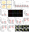 Figure 3
Global Slick deletion attenuates fibrogenesis and preserves LV function after I/R injury. (A) Representative Sirius red staining of myocardial fibrosis (red) in WT and KO hearts at segment 5 (S5) after sham or I45min/R4weeks. Scale bar: 500 μm. (B) Analysis of fibrosis (percentage of total area) from S1 to S8 within the affected area (covering all heart tissue affected by the ligature). Two-way ANOVA with Šidák’s multiple-comparison test, ***P < 0.001. (C) Average cardiac fibrosis per section. Two-way ANOVA with Tukey’s multiple-comparison test showed increased fibrosis in I45min/R4weeks WT compared with sham WT (***P < 0.001) and I45min/R4weeks KO (**P < 0.01), with no difference between sham groups (n = 5 sham WT, n = 5 sham KO, n = 8 I45min/R4weeks WT, n = 7 I45min/R4weeks KO). (D) Representative polarized light images (S5) from Sirius red–stained sections after I45min/R4weeks. Red birefringence indicates collagen I, and green indicates collagen III. Scale bar: 500 μm. (E) Quantification of the collagen I/III (Col1/Col3) ratio. Mann-Whitney U test, *P < 0.05. (F) Diastolic left ventricular anterior wall thickness (LVAW;d) measured by M-mode echocardiography (***P < 0.001; **P < 0.01). (G) LVEF measured by B-mode imaging, *P < 0.05. (H) Cardiac output (CO) measured by B-mode imaging, **P < 0.01. (I) Global longitudinal strain (GLS) measured in the parasternal long axis view using speckle-tracking (*P < 0.05; **P < 0.01). (J and K) Endocardial and epicardial strain analyses in both sham and I45min/R4weeks hearts. (L) Representative B-mode echocardiography images of sham and I45min/R4weeks hearts during systole and diastole. Epicardial and endocardial borders are indicated in yellow. Two-way ANOVA with Tukey’s multiple-comparison test was used for echocardiographic analyses (F–K) (n = 5 sham WT, n = 6 sham KO, n = 5 I45min/R4weeks WT, n = 5 I45min/R4weeks KO).
Consistent with fibrotic remodeling in post-MI hearts, our ECG recordings during the early reperfusion phase (i.e., I45min/R1week) revealed features of impaired ventricular electrophysiology (Supplemental Figure 4, A and B). Compared with the basal group, I45min/R1week-injured hearts exhibited a significantly prolonged QRS interval and a reshaped QRS complex characterized by attenuated R-wave amplitude (Supplemental Figure 4, C and D), indicating slowed ventricular conduction and compromised ventricular depolarization, respectively (13). Moreover, prolonged QTc and JT intervals suggest that ventricular repolarization was impaired as well (64) (Supplemental Figure 4, E and F). Although no significant differences were observed between genotypes in these parameters, WT hearts displayed more pronounced ST-segment depression and greater T-wave inversion than KO hearts (Supplemental Figure 4, G and H). Both alterations are indicative of postischemic electrical remodeling, potentially linked to enhanced fibrotic disruption (13). The observed electrocardiographic abnormalities may in part stem from increased myocardial fibrosis in WT hearts, which likely impairs electrical signal propagation and reduces ventricular compliance, thereby exacerbating electrical instability during the repolarization phase (13).
We next evaluated chamber morphology and function by applying noninvasive echocardiography measurements on I45min/R4weeks hearts. Importantly, there was no difference in heart rate between genotypes or between I/R and sham groups during echocardiographic analysis, thus eliminating an important confounder, as heart rate changes affect cardiac function (Supplemental Figure 4I). Using M-mode analysis, WT hearts subjected to I45min/R4weeks injury displayed a significantly thickened LV anterior wall (LVAW) compared with sham controls. In the diastolic phase, the thickness of the LVAW was significantly reduced when Slick was deleted (Figure 3F), consistent with a milder progression of muscle fibrosis in KO hearts (Figure 3, A–C). Next, B-mode analysis of cardiac cycles in the parasternal long axis view was combined with speckle tracking to objectively quantify global and local myocardial deformation. Using this feasible and reproducible mode, LV ejection fraction (LVEF) and cardiac output, which represent a central measure of LV function, were significantly impaired in WT hearts after I45min/R4weeks, whereas these parameters remained largely intact in KO hearts (Figure 3, G and H). Next, the regional damage to LV segments was probed by tracing the wall motion through the cardiac cycle (Supplemental Figure 4J). Global longitudinal strain, which expresses longitudinal shortening as a proportion of baseline length, was improved throughout the entire LV (Figure 3I) in both the endocardial and epicardial surface area analysis (Figure 3, J and K) in the absence of Slick. This comprehensive assessment revealed that WT hearts subjected to I45min/R4weeks injury exhibited a diminished longitudinal myocardial shortening (magnitude of deformation) and a regional desynchrony regarding the direction of wall motion, indicative of a significant impairment (Figure 3L). However, radial deformation, that is, the thickening and thinning motion of the myocardium in different segments of the anterior and posterior walls of the LV, was not different between Slick-proficient and Slick-deficient post-I/R hearts (data not shown).
To further assess the potential myocardial growth in response to the I45min/R4weeks injury, the heart weight to tibia length ratio (HW/TL) was measured as a hallmark of cardiac hypertrophy (34). There was no significant difference in the HW/TL ratio between the genotypes at baseline and after I45min/R4weeks exposure (Supplemental Figure 4K). Consistently, LV mass (Supplemental Figure 4L) and CM cross-sectional area enlargement were similar in both genotypes after I45min/R4weeks (Supplemental Figure 4, M and N). Together, these results indicate that Slick, in an adverse manner, affects global LV function and multiple wall deformation parameters, including global longitudinal strain, a long-term risk parameter of cardiovascular morbidity and mortality, while the hypertrophic enlargement of post-I45min/R4weeks hearts develops independently of Slick.
Cell-specific ablation of CMF Slick channels attenuates fibrogenesis and LV dysfunction upon I45min/R4weeks injury. So far, our in vitro experiments have provided substantial evidence for the presence and functionality of Slick channels in RCFs and CMFs. Other studies have previously identified functional Slick channels in cardiac mitochondria (30) and demonstrated a key role for Slick as mediator of anesthetic preconditioning in ex vivo Langendorff-perfused hearts (29). The latter results suggest that cardiac Slick activity confers cardioprotection against an acute and transient I/R insult via anesthetic preconditioning, whereas in our experimental model based on a 4-week post-MI follow-up analysis, we observed an increase in cardiac damage in the presence of Slick. Given these potentially opposing actions of Slick on cardiac injury and to explain these apparently contradictory results, we next aimed to clarify in which cell type Slick function would promote fibrogenesis and chamber dysfunction after cardiac I/R in vivo.
We first subjected global Slick KO mice and their WT littermate controls to an open-chest surgery with I30min/R2h. This short-term I30min/R2h protocol allowed us to evaluate whether Slick modulates the acute myocardial cell death that results in the loss of CMs (40), an important pathological feature driving muscle fibrosis. After I30min/R2h, neither the area at risk, defined as cardiac tissue within the vascular territory supplied by the infarct-related (ligated) vessel, nor the infarcted area differed between Slick KO and WT mice (Supplemental Figure 4, O–Q), indicating that Slick has no influence on the acute CM death provoked by I30min/R2h. It is therefore unlikely that the gradually developing differences in cardiac fibrosis between the 2 genotypes are due to a function of Slick in CMs, suggesting instead that Slick contributes to the profibrotic actions of CMFs via cell-intrinsic mechanisms.
To further substantiate Slick’s potential role in CMFs in vivo, we generated a CMF-specific Slick KO (mKO) mouse line that relies on a tamoxifen-inducible Cre-recombinase under the control of the periostin (Postn) promoter and its recognition (loxP) sites flanking exon 22 of the Slick gene (15, 33). This approach ensured that Slick deletion occurred selectively only in the presence of Postn, a matricellular protein predominantly expressed in CMFs under pathophysiological conditions such as I/R injury or angiotensin II infusion (15, 34). On the other hand, nearly all RCF-derived CMFs express Postn, ensuring consistency of the CMF subpopulation analyzed between our in vitro and in vivo studies. The mKO mice (Postni-CreTg/+ Slickfl/fl) and their respective littermate controls (Postni-CreTg/+ Slick+/+ or CTR) were subjected to the identical I45min/R4weeks injury model previously used to assess WT and global Slick KO mice (Figure 4A). After I45min/R4weeks surgery, tamoxifen was administered i.p. for 5 consecutive days to induce Cre-recombination in CMFs, which are highly activated during this time of the progressively developing remodeling response (10). The specificity of Cre-recombinase activity was confirmed by detecting the excised Slick DNA allele exclusively in CMFs (Figure 4B). Additionally, by using validated anti-Slick antibodies, we confirmed that the targeted ablation of the Slick channel protein was restricted to fibrotic heart regions, where Postn-positive CMFs are particularly prevalent (Figure 4C).
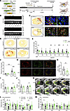 Figure 4
Cell-specific ablation of CMF Slick channels attenuates fibrogenesis and LV dysfunction after I45min/R4weeks injury. (A) CMF-specific Slick KO mutants were generated utilizing a tamoxifen-activated Cre expressed under control of the Postn promoter, ablating Slick in Postn+ CMFs. Mice underwent 45 minutes of closed-chest I/R followed by 1 or 4 weeks of reperfusion. Tamoxifen was administered i.p. for 5 consecutive days starting on postoperative day 1. CMFs were isolated after 1 week for functional analyses. Echocardiography and fibrosis quantification were performed after 4 weeks. (B) PCR validation of tissue-specific recombination. The excised Slick allele (L1) was detected exclusively in CMFs from mKO mice but not in other tissues or CTR samples. (C) Immunofluorescence staining of Slick (red) and periostin (green). Colocalization was observed in fibrotic CTR hearts but absent in mKO hearts after I/R. Scale bars: left, 500 μm; upper right: 10 μm; lower right: 25 μm. (D) Representative Sirius red staining of myocardial fibrosis in CTR and mKO hearts after sham or I45min/R4weeks. (E) Segmental fibrosis quantification. Two-way ANOVA analysis with Šidák’s multiple-comparison test, ***P < 0.001. (F) Overall average fibrosis across S1–S8. Two-way ANOVA with Tukey’s multiple-comparison test, ***P < 0.001 (n = 5 sham CTR, n = 5 sham mKO, n = 10 I45min/R4weeks CTR, and n = 9 I45min/R4weeks mKO). (G) Representative polarized light images (S5). Scale bar: 500 μm. (H) Collagen I/III ratio in I45min/R4weeks hearts. Two-tailed unpaired Student’s t test, **P < 0.01. (I and J) Cardiac output (CO) and stroke volume (SV), assessed by B-mode echocardiography, *P < 0.05. (K) Representative B-mode images during systole and diastole in sham and I45min/R4weeks hearts. (L and M) Radial strain and strain rate analysis in parasternal long axis view. Echocardiographic data (L and M) were evaluated by Tukey’s multiple-comparison test (n = 7 sham CTR, n = 6 sham mKO, n = 10 I45min/R4weeks CTR, n = 9 I45min/R4weeks mKO).
Consistent with previous findings from WT and global Slick KO, a significant increase in fibrosis was observed in CTR hearts, while fibrogenesis provoked in mKO hearts was markedly diminished. This reduction in the amount of fibrosis was evident in all cardiac segments assessed from the apex to the base, that is, close to the ligation site (Figure 4, D–F). mKO hearts exhibited a less rigid collagen matrix compared with CTR hearts, as indicated by polarized light imaging (Figure 4, G and H).
To assess accompanying alterations in cardiac function, we carried out conventional and strain echocardiographic imaging. Again, the heart rate between genotypes did not differ during these measurements (Supplemental Figure 5A). Compared with the sham group, a decline in LVEF was observed in both genotypes that reached significance only for the CTR animals after I45min/R4weeks injury (Supplemental Figure 5B). Although differences in post-I/R LVEF between CTR and mKO hearts were not observed, cardiac output and stroke volume in mKO hearts presented with significantly better values (Figure 4, I and J), indicative of a partially recovered cardiac performance in the absence of Slick in CMF. Because the adverse tissue remodeling upon I45min/R4weeks is an event with locally varying degrees of impairment, we divided the heart into anterior and posterior walls, which were further subdivided into apex, mid, and base regions (Supplemental Figure 4J). LV motion analysis of the anterior wall in which the fibrotic scar predominantly forms due to left coronary artery ligation (i.e., where Postni-Cre–mediated activity in CMFs led to Slick ablation) was impaired in CTR compared with mKO hearts, with respect to endocardial radial strain and strain rates due to the I/R injury. Similar trends were observed in the epicardial layer (data not shown as radial strain parameters across the myocardial wall were largely consistent). This functional decline in myocardial thickening and thinning, which is visualized by the length and direction of vector arrows, was partially preserved in the absence of Slick (Figure 4, K–M). In contrast to WT and global Slick KO post-MI hearts (Figure 3), local longitudinal strain and strain rates in the anterior or posterior walls were not different between CTR and conditional mKO hearts (data not shown). These findings suggest that targeting Slick in CMFs mitigates fibrosis and partially restores cardiac function after I/R injury.
Consistent with our analyses of WT versus global Slick KO, no significant difference in cardiac hypertrophy between CTR and mKO genotypes, as indicated by the comparable LV mass and cross-sectional area assessed by wheat germ agglutinin staining, could be observed (Supplemental Figure 5, C–E). Despite the excessive fibrosis in CTR hearts (Figure 4, D–F), a small but significant decline in the HW/TL ratio in post-I/R CTR compared with mKO hearts was detected (Supplemental Figure 5F). This decrease in the HW/TL ratio might reflect CM loss resulting from harmful overactivation of fibrotic signaling pathways (65). Indeed, a significantly higher number of TUNEL-positive CMs was observed in CTR versus mKO hearts (Supplemental Figure 5, G and H), suggesting that fibrosis and the provoked apoptotic CM cell death correlated closely with each other. Hence, CM survival was improved when Slick-dependent fibrogenesis was attenuated in I45min/R4weeks-exposed hearts.
Given the crucial role of immune cells, particularly T cells and macrophages, in chronic cardiac remodeling after MI (66), we sought to determine whether the chronic immune response is altered in mKO hearts. Indeed, we observed a significant reduction in the number and frequency of CD68+ macrophages (67) in I45min/R4weeks hearts, particularly along the fibrotic scar region. In contrast, CD3+ T cells (68) comprised a smaller fraction of the total immune cell population and showed no significant difference between genotypes (Supplemental Figure 5, I–N).
To further explore whether and how Slick-deficient CMFs influence macrophage function, such as recruitment, activation, proliferation, or maintenance, we assessed the expression of cytokine- or chemokine-related transcripts in both CMFs and whole hearts after I/R (Supplemental Figure 5, O–S). Although no significant changes in transcript levels were detected in CMFs, a notable upregulation of Ccl5 (encoding CCL5), a factor known to mediate the recruitment of proinflammatory macrophages to infarcted myocardium (69), was found in CTR hearts (Supplemental Figure 5R). In addition, CTR hearts exhibited increased expression of proinflammatory cytokines, among which Il1b (encoding IL-1β) was significantly elevated (Supplemental Figure 5, T–V).
Together, these findings suggest that Slick ablation in CMFs attenuates the proinflammatory immune response in post-MI hearts by limiting macrophage recruitment and inflammatory cytokine expression. This effect does not appear to arise from direct changes in CMF chemokine output but may instead reflect a broader suppression of profibrotic signaling and fibrotic-mediated cell death in the infarcted myocardium in mKO hearts.
Loss of Slick attenuates the proliferation capability and diminishes the expression of fibrotic genes in CMFI/R ex vivo. So far, our data from 2 Slick mutant mouse models have suggested that Slick contributes to the profibrotic actions of CMFs in the post-I45min/R4weeks myocardium in vivo. Moreover, loss of Slick in cultured CMFs established from healthy hearts disrupted the [Ca2+]i dynamics of both cell types (Figure 2). To further deepen our understanding of how Slick function might influence CMFs upon I/R injury, we isolated and assessed cardiac fibroblasts after I45min/R1week and refer to these cells as CMFI/R to distinguish them from CMFs obtained in vitro by cultivating RCFs over time. We chose a reperfusion time of 7 days for the isolation of the cells because at this time, CMFs in the myocardium are usually highly active (9).
To confirm the purity of isolated CMFI/R, we assessed the expression of established myofibroblast markers as previously mentioned (Supplemental Figure 1A), which revealed robust expression of Acta2 and Postn. Transcripts corresponding to cell-surface and intracellular markers of other cardiac cell types, however, were not detectable (Supplemental Figure 6A). We also examined the transcript levels of relevant K+ channels that play critical roles in, for example, controlling the electrical activity under I/R conditions, but could not observe compensatory changes in KO cells (Supplemental Figure 6, B–H). Notably, Kcnt2 expression in CMFI/R was nearly 10-fold higher compared with CMFs cultured in vitro, which is consistent with the sequencing data of Qian et al. (32), indicating a strong transcriptional upregulation in response to ischemic stress (Figure 5A). Whole-cell patch-clamp recordings confirmed Slick-dependent outward K+ currents in WT CMFI/R, with KO cells serving as NC (Figure 5, B and C).
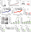 Figure 5
Loss of Slick attenuates proliferation and fibrotic gene expression in CMFI/R. (A) RT-qPCR analysis of Kcnt2 expression in cultured CMFs and in CMFI/R, normalized to Hprt. Mann-Whitney U test, *P < 0.05 (n = 6 hearts for CMF and n = 13 hearts for CMFI/R). (B) I-V relationships of persistent currents in WT and KO CMFI/R. Two-way ANOVA followed by Šidák’s multiple-comparison test, ***P < 0.001 (n = 6 hearts per genotype). (C) Representative outward current traces in WT and KO CMFI/R. (D and E) SOCE traces in WT and KO CMFI/R treated with DMSO (basal) or GSK7975A. (F) ΔSOCE (Rmax – Rmin during Ca2+ re-addition) in CMFI/R. GSK7975A significantly reduced SOCE in WT. Two-way ANOVA followed by Tukey’s multiple-comparison test, *P < 0.05 (n = 5 hearts for basal WT; n = 6 hearts for all other groups). (G) Representative brightfield images (0 h, 72 h) of grid-plate-based proliferation assay. Scale bar: 100 μm. (H) Quantitative analysis of proliferation rate over time. Two-way ANOVA with Šidák’s multiple-comparison test, ***P < 0.001 (n = 7 CTR and n = 11 mKO). (I) MTT assay of CMFI/R. Two-way ANOVA with Šidák’s multiple-comparison test, ***P < 0.001 (n = 6 hearts per genotype). (J and K) Western blot analysis of α-SMA in CMFI/R, normalized to GAPDH. Two-tailed unpaired Student’s t test, ***P < 0.001 (n = 5 hearts per group). (L–N) RT-qPCR analysis of profibrotic transcripts, normalized to Hprt. (L) Acta2 expression. Mann-Whitney U test (*P < 0.05; n = 7 CTR, n = 8 mKO). (M) Tgfb1 expression. Two-tailed unpaired Student’s t test, *P < 0.05 (n = 5 CTR, n = 8 mKO). (N) Col3a1 expression. Two-tailed unpaired Student’s t test (*P < 0.05; n = 7 CTR, n = 8 mKO).
To further validate the alterations in SOCE previously observed in vitro, we applied the same Ca2+ imaging protocol to CMFI/R. Preincubation with the Orai channel inhibitor GSK7975A for 30 minutes significantly reduced basal [Ca2+]i levels in WT CMFI/R but not in KO cells (Supplemental Figure 6I). After SOCE stimulation, GSK7975A treatment led to a pronounced attenuation of Ca2+ influx in WT CMFI/R, while no significant effect was observed in the KO (Figure 5, D–F), supporting a Slick-dependent regulation of SOCE. Importantly, neither ER Ca2+ release nor the gene expression of IP3R or ryanodine receptor subtypes differed significantly between genotypes (Supplemental Figure 6, J–P), except for Ip3r2 encoding IP3R2 (Supplemental Figure 6O). The expression level of Ip3r2, however, was extremely low, approaching the detection threshold, and thus is likely to be biologically negligible. This suggests that the observed differences in [Ca2+]i homeostasis are specific to SOCE alterations. In agreement with the in vitro data, Stim1 and Stim2 levels remained unchanged, whereas the Orai3/Orai1 expression ratio was significantly elevated in Slick-deficient CMFI/R.
Taken together, these findings provide strong evidence that Slick deletion modulates SOCE activity in CMFI/R, consistent with our in vitro observations. This effect is likely mediated by an altered composition of Orai channel subtypes, rather than by changes in upstream ER Ca2+ release mechanisms.
In the next step, we tested the proliferative behavior of Slick-proficient versus Slick-deficient CMFI/R using a grid-based cell-growth assay (34). By tracking individual CMFI/R in the same grid over time, we observed a significant increase in cell numbers. Importantly, at all time points examined, mKO CMFI/R cell counts were markedly reduced compared with CTR (Figure 5, G and H). This difference between both genotypes was further confirmed by evaluating the number of viable CMFI/R over time via the MTT assay (Figure 5I). These findings identified a significant reduction in the proliferative capacity as one possible reason for the attenuated fibrotic response of global and CMF-specific post-I/R hearts lacking Slick.
Additionally, we characterized the activation of CMFI/R by measuring α-SMA. At both transcript and protein levels, α-SMA was increased in CTR versus mKO CMFI/R (Figure 5, J–L). Consistently, we found higher TGF-β and collagen III transcript expression levels in Slick CTR CMFI/R (Figure 5, M and N).
In summary, these findings indicate that Slick function in primary CMFI/R promotes cell proliferation and the fibrotic signaling associated with CMF activation, ultimately leading to accelerated collagen production in vivo. This functional effect is likely mediated through a SOCE-dependent mechanism involving Orai channel remodeling in the presence or absence of Slick.
![Loss of Slick in RCFs and CMFs disrupts [Ca2+]i dynamics.](http://df6sxcketz7bb.cloudfront.net/manuscripts/195000/195805/small/jci.insight.195805.f2.gif "Loss of Slick in RCFs and CMFs disrupts [Ca2+]i dynamics.")
-
Discussion
Our study revealed a role of Slick in post-MI cardiac remodeling that we believe was previously unrecognized. As part of our thorough analysis, we demonstrated Slick expression in cultured CMFs and infarction-derived CMFI/R and identified its role in [K+]cyto efflux, which may consequently affect SOCE-mediated Ca2+ signaling. Furthermore, the absence of Slick in CMFs attenuated cardiac fibrosis, reduced inflammatory activation, limited CM death, and preserved chamber performance after I/R injury. Ex vivo analyses of CMFI/R confirmed diminished proliferation and profibrotic gene expression in Slick-deficient CMFs (Figure 6). Collectively, these findings position myo-/fibroblast Slick channels as key regulators of post-MI fibrogenesis and chamber dysfunction.
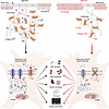 Figure 6
Graphic summary. Slick channels regulate CMF function and post-MI cardiac remodeling. After chronic I/R, WT hearts exhibit increased collagen deposition, inflammatory activity, and CMF activation, contributing to CM loss and LV dysfunction. In contrast, Slick-deficient hearts show reduced fibrosis and inflammation with improved myocardial preservation. At the cellular level, Slick-mediated K+ efflux enhances the electrochemical driving force for SOCE and promotes profibrotic transcriptional programs. Loss of Slick reduces Ca2+ influx and SOCE efficiency, likely through Orai channel remodeling, thereby limiting CMF activation and proliferation and preserving ventricular structure and function after MI. The figure was partly created using BioRender (license TN29GGWQJZ and OF29GGXG83).
Slick channels display pronounced sensitivity to [ATP]i, being markedly blocked at 5 mM ATP (28). Coupled with their activation by elevated [Na+]i, this dual regulation enables integration of metabolic and ionic cues, particularly under ischemic stress characterized by ATP depletion and Na+ accumulation, thereby modulating electrical activity (70). Previous work demonstrated that Slick confers anesthetic preconditioning–mediated cardioprotection against transient hypoxia/reperfusion injury (29). Extending this ex vivo finding, our study identified Slick as a regulator of post-MI cardiac remodeling. In contrast to the mitochondria-centered mechanism described for anesthetic preconditioning (30), we demonstrated that Slick modulates profibrotic signaling in CMFs after I/R injury. These results revealed a context-dependent role of Slick in the heart: While Slick in CMs limits acute ischemic injury, its function in RCFs/CMFs promotes maladaptive remodeling and functional decline during prolonged reperfusion. Overall, fibroblast-driven profibrotic effects of Slick appear to outweigh its early cardioprotective actions in CMs.
Our in vitro analysis of Slick-dependent K+ dynamics and electrophysiological recordings in RCFs and CMFs confirms functional channel expression and electrophysiological relevance in both cell types. To emulate ischemic-associated [Na+]i overload, monensin was applied as a Na+ ionophore (43). This intervention elicited a counteractive Slick-dependent outward current in CMFs (Supplemental Figure 1, M–Q), consistent with a compensatory response to Na+ influx and supporting a role for Slick in preserving K+ homeostasis and maintaining membrane stability under ionic stress. To further probe Slick activity, we applied NFA, a previously reported pharmacological opener of Slick (44). NFA robustly enhanced Slick-mediated outward currents in RCFs, whereas effects in CMFs were modest, and Slick-deficient CMFs displayed reduced outward currents (Figure 1, H–M). This observation may be explained by lower expression of Slick in cultured CMFs compared with RCFs, as indicated by a single-cell RNA-Seq approach (31) and RT-qPCR results (Figure 1B). In addition, NFA inhibits TMEM16A (46, 47), a Ca2+-activated Cl– channel highly expressed in cardiac cells (48, 49) and markedly enriched in CMFs relative to RCFs, with expression levels nearly 200-fold higher in CMFs than in RCFs (31). Thus, it is plausible that TMEM16A inhibition may partially offset Slick-mediated K+ currents in CMFs. This interpretation remains speculative and requires further validation. In addition to NFA, we employed niclosamide, an anthelmintic drug known to modulate various signaling pathways (71, 72), including KNa channels. Although niclosamide has been shown to activate Slack channels in CM sarcolemma (40), its effect on Slick has not been defined. Here, our data indicate that niclosamide also activates Slick channels (Supplemental Figure 2, D–J). Although off-target activation of Slack in RCFs and CMFs cannot be entirely excluded, this is likely minimal given the predominant expression of Slick over Slack in these cells (31). Consistently, RT-qPCR analysis revealed Kcnt1 transcripts (encoding Slack) near the detection limit in both RCFs and CMFs (data not shown), supporting the view that Slick represents the principal KNa channel in RCFs and CMFs.
Previous studies have described distinct subcellular localizations for cardiac Slick channels. Smith et al. identified Slick in CM mitochondria and linked it to anesthetic preconditioning–mediated cardioprotection (30). In contrast, our whole-cell patch-clamp recordings in RCFs and CMFs support functional Slick channels at the plasma membrane (Figure 1, H–M). Consistently, OCR measurements and TMRM-based measurements of Δφmito revealed no differences between Slick-proficient and Slick-deficient CMFs (Supplemental Figure 3, Q–U), arguing against a major mitochondrial contribution in these cells. Moreover, monensin-induced Na+ influx (43) elicited a modest but significant Slick-dependent modulation of FRET-based [K+]cyto signals (Figure 1, C–F). These results indicate that Slick operates at the plasma membrane of RCFs and CMFs to regulate K+ dynamics.
SOCE represents a major source of Ca2+ entry pathway in nonexcitable cells and is triggered by ER Ca2+ depletion (73), while its amplitude is determined by STIM-Orai coupling and the relative expression of Orai isoforms (Orai1, Orai2, and Orai3) (74). Homomeric Orai1 channels exhibit greater efficiency than heteromeric assemblies (75), and shifts in Orai isoform expression modulate SOCE in cancer cells and macrophages (76, 77). Orai3 overexpression leads to higher formation of Orai1/3 heteromers and attenuates SOCE in pulmonary fibroblasts (61). In diseased human hearts, enhanced SOCE and elevated Orai1 expression correlate with fibrotic remodeling (54), and angiotensin II–mediated upregulation of Orai1 and STIM aggravate maladaptive CMF activation and ECM deposition (78). In our study, Slick deficiency markedly attenuated SOCE in CMFs (Figure 2, K and L), coinciding with an increased Orai3/Orai1 ratio, presumably leading to heteromeric channel formation. However, STIM expression remained unchanged (Supplemental Figure 3, K and L), suggesting that altered Orai composition rather than STIM availability underlies the reduced Ca2+ entry and lessened fibrosis after I/R injury.
The reduced SOCE observed in Slick-deficient CMFs may reflect a diminished electrochemical driving force resulting from loss of ISlick current and impaired K+ efflux, as both membrane potential and Ca2+ gradient critically regulate SOCE (79). Consistent with this concept, measurements of membrane potential based on bis-(1,3-dibutylbarbituric acid)trimethine oxonol (DiBAC4(3)) showed that HEK293 cells overexpressing Slick displayed a less depolarized (less positive) plasma membrane than NC cells (Supplemental Figure 2C), supporting this hypothesis. Slick has previously been reported to associate with nonselective voltage-independent Na+ leak channels and transient receptor potential Melastatin 3 channels, contributing to ionic homeostasis through spatial proximity (80, 81). Here, we propose that in CMFs, Slick and Orai channels may similarly reside in close spatial proximity, allowing Slick-mediated K+ efflux to modulate the local membrane potential and thereby influence SOCE. Loss of Slick could disturb this microdomain regulation, potentially favoring adaptive remodeling of Orai isoform composition in response to altered Ca2+ signaling demands. While mechanistically plausible, this hypothesis remains to be experimentally validated.
In vivo, global Slick KO mice subjected to chronic I45min/R4weeks exhibited reduced fibrosis and attenuated LV dysfunction compared with controls, indicating that myocardial Slick promotes adverse remodeling. However, it remained unclear whether these effects reflect altered transient CM death and resulting replacement fibrosis or an intrinsic profibrotic role of Slick in CMFs after MI. To clarify this, we performed acute open-chest I30min/R2h experiments with 2,3,5-triphenyltetrazolium chloride (TTC) staining and observed comparable infarct size in both genotypes (Supplemental Figure 4, M–O), suggesting that Slick does not influence acute CM death under these conditions. Notably, isoflurane was used during closed-chest I45min/R4weeks surgery and has been linked with anesthetic preconditioning (29). In line with this, our unpublished observations indicate reduced anesthetic preconditioning–mediated protection in global KO hearts after transient I/R. However, acute CM damage and chronic fibrotic remodeling involving RCF/CMF represent distinct pathophysiological processes. Our finding suggests that during prolonged reperfusion, the profibrotic actions of Slick in CMFs outweigh any early cardioprotective effects in CMs.
To directly interrogate CMF-specific effects and avoid confounding by CM Slick deletion, we employed a tamoxifen-inducible Postni-Cre mouse line. Periostin (encoded by Postn) serves as a highly specific marker in injury-activated CMFs, being minimally expressed in healthy myocardium yet upregulated nearly 200-fold 7 days after MI (14, 15, 34). This injury-restricted and robust induction makes the Postn gene promoter a suitable driver for CMF-targeted recombination. CMF-specific Slick ablation significantly reduced fibrosis after I45min/R4weeks, supporting a CMF-intrinsic role of Slick in post-MI remodeling. Functional analyses revealed partial preservation of LV performance, particularly within the anterior wall, consistent with the region predominantly affected by left coronary artery ligation.
Because CMFs are highly active during the early reparative phase, when tamoxifen-induced recombination occurred, regional preservation of chamber function likely reflects early suppression of CMF activity in this area (Figure 4, L and M). Given the limited 5-day tamoxifen treatment window (14), incomplete recombination of Slick-proficient CMFs may have contributed to residual fibrosis during prolonged reperfusion, explaining the more region-restricted improvement in mKO compared with global KO hearts. Specifically, the mKO hearts exhibited greater improvement in radial chamber function, whereas global Slick KO hearts showed a broader improvement in overall cardiac motion (i.e., longitudinal contractility). Examination of CMFI/R after 1 week revealed reduced α-SMA expression and diminished proliferative capacity in mKO cells. These in vitro features align with attenuated fibrotic remodeling and the improved functional outcome observed in the absence of global or CMF-specific Slick.
CMFs in post-MI hearts represent a heterogeneous population of diverse origins. The majority arise from RCF-derived CMFs, originating from embryonic epicardium (12); additionally contributions from bone marrow–derived fibroblast-like cells and endothelial-to-mesenchymal transition must be taken into account (11). To define Slick expression and function in a controlled setting, we employed cultured RCF-derived CMFs, representing the dominant injury-responsive subpopulation. This approach minimized lineage heterogeneity and enabled more precise interpretation of Slick-dependent mechanisms in these cells. Indeed, Slick was predominantly expressed in RCF-derived CMFs relative to other K+ channels (Figure 1B and Supplemental Figure 1, B–H) and exerted a clear modulatory effect on SOCE dynamics (Figure 2).
Although RCF-derived CMFs constitute the predominant subpopulation in post-MI hearts in vitro, the in vivo MI milieu entails complex and integrated profibrotic signaling pathways that cannot be fully recapitulated in vitro. We therefore isolated CMFs from I/R-injured hearts and observed higher expression of Kcnt2 together with coordinated upregulation of profibrotic genes (Figure 5A and Supplemental Figure 6A), strengthening the association between Slick and post-MI fibrogenesis. Notably, the SOCE regulation and Orai remodeling identified in cultured CMFs were reproducible in CMFI/R (Figure 5, D–F), supporting a conserved mechanism under I/R conditions. To delineate CMF-intrinsic effects, we further employed Postni-Cre mice, in which nearly all RCF-derived CMFs express Postn (13). The substantial overlap between RCF-derived and Postn+ CMFs enabled interpretation of Slick function within the dominant CMF lineage analyzed in this study.
Finally, cardiac K+ channels can perturb action potential configuration and thereby modulate arrhythmogenic risk (36). Given the electrical coupling between fibroblasts and CMs, Slick activity residing in RCFs/CMFs could potentially alter adjacent sarcolemmal depolarization and predispose to arrhythmias. However, ECG recordings revealed no electrophysiological differences between WT and global KO mice. After I/R injury, both genotypes exhibited prolonged LV repolarization and impaired conduction, reflected by extended QRS duration, reduced R-wave amplitude, and prolonged QT and JT intervals (Supplemental Figure 4, C–F), yet these changes were comparable between groups. Thus, the profibrotic effects of Slick in post-MI remodeling are unlikely to be primarily mediated by electrical abnormalities.
In conclusion, our study identifies Slick as a key driver of RCF/CMF-mediated fibrogenesis after MI. CMF-specific Slick deletion attenuates profibrotic activation and maladaptive remodeling following chronic I/R injury. Mechanistically, Slick-dependent K+ efflux enhances SOCE-mediated Ca2+ signaling, thereby promoting key fibroblast functions. Notably, although Slick confers protection against acute CM death during early I/R injury, its profibrotic actions in CMFs dominate during chronic remodeling. These findings highlight the importance of temporal precision when considering Slick as a potential therapeutic target (Figure 6).
This study has several limitations. Although both male and female mice were used for in vitro experiments, the in vivo I/R model was restricted to males. Thus, the profibrotic role of Slick in females remains to be defined. Given the lower incidence of ischemic events in females and the confounding effect of tamoxifen on estrogen receptor signaling (82), we focused on male mice to ensure experimental consistency and interpretability. Future studies using female mouse models and tamoxifen-independent recombination systems will be required to address potential sex-specific effects. In addition, our findings are currently limited to murine models. Validation in human samples will be necessary to determine the clinical relevance of Slick expression and potential genetic variation in ischemic heart disease.


