Research ArticleInflammationTherapeutics
Open Access | ![]() 10.1172/jci.insight.191463
10.1172/jci.insight.191463
Identification of HIF1A as a therapeutic target during SARS-CoV-2–associated lung injury
Bentley Bobrow,1 Samuel D. Luber,1 Paul Potnuru,2,3 Katherine Figarella,2 Jieun Kim,2 Yanyu Wang,2 In Hyuk Bang,2 David Robinson,1 Paulina B. Sergot,1 Steven K. Burke,4 Tingting Mills,5 Constanza de Dios,6 Robert Suchting,6 George W. Williams,2 Adit A. Ginde,7 Yafen Liang,2 Hongfang Liu,8 Charles Green,9 Marie-Francoise Doursout,2 Alparslan Turan,2,3 Daniel I. Sessler,2,3 Xiaoyi Yuan,2 and Holger K. Eltzschig2,3
1Department of Emergency Medicine, McGovern Medical School,
2Department of Anesthesiology, Critical Care and Pain Medicine, McGovern Medical School, and
3Center for Outcomes Research, Department of Anesthesiology, Critical Care and Pain Medicine, McGovern Medical School, University of Texas Health Science Center at Houston, Houston, Texas, USA.
4Akebia Therapeutics, Inc., Cambridge, Massachusetts, USA.
5Department of Biochemistry, McGovern Medical School, and
6Department of Psychiatry and Behavioral Sciences, McGovern Medical School, University of Texas Health Science Center at Houston, Houston, Texas, USA.
7Department of Emergency Medicine, University of Colorado School of Medicine, Aurora, Colorado, USA.
8Department of Health Data Science and Artificial Intelligence, D. Bradley McWilliams School of Biomedical Informatics, and
9Institute for Clinical Research and Learning Health Care, Department of Pediatrics, UTHealth Houston; Houston, Texas, USA.
Address correspondence to: Holger K. Eltzschig, 6431 Fannin St. MSB 5.020, Houston, Texas, 77030, USA. Phone: 713.500.6222; Email: holger.eltzschig@uth.tmc.edu. Or to: Xiaoyi Yuan, 6431 Fannin St. MSB 5.156, Houston, Texas, 77030, USA. Phone: 713.500.6307; Email: xiaoyi.yuan@uth.tmc.edu.
Authorship note: BB, SL, and PPP have been designated as co–first authors.
Find articles by Bobrow, B. in: PubMed | Google Scholar
1Department of Emergency Medicine, McGovern Medical School,
2Department of Anesthesiology, Critical Care and Pain Medicine, McGovern Medical School, and
3Center for Outcomes Research, Department of Anesthesiology, Critical Care and Pain Medicine, McGovern Medical School, University of Texas Health Science Center at Houston, Houston, Texas, USA.
4Akebia Therapeutics, Inc., Cambridge, Massachusetts, USA.
5Department of Biochemistry, McGovern Medical School, and
6Department of Psychiatry and Behavioral Sciences, McGovern Medical School, University of Texas Health Science Center at Houston, Houston, Texas, USA.
7Department of Emergency Medicine, University of Colorado School of Medicine, Aurora, Colorado, USA.
8Department of Health Data Science and Artificial Intelligence, D. Bradley McWilliams School of Biomedical Informatics, and
9Institute for Clinical Research and Learning Health Care, Department of Pediatrics, UTHealth Houston; Houston, Texas, USA.
Address correspondence to: Holger K. Eltzschig, 6431 Fannin St. MSB 5.020, Houston, Texas, 77030, USA. Phone: 713.500.6222; Email: holger.eltzschig@uth.tmc.edu. Or to: Xiaoyi Yuan, 6431 Fannin St. MSB 5.156, Houston, Texas, 77030, USA. Phone: 713.500.6307; Email: xiaoyi.yuan@uth.tmc.edu.
Authorship note: BB, SL, and PPP have been designated as co–first authors.
Find articles by Luber, S. in: PubMed | Google Scholar
1Department of Emergency Medicine, McGovern Medical School,
2Department of Anesthesiology, Critical Care and Pain Medicine, McGovern Medical School, and
3Center for Outcomes Research, Department of Anesthesiology, Critical Care and Pain Medicine, McGovern Medical School, University of Texas Health Science Center at Houston, Houston, Texas, USA.
4Akebia Therapeutics, Inc., Cambridge, Massachusetts, USA.
5Department of Biochemistry, McGovern Medical School, and
6Department of Psychiatry and Behavioral Sciences, McGovern Medical School, University of Texas Health Science Center at Houston, Houston, Texas, USA.
7Department of Emergency Medicine, University of Colorado School of Medicine, Aurora, Colorado, USA.
8Department of Health Data Science and Artificial Intelligence, D. Bradley McWilliams School of Biomedical Informatics, and
9Institute for Clinical Research and Learning Health Care, Department of Pediatrics, UTHealth Houston; Houston, Texas, USA.
Address correspondence to: Holger K. Eltzschig, 6431 Fannin St. MSB 5.020, Houston, Texas, 77030, USA. Phone: 713.500.6222; Email: holger.eltzschig@uth.tmc.edu. Or to: Xiaoyi Yuan, 6431 Fannin St. MSB 5.156, Houston, Texas, 77030, USA. Phone: 713.500.6307; Email: xiaoyi.yuan@uth.tmc.edu.
Authorship note: BB, SL, and PPP have been designated as co–first authors.
Find articles by Potnuru, P. in: PubMed | Google Scholar
1Department of Emergency Medicine, McGovern Medical School,
2Department of Anesthesiology, Critical Care and Pain Medicine, McGovern Medical School, and
3Center for Outcomes Research, Department of Anesthesiology, Critical Care and Pain Medicine, McGovern Medical School, University of Texas Health Science Center at Houston, Houston, Texas, USA.
4Akebia Therapeutics, Inc., Cambridge, Massachusetts, USA.
5Department of Biochemistry, McGovern Medical School, and
6Department of Psychiatry and Behavioral Sciences, McGovern Medical School, University of Texas Health Science Center at Houston, Houston, Texas, USA.
7Department of Emergency Medicine, University of Colorado School of Medicine, Aurora, Colorado, USA.
8Department of Health Data Science and Artificial Intelligence, D. Bradley McWilliams School of Biomedical Informatics, and
9Institute for Clinical Research and Learning Health Care, Department of Pediatrics, UTHealth Houston; Houston, Texas, USA.
Address correspondence to: Holger K. Eltzschig, 6431 Fannin St. MSB 5.020, Houston, Texas, 77030, USA. Phone: 713.500.6222; Email: holger.eltzschig@uth.tmc.edu. Or to: Xiaoyi Yuan, 6431 Fannin St. MSB 5.156, Houston, Texas, 77030, USA. Phone: 713.500.6307; Email: xiaoyi.yuan@uth.tmc.edu.
Authorship note: BB, SL, and PPP have been designated as co–first authors.
Find articles by Figarella, K. in: PubMed | Google Scholar
1Department of Emergency Medicine, McGovern Medical School,
2Department of Anesthesiology, Critical Care and Pain Medicine, McGovern Medical School, and
3Center for Outcomes Research, Department of Anesthesiology, Critical Care and Pain Medicine, McGovern Medical School, University of Texas Health Science Center at Houston, Houston, Texas, USA.
4Akebia Therapeutics, Inc., Cambridge, Massachusetts, USA.
5Department of Biochemistry, McGovern Medical School, and
6Department of Psychiatry and Behavioral Sciences, McGovern Medical School, University of Texas Health Science Center at Houston, Houston, Texas, USA.
7Department of Emergency Medicine, University of Colorado School of Medicine, Aurora, Colorado, USA.
8Department of Health Data Science and Artificial Intelligence, D. Bradley McWilliams School of Biomedical Informatics, and
9Institute for Clinical Research and Learning Health Care, Department of Pediatrics, UTHealth Houston; Houston, Texas, USA.
Address correspondence to: Holger K. Eltzschig, 6431 Fannin St. MSB 5.020, Houston, Texas, 77030, USA. Phone: 713.500.6222; Email: holger.eltzschig@uth.tmc.edu. Or to: Xiaoyi Yuan, 6431 Fannin St. MSB 5.156, Houston, Texas, 77030, USA. Phone: 713.500.6307; Email: xiaoyi.yuan@uth.tmc.edu.
Authorship note: BB, SL, and PPP have been designated as co–first authors.
Find articles by Kim, J. in: PubMed | Google Scholar
1Department of Emergency Medicine, McGovern Medical School,
2Department of Anesthesiology, Critical Care and Pain Medicine, McGovern Medical School, and
3Center for Outcomes Research, Department of Anesthesiology, Critical Care and Pain Medicine, McGovern Medical School, University of Texas Health Science Center at Houston, Houston, Texas, USA.
4Akebia Therapeutics, Inc., Cambridge, Massachusetts, USA.
5Department of Biochemistry, McGovern Medical School, and
6Department of Psychiatry and Behavioral Sciences, McGovern Medical School, University of Texas Health Science Center at Houston, Houston, Texas, USA.
7Department of Emergency Medicine, University of Colorado School of Medicine, Aurora, Colorado, USA.
8Department of Health Data Science and Artificial Intelligence, D. Bradley McWilliams School of Biomedical Informatics, and
9Institute for Clinical Research and Learning Health Care, Department of Pediatrics, UTHealth Houston; Houston, Texas, USA.
Address correspondence to: Holger K. Eltzschig, 6431 Fannin St. MSB 5.020, Houston, Texas, 77030, USA. Phone: 713.500.6222; Email: holger.eltzschig@uth.tmc.edu. Or to: Xiaoyi Yuan, 6431 Fannin St. MSB 5.156, Houston, Texas, 77030, USA. Phone: 713.500.6307; Email: xiaoyi.yuan@uth.tmc.edu.
Authorship note: BB, SL, and PPP have been designated as co–first authors.
Find articles by Wang, Y. in: PubMed | Google Scholar
1Department of Emergency Medicine, McGovern Medical School,
2Department of Anesthesiology, Critical Care and Pain Medicine, McGovern Medical School, and
3Center for Outcomes Research, Department of Anesthesiology, Critical Care and Pain Medicine, McGovern Medical School, University of Texas Health Science Center at Houston, Houston, Texas, USA.
4Akebia Therapeutics, Inc., Cambridge, Massachusetts, USA.
5Department of Biochemistry, McGovern Medical School, and
6Department of Psychiatry and Behavioral Sciences, McGovern Medical School, University of Texas Health Science Center at Houston, Houston, Texas, USA.
7Department of Emergency Medicine, University of Colorado School of Medicine, Aurora, Colorado, USA.
8Department of Health Data Science and Artificial Intelligence, D. Bradley McWilliams School of Biomedical Informatics, and
9Institute for Clinical Research and Learning Health Care, Department of Pediatrics, UTHealth Houston; Houston, Texas, USA.
Address correspondence to: Holger K. Eltzschig, 6431 Fannin St. MSB 5.020, Houston, Texas, 77030, USA. Phone: 713.500.6222; Email: holger.eltzschig@uth.tmc.edu. Or to: Xiaoyi Yuan, 6431 Fannin St. MSB 5.156, Houston, Texas, 77030, USA. Phone: 713.500.6307; Email: xiaoyi.yuan@uth.tmc.edu.
Authorship note: BB, SL, and PPP have been designated as co–first authors.
Find articles by Bang, I. in: PubMed | Google Scholar
1Department of Emergency Medicine, McGovern Medical School,
2Department of Anesthesiology, Critical Care and Pain Medicine, McGovern Medical School, and
3Center for Outcomes Research, Department of Anesthesiology, Critical Care and Pain Medicine, McGovern Medical School, University of Texas Health Science Center at Houston, Houston, Texas, USA.
4Akebia Therapeutics, Inc., Cambridge, Massachusetts, USA.
5Department of Biochemistry, McGovern Medical School, and
6Department of Psychiatry and Behavioral Sciences, McGovern Medical School, University of Texas Health Science Center at Houston, Houston, Texas, USA.
7Department of Emergency Medicine, University of Colorado School of Medicine, Aurora, Colorado, USA.
8Department of Health Data Science and Artificial Intelligence, D. Bradley McWilliams School of Biomedical Informatics, and
9Institute for Clinical Research and Learning Health Care, Department of Pediatrics, UTHealth Houston; Houston, Texas, USA.
Address correspondence to: Holger K. Eltzschig, 6431 Fannin St. MSB 5.020, Houston, Texas, 77030, USA. Phone: 713.500.6222; Email: holger.eltzschig@uth.tmc.edu. Or to: Xiaoyi Yuan, 6431 Fannin St. MSB 5.156, Houston, Texas, 77030, USA. Phone: 713.500.6307; Email: xiaoyi.yuan@uth.tmc.edu.
Authorship note: BB, SL, and PPP have been designated as co–first authors.
Find articles by Robinson, D. in: PubMed | Google Scholar
1Department of Emergency Medicine, McGovern Medical School,
2Department of Anesthesiology, Critical Care and Pain Medicine, McGovern Medical School, and
3Center for Outcomes Research, Department of Anesthesiology, Critical Care and Pain Medicine, McGovern Medical School, University of Texas Health Science Center at Houston, Houston, Texas, USA.
4Akebia Therapeutics, Inc., Cambridge, Massachusetts, USA.
5Department of Biochemistry, McGovern Medical School, and
6Department of Psychiatry and Behavioral Sciences, McGovern Medical School, University of Texas Health Science Center at Houston, Houston, Texas, USA.
7Department of Emergency Medicine, University of Colorado School of Medicine, Aurora, Colorado, USA.
8Department of Health Data Science and Artificial Intelligence, D. Bradley McWilliams School of Biomedical Informatics, and
9Institute for Clinical Research and Learning Health Care, Department of Pediatrics, UTHealth Houston; Houston, Texas, USA.
Address correspondence to: Holger K. Eltzschig, 6431 Fannin St. MSB 5.020, Houston, Texas, 77030, USA. Phone: 713.500.6222; Email: holger.eltzschig@uth.tmc.edu. Or to: Xiaoyi Yuan, 6431 Fannin St. MSB 5.156, Houston, Texas, 77030, USA. Phone: 713.500.6307; Email: xiaoyi.yuan@uth.tmc.edu.
Authorship note: BB, SL, and PPP have been designated as co–first authors.
Find articles by Sergot, P. in: PubMed | Google Scholar
1Department of Emergency Medicine, McGovern Medical School,
2Department of Anesthesiology, Critical Care and Pain Medicine, McGovern Medical School, and
3Center for Outcomes Research, Department of Anesthesiology, Critical Care and Pain Medicine, McGovern Medical School, University of Texas Health Science Center at Houston, Houston, Texas, USA.
4Akebia Therapeutics, Inc., Cambridge, Massachusetts, USA.
5Department of Biochemistry, McGovern Medical School, and
6Department of Psychiatry and Behavioral Sciences, McGovern Medical School, University of Texas Health Science Center at Houston, Houston, Texas, USA.
7Department of Emergency Medicine, University of Colorado School of Medicine, Aurora, Colorado, USA.
8Department of Health Data Science and Artificial Intelligence, D. Bradley McWilliams School of Biomedical Informatics, and
9Institute for Clinical Research and Learning Health Care, Department of Pediatrics, UTHealth Houston; Houston, Texas, USA.
Address correspondence to: Holger K. Eltzschig, 6431 Fannin St. MSB 5.020, Houston, Texas, 77030, USA. Phone: 713.500.6222; Email: holger.eltzschig@uth.tmc.edu. Or to: Xiaoyi Yuan, 6431 Fannin St. MSB 5.156, Houston, Texas, 77030, USA. Phone: 713.500.6307; Email: xiaoyi.yuan@uth.tmc.edu.
Authorship note: BB, SL, and PPP have been designated as co–first authors.
Find articles by Burke, S. in: PubMed | Google Scholar
1Department of Emergency Medicine, McGovern Medical School,
2Department of Anesthesiology, Critical Care and Pain Medicine, McGovern Medical School, and
3Center for Outcomes Research, Department of Anesthesiology, Critical Care and Pain Medicine, McGovern Medical School, University of Texas Health Science Center at Houston, Houston, Texas, USA.
4Akebia Therapeutics, Inc., Cambridge, Massachusetts, USA.
5Department of Biochemistry, McGovern Medical School, and
6Department of Psychiatry and Behavioral Sciences, McGovern Medical School, University of Texas Health Science Center at Houston, Houston, Texas, USA.
7Department of Emergency Medicine, University of Colorado School of Medicine, Aurora, Colorado, USA.
8Department of Health Data Science and Artificial Intelligence, D. Bradley McWilliams School of Biomedical Informatics, and
9Institute for Clinical Research and Learning Health Care, Department of Pediatrics, UTHealth Houston; Houston, Texas, USA.
Address correspondence to: Holger K. Eltzschig, 6431 Fannin St. MSB 5.020, Houston, Texas, 77030, USA. Phone: 713.500.6222; Email: holger.eltzschig@uth.tmc.edu. Or to: Xiaoyi Yuan, 6431 Fannin St. MSB 5.156, Houston, Texas, 77030, USA. Phone: 713.500.6307; Email: xiaoyi.yuan@uth.tmc.edu.
Authorship note: BB, SL, and PPP have been designated as co–first authors.
Find articles by
Mills, T.
in:
PubMed
|
Google Scholar
|

1Department of Emergency Medicine, McGovern Medical School,
2Department of Anesthesiology, Critical Care and Pain Medicine, McGovern Medical School, and
3Center for Outcomes Research, Department of Anesthesiology, Critical Care and Pain Medicine, McGovern Medical School, University of Texas Health Science Center at Houston, Houston, Texas, USA.
4Akebia Therapeutics, Inc., Cambridge, Massachusetts, USA.
5Department of Biochemistry, McGovern Medical School, and
6Department of Psychiatry and Behavioral Sciences, McGovern Medical School, University of Texas Health Science Center at Houston, Houston, Texas, USA.
7Department of Emergency Medicine, University of Colorado School of Medicine, Aurora, Colorado, USA.
8Department of Health Data Science and Artificial Intelligence, D. Bradley McWilliams School of Biomedical Informatics, and
9Institute for Clinical Research and Learning Health Care, Department of Pediatrics, UTHealth Houston; Houston, Texas, USA.
Address correspondence to: Holger K. Eltzschig, 6431 Fannin St. MSB 5.020, Houston, Texas, 77030, USA. Phone: 713.500.6222; Email: holger.eltzschig@uth.tmc.edu. Or to: Xiaoyi Yuan, 6431 Fannin St. MSB 5.156, Houston, Texas, 77030, USA. Phone: 713.500.6307; Email: xiaoyi.yuan@uth.tmc.edu.
Authorship note: BB, SL, and PPP have been designated as co–first authors.
Find articles by
de Dios, C.
in:
PubMed
|
Google Scholar
|
1Department of Emergency Medicine, McGovern Medical School,
2Department of Anesthesiology, Critical Care and Pain Medicine, McGovern Medical School, and
3Center for Outcomes Research, Department of Anesthesiology, Critical Care and Pain Medicine, McGovern Medical School, University of Texas Health Science Center at Houston, Houston, Texas, USA.
4Akebia Therapeutics, Inc., Cambridge, Massachusetts, USA.
5Department of Biochemistry, McGovern Medical School, and
6Department of Psychiatry and Behavioral Sciences, McGovern Medical School, University of Texas Health Science Center at Houston, Houston, Texas, USA.
7Department of Emergency Medicine, University of Colorado School of Medicine, Aurora, Colorado, USA.
8Department of Health Data Science and Artificial Intelligence, D. Bradley McWilliams School of Biomedical Informatics, and
9Institute for Clinical Research and Learning Health Care, Department of Pediatrics, UTHealth Houston; Houston, Texas, USA.
Address correspondence to: Holger K. Eltzschig, 6431 Fannin St. MSB 5.020, Houston, Texas, 77030, USA. Phone: 713.500.6222; Email: holger.eltzschig@uth.tmc.edu. Or to: Xiaoyi Yuan, 6431 Fannin St. MSB 5.156, Houston, Texas, 77030, USA. Phone: 713.500.6307; Email: xiaoyi.yuan@uth.tmc.edu.
Authorship note: BB, SL, and PPP have been designated as co–first authors.
Find articles by Suchting, R. in: PubMed | Google Scholar
1Department of Emergency Medicine, McGovern Medical School,
2Department of Anesthesiology, Critical Care and Pain Medicine, McGovern Medical School, and
3Center for Outcomes Research, Department of Anesthesiology, Critical Care and Pain Medicine, McGovern Medical School, University of Texas Health Science Center at Houston, Houston, Texas, USA.
4Akebia Therapeutics, Inc., Cambridge, Massachusetts, USA.
5Department of Biochemistry, McGovern Medical School, and
6Department of Psychiatry and Behavioral Sciences, McGovern Medical School, University of Texas Health Science Center at Houston, Houston, Texas, USA.
7Department of Emergency Medicine, University of Colorado School of Medicine, Aurora, Colorado, USA.
8Department of Health Data Science and Artificial Intelligence, D. Bradley McWilliams School of Biomedical Informatics, and
9Institute for Clinical Research and Learning Health Care, Department of Pediatrics, UTHealth Houston; Houston, Texas, USA.
Address correspondence to: Holger K. Eltzschig, 6431 Fannin St. MSB 5.020, Houston, Texas, 77030, USA. Phone: 713.500.6222; Email: holger.eltzschig@uth.tmc.edu. Or to: Xiaoyi Yuan, 6431 Fannin St. MSB 5.156, Houston, Texas, 77030, USA. Phone: 713.500.6307; Email: xiaoyi.yuan@uth.tmc.edu.
Authorship note: BB, SL, and PPP have been designated as co–first authors.
Find articles by Williams, G. in: PubMed | Google Scholar
1Department of Emergency Medicine, McGovern Medical School,
2Department of Anesthesiology, Critical Care and Pain Medicine, McGovern Medical School, and
3Center for Outcomes Research, Department of Anesthesiology, Critical Care and Pain Medicine, McGovern Medical School, University of Texas Health Science Center at Houston, Houston, Texas, USA.
4Akebia Therapeutics, Inc., Cambridge, Massachusetts, USA.
5Department of Biochemistry, McGovern Medical School, and
6Department of Psychiatry and Behavioral Sciences, McGovern Medical School, University of Texas Health Science Center at Houston, Houston, Texas, USA.
7Department of Emergency Medicine, University of Colorado School of Medicine, Aurora, Colorado, USA.
8Department of Health Data Science and Artificial Intelligence, D. Bradley McWilliams School of Biomedical Informatics, and
9Institute for Clinical Research and Learning Health Care, Department of Pediatrics, UTHealth Houston; Houston, Texas, USA.
Address correspondence to: Holger K. Eltzschig, 6431 Fannin St. MSB 5.020, Houston, Texas, 77030, USA. Phone: 713.500.6222; Email: holger.eltzschig@uth.tmc.edu. Or to: Xiaoyi Yuan, 6431 Fannin St. MSB 5.156, Houston, Texas, 77030, USA. Phone: 713.500.6307; Email: xiaoyi.yuan@uth.tmc.edu.
Authorship note: BB, SL, and PPP have been designated as co–first authors.
Find articles by Ginde, A. in: PubMed | Google Scholar
1Department of Emergency Medicine, McGovern Medical School,
2Department of Anesthesiology, Critical Care and Pain Medicine, McGovern Medical School, and
3Center for Outcomes Research, Department of Anesthesiology, Critical Care and Pain Medicine, McGovern Medical School, University of Texas Health Science Center at Houston, Houston, Texas, USA.
4Akebia Therapeutics, Inc., Cambridge, Massachusetts, USA.
5Department of Biochemistry, McGovern Medical School, and
6Department of Psychiatry and Behavioral Sciences, McGovern Medical School, University of Texas Health Science Center at Houston, Houston, Texas, USA.
7Department of Emergency Medicine, University of Colorado School of Medicine, Aurora, Colorado, USA.
8Department of Health Data Science and Artificial Intelligence, D. Bradley McWilliams School of Biomedical Informatics, and
9Institute for Clinical Research and Learning Health Care, Department of Pediatrics, UTHealth Houston; Houston, Texas, USA.
Address correspondence to: Holger K. Eltzschig, 6431 Fannin St. MSB 5.020, Houston, Texas, 77030, USA. Phone: 713.500.6222; Email: holger.eltzschig@uth.tmc.edu. Or to: Xiaoyi Yuan, 6431 Fannin St. MSB 5.156, Houston, Texas, 77030, USA. Phone: 713.500.6307; Email: xiaoyi.yuan@uth.tmc.edu.
Authorship note: BB, SL, and PPP have been designated as co–first authors.
Find articles by Liang, Y. in: PubMed | Google Scholar
1Department of Emergency Medicine, McGovern Medical School,
2Department of Anesthesiology, Critical Care and Pain Medicine, McGovern Medical School, and
3Center for Outcomes Research, Department of Anesthesiology, Critical Care and Pain Medicine, McGovern Medical School, University of Texas Health Science Center at Houston, Houston, Texas, USA.
4Akebia Therapeutics, Inc., Cambridge, Massachusetts, USA.
5Department of Biochemistry, McGovern Medical School, and
6Department of Psychiatry and Behavioral Sciences, McGovern Medical School, University of Texas Health Science Center at Houston, Houston, Texas, USA.
7Department of Emergency Medicine, University of Colorado School of Medicine, Aurora, Colorado, USA.
8Department of Health Data Science and Artificial Intelligence, D. Bradley McWilliams School of Biomedical Informatics, and
9Institute for Clinical Research and Learning Health Care, Department of Pediatrics, UTHealth Houston; Houston, Texas, USA.
Address correspondence to: Holger K. Eltzschig, 6431 Fannin St. MSB 5.020, Houston, Texas, 77030, USA. Phone: 713.500.6222; Email: holger.eltzschig@uth.tmc.edu. Or to: Xiaoyi Yuan, 6431 Fannin St. MSB 5.156, Houston, Texas, 77030, USA. Phone: 713.500.6307; Email: xiaoyi.yuan@uth.tmc.edu.
Authorship note: BB, SL, and PPP have been designated as co–first authors.
Find articles by Liu, H. in: PubMed | Google Scholar
1Department of Emergency Medicine, McGovern Medical School,
2Department of Anesthesiology, Critical Care and Pain Medicine, McGovern Medical School, and
3Center for Outcomes Research, Department of Anesthesiology, Critical Care and Pain Medicine, McGovern Medical School, University of Texas Health Science Center at Houston, Houston, Texas, USA.
4Akebia Therapeutics, Inc., Cambridge, Massachusetts, USA.
5Department of Biochemistry, McGovern Medical School, and
6Department of Psychiatry and Behavioral Sciences, McGovern Medical School, University of Texas Health Science Center at Houston, Houston, Texas, USA.
7Department of Emergency Medicine, University of Colorado School of Medicine, Aurora, Colorado, USA.
8Department of Health Data Science and Artificial Intelligence, D. Bradley McWilliams School of Biomedical Informatics, and
9Institute for Clinical Research and Learning Health Care, Department of Pediatrics, UTHealth Houston; Houston, Texas, USA.
Address correspondence to: Holger K. Eltzschig, 6431 Fannin St. MSB 5.020, Houston, Texas, 77030, USA. Phone: 713.500.6222; Email: holger.eltzschig@uth.tmc.edu. Or to: Xiaoyi Yuan, 6431 Fannin St. MSB 5.156, Houston, Texas, 77030, USA. Phone: 713.500.6307; Email: xiaoyi.yuan@uth.tmc.edu.
Authorship note: BB, SL, and PPP have been designated as co–first authors.
Find articles by Green, C. in: PubMed | Google Scholar
1Department of Emergency Medicine, McGovern Medical School,
2Department of Anesthesiology, Critical Care and Pain Medicine, McGovern Medical School, and
3Center for Outcomes Research, Department of Anesthesiology, Critical Care and Pain Medicine, McGovern Medical School, University of Texas Health Science Center at Houston, Houston, Texas, USA.
4Akebia Therapeutics, Inc., Cambridge, Massachusetts, USA.
5Department of Biochemistry, McGovern Medical School, and
6Department of Psychiatry and Behavioral Sciences, McGovern Medical School, University of Texas Health Science Center at Houston, Houston, Texas, USA.
7Department of Emergency Medicine, University of Colorado School of Medicine, Aurora, Colorado, USA.
8Department of Health Data Science and Artificial Intelligence, D. Bradley McWilliams School of Biomedical Informatics, and
9Institute for Clinical Research and Learning Health Care, Department of Pediatrics, UTHealth Houston; Houston, Texas, USA.
Address correspondence to: Holger K. Eltzschig, 6431 Fannin St. MSB 5.020, Houston, Texas, 77030, USA. Phone: 713.500.6222; Email: holger.eltzschig@uth.tmc.edu. Or to: Xiaoyi Yuan, 6431 Fannin St. MSB 5.156, Houston, Texas, 77030, USA. Phone: 713.500.6307; Email: xiaoyi.yuan@uth.tmc.edu.
Authorship note: BB, SL, and PPP have been designated as co–first authors.
Find articles by Doursout, M. in: PubMed | Google Scholar
1Department of Emergency Medicine, McGovern Medical School,
2Department of Anesthesiology, Critical Care and Pain Medicine, McGovern Medical School, and
3Center for Outcomes Research, Department of Anesthesiology, Critical Care and Pain Medicine, McGovern Medical School, University of Texas Health Science Center at Houston, Houston, Texas, USA.
4Akebia Therapeutics, Inc., Cambridge, Massachusetts, USA.
5Department of Biochemistry, McGovern Medical School, and
6Department of Psychiatry and Behavioral Sciences, McGovern Medical School, University of Texas Health Science Center at Houston, Houston, Texas, USA.
7Department of Emergency Medicine, University of Colorado School of Medicine, Aurora, Colorado, USA.
8Department of Health Data Science and Artificial Intelligence, D. Bradley McWilliams School of Biomedical Informatics, and
9Institute for Clinical Research and Learning Health Care, Department of Pediatrics, UTHealth Houston; Houston, Texas, USA.
Address correspondence to: Holger K. Eltzschig, 6431 Fannin St. MSB 5.020, Houston, Texas, 77030, USA. Phone: 713.500.6222; Email: holger.eltzschig@uth.tmc.edu. Or to: Xiaoyi Yuan, 6431 Fannin St. MSB 5.156, Houston, Texas, 77030, USA. Phone: 713.500.6307; Email: xiaoyi.yuan@uth.tmc.edu.
Authorship note: BB, SL, and PPP have been designated as co–first authors.
Find articles by Turan, A. in: PubMed | Google Scholar
1Department of Emergency Medicine, McGovern Medical School,
2Department of Anesthesiology, Critical Care and Pain Medicine, McGovern Medical School, and
3Center for Outcomes Research, Department of Anesthesiology, Critical Care and Pain Medicine, McGovern Medical School, University of Texas Health Science Center at Houston, Houston, Texas, USA.
4Akebia Therapeutics, Inc., Cambridge, Massachusetts, USA.
5Department of Biochemistry, McGovern Medical School, and
6Department of Psychiatry and Behavioral Sciences, McGovern Medical School, University of Texas Health Science Center at Houston, Houston, Texas, USA.
7Department of Emergency Medicine, University of Colorado School of Medicine, Aurora, Colorado, USA.
8Department of Health Data Science and Artificial Intelligence, D. Bradley McWilliams School of Biomedical Informatics, and
9Institute for Clinical Research and Learning Health Care, Department of Pediatrics, UTHealth Houston; Houston, Texas, USA.
Address correspondence to: Holger K. Eltzschig, 6431 Fannin St. MSB 5.020, Houston, Texas, 77030, USA. Phone: 713.500.6222; Email: holger.eltzschig@uth.tmc.edu. Or to: Xiaoyi Yuan, 6431 Fannin St. MSB 5.156, Houston, Texas, 77030, USA. Phone: 713.500.6307; Email: xiaoyi.yuan@uth.tmc.edu.
Authorship note: BB, SL, and PPP have been designated as co–first authors.
Find articles by Sessler, D. in: PubMed | Google Scholar
1Department of Emergency Medicine, McGovern Medical School,
2Department of Anesthesiology, Critical Care and Pain Medicine, McGovern Medical School, and
3Center for Outcomes Research, Department of Anesthesiology, Critical Care and Pain Medicine, McGovern Medical School, University of Texas Health Science Center at Houston, Houston, Texas, USA.
4Akebia Therapeutics, Inc., Cambridge, Massachusetts, USA.
5Department of Biochemistry, McGovern Medical School, and
6Department of Psychiatry and Behavioral Sciences, McGovern Medical School, University of Texas Health Science Center at Houston, Houston, Texas, USA.
7Department of Emergency Medicine, University of Colorado School of Medicine, Aurora, Colorado, USA.
8Department of Health Data Science and Artificial Intelligence, D. Bradley McWilliams School of Biomedical Informatics, and
9Institute for Clinical Research and Learning Health Care, Department of Pediatrics, UTHealth Houston; Houston, Texas, USA.
Address correspondence to: Holger K. Eltzschig, 6431 Fannin St. MSB 5.020, Houston, Texas, 77030, USA. Phone: 713.500.6222; Email: holger.eltzschig@uth.tmc.edu. Or to: Xiaoyi Yuan, 6431 Fannin St. MSB 5.156, Houston, Texas, 77030, USA. Phone: 713.500.6307; Email: xiaoyi.yuan@uth.tmc.edu.
Authorship note: BB, SL, and PPP have been designated as co–first authors.
Find articles by Yuan, X. in: PubMed | Google Scholar
1Department of Emergency Medicine, McGovern Medical School,
2Department of Anesthesiology, Critical Care and Pain Medicine, McGovern Medical School, and
3Center for Outcomes Research, Department of Anesthesiology, Critical Care and Pain Medicine, McGovern Medical School, University of Texas Health Science Center at Houston, Houston, Texas, USA.
4Akebia Therapeutics, Inc., Cambridge, Massachusetts, USA.
5Department of Biochemistry, McGovern Medical School, and
6Department of Psychiatry and Behavioral Sciences, McGovern Medical School, University of Texas Health Science Center at Houston, Houston, Texas, USA.
7Department of Emergency Medicine, University of Colorado School of Medicine, Aurora, Colorado, USA.
8Department of Health Data Science and Artificial Intelligence, D. Bradley McWilliams School of Biomedical Informatics, and
9Institute for Clinical Research and Learning Health Care, Department of Pediatrics, UTHealth Houston; Houston, Texas, USA.
Address correspondence to: Holger K. Eltzschig, 6431 Fannin St. MSB 5.020, Houston, Texas, 77030, USA. Phone: 713.500.6222; Email: holger.eltzschig@uth.tmc.edu. Or to: Xiaoyi Yuan, 6431 Fannin St. MSB 5.156, Houston, Texas, 77030, USA. Phone: 713.500.6307; Email: xiaoyi.yuan@uth.tmc.edu.
Authorship note: BB, SL, and PPP have been designated as co–first authors.
Find articles by Eltzschig, H. in: PubMed | Google Scholar
Published June 17, 2025 - More info
JCI Insight. 2025;10(14):e191463. https://doi.org/10.1172/jci.insight.191463.
© 2025 Bobrow et al. This work is licensed under the Creative Commons Attribution 4.0 International License. To view a copy of this license, visit http://creativecommons.org/licenses/by/4.0/.
Received: January 24, 2025; Accepted: June 6, 2025
-
Results
HIF stabilizer vadadustat provides lung protection during SARS-CoV-2 infection in mice
Previous experimental studies implicate HIF stabilizers in lung protection during ARDS or pathogen-associated lung injury (28). We thus initially pursued experimental studies using murine models to examine the functional role of the FDA-approved HIF stabilizer vadadustat during viral pneumonia with SARS-CoV-2. Specifically, we examined the effect of vadadustat treatment on HIF stabilization in the lungs of mice.
We used previously described HIF reporter mice with transgenic expression of the human HIFA oxygen-dependent degradation domain linked to a luciferase reporter (ODD-luc mice) (29). There were significant increases in luciferase activity in the lungs of ODD-luc mice 2 hours after intraperitoneal (i.p.) injection of vadadustat (Figure 1, A–C). Luciferase activity in the lungs decreased 4 hours after the i.p. injection, consistent with the reported half-life of vadadustat (30, 31). To identify which isoform of HIFA is stabilized by vadadustat treatment, we performed Western blot analyses of lung tissues 2 hours after injection and observed significant stabilization of HIF1A and HIF2A (Figure 1, D–G). To investigate the activation of HIF signaling, we assessed the expression level of several HIF target genes and observed an increase in erythropoietin in the lung after vadadustat treatment (Supplemental Figure 1; supplemental material available online with this article; https://doi.org/10.1172/jci.insight.191463DS1).
 Figure 1
The HIF stabilizer vadadustat provided lung protection during SARS-CoV-2 infection in mice. (A) Schematic diagram of vadadustat treatment in HIF reporter ODD-luc mice. (B) Representative ex vivo bioluminescence imaging of luciferase activity using IVIS imager at 2 hours after vadadustat i.p. injection. (C) Quantification of bioluminescence intensity at 2 hours and 4 hours after vadadustat i.p. injection. Data are presented as mean ± SD. (D) Schematic diagram of vadadustat treatment in C57BL/6 mice. (E) Hif1A or Hif2A immunoblotting was performed on protein isolated from whole lung tissue after treatment with vadadustat. Each column represents 1 animal. (F and G) Quantification of Hif1a and Hif2a protein after treatment with vadadustat for 3 days. Data are presented as mean ± SD. (H) Schematic diagram of WA1 infection (280 PFU) in K18-hACE2 mice treated with vadadustat. (I) Kaplan-Meier plots of K18-hACE2 mice with vehicle or vadadustat treatment. P values were calculated with the Mantel-Cox test. (J) Blinded histological injury scores of the lungs were quantified as described in the Methods. Data are represented as mean ± SEM. (K) Representative H&E staining images of lung tissue from vehicle- and vadadustat-treated mice. Scale bars: 50 μm. (L) Schematic diagram of MA10 infection (200 PFU) in BALB/c mice treated with vadadustat. (M) Kaplan-Meier plots of BALB/c mice with vehicle or vadadustat treatment. P values were calculated with the Mantel-Cox test. *P < 0.05, **P < 0.01, ***P < 0.001 by 1-way ANOVA with Dunnett’s multiple-comparison test (C) or 2-tailed Student’s t test (F, G, and J).
To assess the potential therapeutic role of vadadustat treatment during SARS-CoV-2 pneumonia, we used mice with transgenic overexpression of human angiotensin-converting enzyme 2 (K18-hACE2 mice) (32, 33) and infected them with 280 PFU of SARS-CoV-2 (variant WA1) (34). To investigate vadadustat as a treatment for SARS-CoV-2 infection, instead of a preventative strategy, daily vadadustat treatment began 3 days after the infection (Figure 1H). Mice treated with vadadustat showed significant survival improvement compared with mice given vehicle (Figure 1I). Mortality in the vehicle group was 80%, whereas 18% of mice died in the treatment group (P = 0.022). We next investigated the impact of vadadustat treatment on lung injury. Pathological lung injury scores were attenuated in mice treated with vadadustat after SARS-CoV-2 infection (Figure 1, J and K, and Supplemental Figure 2). To further investigate the HIF stabilizer vadadustat in recovery from SARS-CoV-2 infection, we carried out a vadadustat treatment study in murine-adapted virus (MA10) (35) in BALB/c mice. Similarly, mortality in the vadadustat-treated group was significantly lower compared with the vehicle-treated group, with animals starting recovery on day 5 after infection (Figure 1, L and M, and Supplemental Figure 3). Taken together, our results indicate that treatment with the FDA-approved HIF stabilizer vadadustat stabilizes HIFs in the lungs of mice and improves clinically meaningful outcomes of SARS-CoV-2 infections of mice in vivo.
Epithelia-expressed Hif1a mediates lung protection during SARS-CoV-2 pneumonia
Both dominant isoforms of HIFA (HIF1A and HIF2A) have been implicated in lung protection during ARDS (31, 36, 37). For example, there appear to be functional roles for HIF1A in optimizing alveolar-epithelial metabolism during ARDS (15). There is also a functional role of HIF2A in promoting endothelial barrier function and dampening lung edema during noninfectious ARDS (37). However, the roles of HIF1A and HIF2A during viral pneumonia, such as SARS-CoV-2 infections, remain unclear.
We, therefore, infected mice with induced global deletion of Hif1a (38) or Hif2a with SARS-CoV-2 (MA10, 3 × 10⁴ PFU) and tracked the disease progression for 7 days (Figure 2A). Both experimental groups were treated with tamoxifen for 5 days and rested for 7 days prior to infection. Interestingly, mortality was increased in mice with induced global deletion of HIF1A (Hif1afl/fl UBCCreER) compared with control Hif1afl/fl mice (Figure 2B). However, response rates were similar in Hif2afl/fl UBCCreER and control mice (Figure 2C). HIF1A thus provides lung protection during murine SARS-CoV-2 infection, whereas HIF2A does not.
 Figure 2
Selective role of HIF1A-mediated lung protection during SARS-CoV-2 infection. (A) Mice were inoculated with 3 × 10⁴ PFU of the murine-adapted SARS-CoV-2 strain (MA10) via oropharyngeal aspiration, and clinical outcomes were monitored over 7 days. (B and C) The survival rate in SARS-CoV-2–infected mice with whole-body deletion of Hif1a (Hif1afl/fl UBCCreER) or (C) Hif2a-deleted mice (Hif2afl/fl UBCCreER) compared to their respective Hiffl/fl litter mates. P values were obtained using the Mantel-Cox test. (D) Mice with a specific deletion of Hif1a in alveolar epithelial cells (Hif1afl/fl SPCCreER) and their Cre-inducible counterpart (SPCCreER) were infected with 3 × 10³ PFU of the MA10 strain via oropharyngeal aspiration or mock infected, monitored for clinical outcomes and euthanized on day 4 to harvest BALF and lung tissue. (E) Albumin concentration in BALF was measured by ELISA. Data are represented as mean ± SEM. Two-tailed Student’s t test. (F and G) Viral load in BALF and lung tissue was detected by plaque assay. Gaussian distribution was assayed using the Shapiro-Wilk test. Unpaired 2-tailed Student’s t test or Mann-Whitney U test was applied to parametric or nonparametric data, respectively. (H) The lungs of infected SPCCreER and Hif1afl/fl SPCCreER mice 4 days after infection were collected, fixed, and paraffin embedded. H&E staining was performed, and images were taken at ×10 magnification (n = 5 or 8, respectively; representative images are shown). Scale bars: 200 μm. (I) The lung injury score was performed blindly. In the bar-and-whisker plots, the bounds of the boxes represent the 25%–75% interquartile range, the lines within the boxes represent the median, the whiskers represent data min/max, and there are no outlying values. Two-tailed Student’s t test. (J) Inflammatory molecules were measured using a multiplex array in the BALF from SPCCreER and Hif1afl/fl SPCCreER SARS-CoV-2– or mock-infected mice. Volcano plot resulting from an unpaired 2-tailed Student’s t test with Welch’s correction comparing both groups. Molecules that were highly differentially secreted are emphasized in red. The column graphs represent individual results for IL-6, G-CSF, and IP-10 (n = 10–12). Unpaired 2-tailed Student’s t tests with Welch’s correction or Mann-Whitney U test was applied to parametric or nonparametric data. Normality was established using the Shapiro-Wilk test. *P < 0.05, **P < 0.01, ***P < 0.001.
Since type II alveolar epithelial cells are the primary target for SARS-CoV-2 infection in the lungs (3), we examined lung inflammation and injury in mice with an inducible deletion of Hif1a specifically in alveolar epithelial cells (Hif1afl/fl SPCCreER) (13) (Figure 2D). Four days after mock or SARS-CoV-2 infection, there was increased albumin leakage into bronchoalveolar lavage fluid (BALF) synonymous with pulmonary edema in the infection group, while baseline levels were similar between Hif1afl/fl SPCCreER and SPCCreER control mice (Figure 2E). Previous studies suggest that HIFs promote pathogen clearance (39, 40), including during viral pneumonia (23, 24). We, therefore, performed plaque assays on BALF and lung tissue to assess the SARS-CoV-2 viral load. Indeed, the viral load in Hif1afl/fl SPCCreER was significantly higher than in SPCCreER mice in both BALF samples and the lung tissue, suggesting that HIF1A is essential for controlling viral replication (Figure 2, F and G).
Consistent with higher viral loads, histologic lung injury was exaggerated in Hif1afl/fl SPCCreER mice, while baseline levels were similar between Hif1afl/fl SPCCreER and SPCCreER control mice (Figure 2, H and I). Several studies suggest an association between higher levels of inflammatory cytokines/chemokines and more severe disease outcomes during COVID-19 (41, 42). To assess lung inflammation, we used a multiplex platform (43, 44) to simultaneously assess the level of key cytokine/chemokine/growth factors in the BALF. IL-6, G-CSF, and IFN-γ–inducible protein-10 (IP-10) were significantly increased in Hif1afl/fl SPCCreER mice during SARS-CoV-2 infection, with no major changes in the mock-infected groups (Figure 2J), suggesting heightened pulmonary inflammation, which is consistent with observed exaggerated injury.
Vadadustat activates HIFs and improves outcomes in SARS-CoV-2–associated lung injury in patients
Inspired by preclinical findings suggesting a protective role of HIF activation in SARS-CoV-2 infection, we conducted a randomized, double-blind, placebo-controlled phase II trial in hypoxemic (oxygen saturation [SpO2] ≤ 94%) patients hospitalized for SARS-CoV-2 infection at 5 US sites (ClinicalTrials.gov NCT04478071). Between August 2020 and March 2022, we randomized 227 patients to placebo and 221 patients to receive vadadustat 900 mg daily for up to 14 days.
Our final intent-to-treat analysis included all 448 randomized patients allocated to a study group (Figure 3). Demographic, morphometric, and clinical characteristics were well balanced (Table 1).
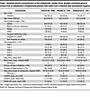 Table 1
Baseline patient characteristics in the randomized, double-blind, placebo-controlled phase II clinical trial of vadadustat in hospitalized patients with SARS-CoV-2 infection and concomitant hypoxia
Safety of vadadustat treatment in hospitalized patients with SARS-CoV-2. Previous studies of vadadustat have primarily evaluated treatment of anemia in outpatients with chronic kidney disease. Our trial differed in evaluating vadadustat in critically ill patients, including those with ARDS, sepsis, and other forms of severe organ injury. This shift in patient population raised salient safety concerns, particularly the risk of serious adverse events, including thromboembolic complications, which are already common in critically ill patients. However, our safety analysis revealed that adverse events were comparable in patients randomized to vadadustat or placebo (relative risk [RR] = 1.02, 95% Bayesian credible interval [CrI] = 0.94 to 1.11; posterior probability RR < 1 = 34.5%) and across all organ systems, adverse event grades, and treatment relatedness (Figure 4). The absence of significant differences in adverse events between vadadustat and placebo groups provides strong reassurance of vadadustat’s safety profile in hospitalized and critically ill patients.
 Figure 4
Vadadustat demonstrated a reassuring safety profile in hospitalized patients with SARS-CoV-2–associated lung injury. Forest plot of the favorable safety profile of vadadustat (900 mg daily) compared with placebo. Each category presents the estimated relative risk (RR) and corresponding 95% Bayesian credible interval (CrI), with dots indicating point estimates and error bars representing the CrIs. The overall pooled safety estimate, shown by the diamond, demonstrated vadadustat’s comparable safety relative to placebo across all organ systems, adverse event grades, and relatedness categories. Importantly, no increased risk was observed for thromboembolic events or serious adverse events (grade 3 and higher). These findings provide strong reassurance regarding the safety of vadadustat in hospitalized patients.
Pharmacological HIF activation. To assess whether treatment with the HIF stabilizer vadadustat increases HIF activity, we assessed HIF target gene expression. Since HIF stabilizers are used for renal anemia (45, 46), we chose the known target gene erythropoietin to indicate HIF activity (31). Consistent with the effect of vadadustat treatment on HIF stabilization, we observed elevated erythropoietin protein expression in treated patients. Erythropoietin expression increased 3-fold more in patients given vadadustat (β = 3, 95% CrI = 1.94 to 4.03) than placebo (β = 0.85, 95% CrI = 0.61 to 1.08; Figure 5A). Vadadustat dosing in our trial was thus sufficient to substantially augment HIF activity.
 Figure 5
Efficacy of vadadustat in hospitalized patients with SARS-CoV-2 infection and concomitant hypoxia. (A) Erythropoietin level in the plasma measured by ELISA showing changes over 14 days. Mean slopes with 95% Bayesian credible intervals (CrIs) are shown, highlighting a 3-fold increase in erythropoietin levels in the vadadustat group (β = 3, 95% CrI = 1.94 to 4.03) compared with placebo (β = 0.85, 95% CrI = 0.61 to 1.08) with a >99% posterior probability (PP) of treatment and time interaction. (B) Proportion of patients with severe lung injury requiring high oxygen support (NIAID-OS score ≥ 6) on day 14 (primary outcome). The vadadustat group showed a reduced absolute probability (13.3%) compared with placebo (16.9%), with an absolute risk difference (ARD) of –3.6% (95% CrI = –8.4% to 0.9%). PPs of benefit for ARD < 0% and ARD ≤ –2.5% are 94% and 69%, respectively. (C) Proportion of patients with NIAID-OS score ≥ 6 on day 7 (key secondary outcome). Vadadustat treatment demonstrated a higher likelihood of clinical improvement compared with placebo, with an ARD of –4.2% (95% CrI = –9.0% to –0.1%) and a PP of benefit (ARD < 0%) of 97%. (D) Inflammatory mediators in the plasma measured by MILLIPLEX Multiplex Assays (IL-17E, IP-10, M-CSF, and TNF-α) between day 0 and day 7. Vadadustat treatment resulted in greater reductions in systemic inflammation compared with placebo, with significant differences noted for each marker. P values were obtained by Mann-Whitney U test. In the bar-and-whisker plots, the bounds of the boxes represent the 25%–75% interquartile range, the lines within the boxes represent the median, the whiskers represent data min/max, and there are no outlying values. (E) Subgroup analysis by FiO2 levels upon hospital admission. PPs of clinical benefit for vadadustat (relative to placebo) were highest in patients with baseline FiO2 ≥ 80% (PP > 99%), followed by lower probabilities for FiO2 60%–79% (PP = 92%), 40%–59% (PP = 95%), and <40% (PP = 66%).
Efficacy endpoints. The primary outcome of clinically severe lung injury requiring high-level supplemental oxygen support (National Institute of Allergy and Infectious Diseases Ordinal Scale [NIAID-OS] score ≥ 6) on day 14 occurred in 43 patients in the vadadustat group (estimated probability, 13.3%) compared with 53 patients in the placebo group (estimated probability, 16.9%), representing an absolute risk difference (ARD) of –3.6% (95% CrI = –8.4% to 0.9%) (Figure 5B). The number of patients at each NIAID-OS score is reported in Supplemental Table 1. There was a 69% posterior probability that vadadustat reduced the absolute risk of the primary outcome by 2.5%, which did not meet our strict prespecified criterion for superiority (≥85% posterior probability of ARD ≤ 2.5%). However, there was a 94% posterior probability that vadadustat improved the primary outcome to some degree (ARD < 0%) compared with placebo (Supplemental Table 1 and Supplemental Figure 4A). Additionally, there was a 97.3% posterior probability that vadadustat improved the key secondary outcome of clinically severe lung injury requiring high-level supplemental oxygen support (NIAID-OS score ≥ 6) on day 7 (Supplemental Table 1 and Supplemental Figure 4B). This key secondary outcome occurred more often in the placebo group (estimated probability, 29.7%) than in the vadadustat group (estimated probability, 25.4%), resulting in an ARD of –4.2% (95% CrI = –9.0% to 0.1%; Figure 5C). The additional secondary outcomes are reported in Supplemental Table 2. Vadadustat reduced systemic inflammation on day 7, with substantial decreases in IL-17E, IP-10, M-CSF, and TNF-α (Figure 5D).
Subgroup analysis. We used Bayesian analyses to explore the probability of benefit from vadadustat (relative to placebo) in clinically important subgroups determined by baseline fraction of inspired oxygen (FiO2) ranges upon hospital admission, baseline Modified Sequential Organ Failure Assessment (mSOFA) respiratory score, and baseline SpO2. The strongest interaction was for FiO2 requirement upon hospital admission (posterior probability = 91%), with a benefit being most apparent in patients with high FiO2 requirements at hospital admission (Figure 5E). Specifically, there was a greater than 99% chance that treatment with vadadustat was associated with a clinical benefit on reducing lung injury severity in patients with baseline FiO2 of 80% or higher.
Lower probabilities of benefit were observed in patients with lower oxygen requirements (FiO2 60%–79%: 92%; FiO2 40%–59%: 95%; FiO2 < 40%: 66%). Modest evidence for treatment heterogeneity was observed for baseline SpO2 (posterior probability = 66%) (Supplemental Figure 5, A and B) and baseline mSOFA respiratory score (posterior probability = 72%) (Supplemental Figure 5, C and D). These results suggest that vadadustat treatment offers the most benefit in patients who have severe hypoxia and a high oxygen requirement.


