

Abstract
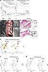
The precise mechanisms underlying the pathogenesis of idiopathic inflammatory myopathy (IIM) remain undefined. However, there has been increasing recognition that tissue-resident memory T cells (TRMs) play an important role in the pathogenesis of systemic autoimmune disease. In IIM, TRM-associated transcriptional signatures have been reported but on a very limited basis. By using multimodal single-cell RNA-sequencing analysis in our established murine model of histidyl-tRNA synthetase–induced myositis, we identified a prominent population of CD4+ TRMs in inflamed skeletal muscle. Muscle CD4+ TRMs exhibited high expression of genes encoding Cd69, Cxcr6, Runx3, and Prdm1, alongside low expression of Klf2, Ccr7, Sell, S1pr1, and Tcf7 — a profile that is generally consistent with previous reports of TRM gene signature and that we validate through comparison with transcriptomic profiles of human muscle tissue. Detailed pathway analysis in our model indicates that muscle CD4+ TRMs contribute to innate immune regulatory pathways enriched for TNF and IFN-γ signaling. Furthermore, analysis of TCR clonotype distribution and CDR3 sequence similarity revealed pronounced clonal expansion of CD4+ TRMs relative to other T cell subsets — a pattern that remained stable from 2 to 6 weeks after immunization. Collectively, these results suggest a potential role for CD4+ TRMs in the pathogenesis of autoimmune myositis.
Authors
Decheng Li, Daniel P. Reay, Iago Pinal-Fernandez, Maria Casal-Dominguez, Andrew L. Mammen, Sarah L. Gaffen, Timothy B. Oriss, Dana P. Ascherman
Abstract
BACKGROUND. Previous studies identified plasma proteins associated with chronic graft-versus-host disease (cGVHD). The goal of this cross-sectional study was to evaluate whether plasma biomarkers were associated with specific organ manifestations to help guide treatment choice. METHODS. Plasma proteins were measured in patients with cGVHD (N = 695) from Chronic GVHD Consortium studies. Correlations of plasma protein levels with individual organ involvement were tested with a P value of 0.05 or less considered significant after Benjamini-Hochberg adjustment and adjustment for 5 baseline clinical variables. RESULTS. Median time from cGVHD diagnosis to blood draw was 0.9 months (IQR 0.1–9.5). Donors were 50% HLA-matched unrelated, 32% matched related, and the remainder were umbilical cord blood, haploidentical, or mismatched unrelated donors. Methotrexate and calcineurin inhibitor prophylaxis for acute GVHD was used in 53% of patients. Overall, 326 (47%) had moderate and 244 (35%) had severe cGVHD with the following organ involvement at time of blood draw: skin (67%), mouth (60%), eye (49%), joint (34%), GI (31%), lung (23%), and liver (17%). After adjustment for batch effects and patient and transplant characteristics, 14 plasma proteins were associated with organ involvement with independent AUCs of 0.7–0.8. All organs except the eye were associated with at least 1 biomarker. However, no combination of plasma proteins improved model fit after adjusting for patient and transplant clinical variables. CONCLUSION. Correlations between plasma proteins and organ involvement were identified but are not actionable. Our future investigations will focus on more granular and immediately proximal determinants of cGVHD biology in both blood and tissue.
Authors
Stephanie J. Lee, Corey Cutler, Ningxin Ma, Timothy W. Randolph, George L. Chen, Joseph Pidala, Betty K. Hamilton, Carrie L. Kitko, Sally Arai, Najla El Jurdi, Lynn Onstad, Motoko Koyama, Catherine J. Lee, Sophie Paczesny, Geoffrey R. Hill
Abstract
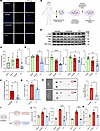
Estrogen can promote aggressive tumor phenotypes in estrogen receptor–positive (ER+) breast cancer; however, ER– cell lines are not widely considered estrogen responsive. Noncanonical estrogen-stimulated pathways such as the membrane-bound G protein–coupled estrogen receptor (GPR30) can mediate migratory and proliferative phenotypes in breast cancer and are postulated to promote resistance to aromatase therapies. Moreover, dysregulation of UDP-glucose 6-dehydrogenase (UGDH), a ubiquitously expressed enzyme critical to the metabolism of UDP-glucuronic acid into extracellular matrix precursors and hormone regulation, is associated with tumorigenesis. Here, we illustrated the impact of estrogen stimulation on tumor phenotypes in ER+ and ER– cell models in vitro and in vivo. We then demonstrated UGDH’s association with metastatic breast cancer via single-cell sequencing of patient specimens. Genetic knockdown of UGDH blunted estrogen-stimulated tumor phenotypes in vitro, ex vivo, and in vivo using both ER+ and ER– breast cancer lines. Finally, we demonstrated that UGDH knockdown blunted noncanonical estrogen stimulation through GPR30. Ultimately, our study validated prior studies demonstrating estrogen-responsive malignant phenotypes in ER– breast cancer and demonstrated that estrogen-stimulated breast cancer progression can be mediated through noncanonical pathways (e.g., UGDH/GPR30), regardless of ER status.
Authors
Meghan J. Price, Annee D. Nguyen, Corinne H. Strawser, Trupti Trivedi, Cesar C.D. Baeta, Catherine Lavau, Jovita K. Byemerwa, Debarati Mukherjee, Suzanne E. Wardell, Sandeep Artham, Vardhman Kumar, Shyni Varghese, C. Rory. Goodwin

Abstract
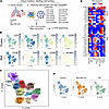
In rheumatoid arthritis (RA), CD4+ T cells specific for citrullinated antigens (cit-antigens) are key drivers of disease, but knowledge about epitopes and phenotypes remains limited. We characterized the frequency and phenotype of cit-specific CD4+ T cells in peripheral blood using HLA class II tetramers combined with computational analysis of phenotypic clusters to simultaneously detect peptides derived from 5 cit-antigens (aggrecan, vimentin, fibrinogen, cartilage intermediate layer protein, and α-enolase) previously implicated in RA pathogenesis. In a cross-sectional cohort, cit-aggrecan–, cit-vimentin–, and cit-fibrinogen–specific T cells were more frequent in participants with RA than healthy volunteers, associated with active disease, and had Th1-like and stem-like lineages in RA. In a longitudinal cohort investigating response to therapy, the frequency of cit-aggrecan–, cit-vimentin–, and cit-fibrinogen–specific CD4+ T cells was significantly higher at baseline and further elevated in responders. Furthermore, the frequency of cit-specific Th1-like cells in responders decreased over time. In contrast, the frequency of Th1-like cells in non-responders increased over time. Collectively, these findings demonstrate that cit-specific CD4+ T cells are expanded in RA and target a broad number of antigens across a breadth of phenotypes. Furthermore, the predominant antigen specificities associate with disease activity and exhibit dynamic changes in phenotype that reflect response to therapy.
Authors
Cliff Rims, Hannah A. DeBerg, Sylvia E. Posso, Virginia S. Muir, Hannes Uchtenhagen, Anne M. Hocking, Heather Bukiri, Jeffrey Carlin, Bernard Ng, Peter S. Linsley, Eddie A. James, Jane H. Buckner
Abstract
DNA damage and the cGAS/STING innate immunity pathway have been associated with fibrosis in systemic sclerosis (SSc), but a cause-and-effect role has not been established. Here we report the effects of TY1, a noncoding RNA drug of the exomer class that suppresses DNA damage and thereby inhibits cGAS/STING, in human SSc cells and in 2 preclinical models of SSc. Macrophages from patients with SSc exhibited high levels of phosphorylated DNA damage, cGAS, 2’3’-cGAMP, STING, and IFNs, all of which decreased after exposure to TY1. In mice that had been injected s.c. with bleomycin to model SSc, exercise tolerance, cardiac function, lung hydroxyproline, and skin thickness reverted to normal levels after oral administration of TY1. Similar therapeutic benefits were evident in the genetic tsk-1 mouse model of SSc. TY1 attenuated fibrosis and/or fibrotic gene expression in both mouse models of SSc and in human SSc skin fibroblasts. Our findings support the hypothesis that cGAS/STING, activated by DNA damage, is a key driver of fibrosis in SSc.
Authors
Xaviar M. Jones, Salwa Soussi, Alessandra Ciullo, Kara Tsi, Weixin Liu, Liang Li, Mario Fournier, Thassio Mesquita, Alberto M. Marchevsky, Nunzio Bottini, Francesco Boin, Ahmed G.E. Ibrahim, Eduardo Marbán

Abstract
Despite widespread vaccination, Bordetella pertussis (Bp) cases are resurging globally. Although CD4+ T cells are known to be essential for sustained protection, the antigens they recognize are not fully characterized, hindering vaccine refinement. Using immunopeptidomics, bioinformatics, and functional T cell assays, we identified high-affinity epitopes from reference and clinical Bp strains presented on MHC-II I-Ab. A subset of these epitopes stimulated systemic and mucosal CD4+ T cells of mice immunized with heat-killed Bp, and peripheral blood T cells from humans vaccinated with the whole-cell pertussis vaccine. Mice immunized with a subunit vaccine comprising two recombinant proteins identified in our screen were subsequently challenged with Bp. Bacterial burden was nearly eliminated from the lower respiratory tract and significantly reduced in the upper respiratory tract. Th1/Th17-polarized CD4+ tissue-resident memory T cells (Trms) were induced in nasal and pulmonary tissues. Depleting memory CD4+ T cells before challenge abolished protection, confirming that antigen-specific CD4+ T cells are critical for clearing Bp from the respiratory tract. Our integrated antigen identification and T cell assay approach revealed previously untested Bp antigens that elicit protective CD4+ T cell–mediated immunity, suggesting that incorporating them into new vaccines may help curb the resurgence of pertussis.
Authors
Mohamed M. Shamseldin, Jesse M. Hall, Griffin M. Lawrence, Gabrielle M. Hernandez, Yue Liu, Alexa R. Aubrey, Eva K. Verzani, Rajendar Deora, Amy Lovett-Racke, Jennifer G. Abelin, Purnima Dubey

Abstract
BACKGROUND. Loss of the Y chromosome (LOY) is a frequent event in male tumors and has been linked to cancer progression. However, the degree of mosaic LOY (mLOY) within normal tissues from men with or without cancer remains uncharacterized. METHODS. Here we used a FISH-based assay targeting X- and Y-chromosome centromeres to perform a pan-organ analysis of mLOY in 1,000 male tissue samples from 405 individuals representing 11 organs. Automated image processing generated a quantitative FISH-based mLOY score (YchrFISH) that we validated against a transcriptomic surrogate of Y-chromosome dosage from RNA-seq data. RESULTS. mLOY burden varied by tumor type, with highest degree in colorectal carcinoma. Across tissue groups, YchrFISH scores declined progressively from normal tissues of cancer-free men to histologically normal tissues adjacent to cancer and carcinoma (P < 0.0001). Paired analyses confirmed consistently greater mLOY in malignant compared with tumor-adjacent histologically normal tissue in different organs. Spatially resolved RNA-seq maps of bladders removed for cancer demonstrated a transcriptional gradient of Y-chromosome loss from normal urothelium through intraepithelial neoplasia to invasive carcinoma. CONCLUSION. mLOY gradients exist across histologically normal and malignant tissues, consistent with the concept of field cancerization. Our findings support epithelial mLOY as a biomarker of early malignant transformation and, to our knowledge, a previously unrecognized hallmark of male oncogenesis. FUNDING. NIH grants R35CA294022, P01CA163227, and P50CA97186 (the Pacific Northwest Prostate Cancer SPORE) and the Institute for Prostate Cancer Research.
Authors
Arkadiusz Gertych, Huihui Ye, Xingyu Chen, Eric Vail, V. Krishnan Ramanujan, Lauren Brady, Lawrence D. True, Peter S. Nelson, Peter R. Carroll, Dan Theodorescu

Abstract
Foxp3 deficiency causes a profound loss of immune tolerance, unleashing autoreactive T and B cells, lymphoproliferation, cytokine-driven inflammation, and autoantibody production. This autoimmune pathology is fueled by increased glutamine usage, but it remains unresolved whether glutamine is necessary to produce energy or for intermediate metabolite biosynthesis responsible for immunomodulation. Here, we demonstrate that glutamine utilization for biosynthetic pathways supported autoimmune inflammation in the settings of Foxp3 deficiency and dextran sodium sulfate–induced colitis. By employing a model of autoimmunity driven by Treg-specific loss of Foxp3, we showed that this effect is independent of pathogenic Foxp3-deficient Treg reprogramming. Mechanistically, glutamine biosynthetic pathways sustained conventional T cell activation and proinflammatory cytokine production by preventing inosine accumulation and signaling, thus implicating adenosine pathway modulation in autoreactive T cell dysregulation. Conversely, autoreactive B cell activation and autoantibody production relied on glutamine-dependent asparagine availability, which we identified as a targetable vulnerability for autoantibody formation. These findings highlighted glutamine-driven biosynthetic processes as critical drivers of autoimmunity and revealed distinct metabolic vulnerabilities in autoreactive T and B cells that could be targeted for therapeutic intervention.
Authors
Mohammad Adeel Zafar, Charlotte N. Hill Machado, Jyotirmaya Behera, Yuelin Zhong, Xiao Li, Shakchhi Joshi, Yassine El Fazaa, Virginia Camacho, Peter Georgiev, Kiran Kurmi, Marcia Haigis, Louis-Marie Charbonnier
Abstract
Idiopathic pulmonary fibrosis (IPF) is a fatal interstitial lung disease characterized by progressive scarring and respiratory failure. While T cells are elevated in IPF lungs, their contributions to fibrosis beyond inflammation remain poorly understood. Here, we performed multiplex imaging and single-cell RNA and protein profiling on about 90,000 CD3+ T cells from control and fibrotic lungs, revealing 11 distinct subsets of CD4+ and CD8+ T cells, including a rare CD56+ regulatory T cell. In addition to increased T cell numbers in severely fibrotic lungs compared with non-diseased controls, we observed CD4+ and CD8+ T cells localized near epithelial cells and in niches of abnormal epithelium. CXCR4/MIF signaling emerged as a central axis mediating T cell–epithelial interactions, while epidermal growth factor receptor (EGFR) and TGF-β pathways dominated in multiple T cell subsets. Our findings support the concept that T cells in IPF adopt nonclassical activation patterns that are driven by epithelial interactions within the fibrotic microenvironment. These studies provide a foundation for exploring alternative therapeutic strategies in IPF lungs by modulating T cell behavior and communication networks.
Authors
Ana P.M. Serezani, Julia M.R. Bazzano, Bruno D. Pascoalino, Ludmilla da Silva, Abigail J. Dietrich, Chase J. Taylor, Taylor Sherrill, Annika Vannan, Carla L. Calvi, Paula I. Gonzalez-Ericsson, Erin M. Wilfong, Matthew Bacchetta, Ciara M. Shaver, Lorraine B. Ware, Margaret L. Salisbury, Luc Van Kaer, Nicholas E. Banovich, Jonathan A. Kropski, Timothy S. Blackwell
Abstract
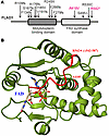
The FLAD1 gene codes for flavin adenine dinucleotide (FAD) synthase. FAD is a cofactor for many redox enzymes involved in vital processes from respiration to signal transduction. In this work, we described a clinical case of 2 siblings carrying compound heterozygous mutations in the FLAD1 gene resulting in the substitutions A418V and R542* at the protein level. The patients demonstrate adrenal insufficiency, which has not previously been associated with FLAD1 protein defects. To verify that adrenal insufficiency is caused by FLAD1 mutations, we created a personalized mouse model carrying the mutations found in the patients. The mutation in the FLAD1 gene, leading to the A418V substitution, appeared viable in the homozygous state, with minimal difference from the WT. The FLAD1 gene mutation leading to the R542* truncation is lethal when homozygous. The mouse model of the compound heterozygous FLAD1A418V/R542* mutations recapitulated the physiological, biochemical, and endocrine manifestations of FLAD1 mutations in patients. The mouse model created demonstrates the causal effect of FLAD1 mutations on the described pathology and potentially paves the way for understanding the disease’s molecular mechanism and developing better therapies.
Authors
Olga A. Averina, Natalia Yu. Kalinchenko, Vitaly A. Ioutsi, Andrey V. Pirogov, Alexander V. Bogachev, Oleg A. Permyakov, Vitaly S. Buev, Ekaterina A. Guseva, Anastasia V. Priymak, Olga A. Bazhanova, Mariia A. Emelianova, Olga O. Grigoryeva, Galina V. Baydakova, Maxim A. Abakumov, Vasily N. Manskikh, Olga A. Dontsova, Petr V. Sergiev, Anatoly N. Tiulpakov
No posts were found with this tag.


