
Research ArticleAIDS/HIVInflammationMicrobiology
Open Access |  10.1172/jci.insight.205379
Multi-omics links microbial dysbiosis, systemic inflammation, and metabolomic disruptions to SNAE risk in treated HIV
Christopher M. Basting,1 Jodi Anderson,2 Kevin Escandón,2 Garritt Wieking,2 Candace Guerrero,1 Jarrett Reichel,2 Ross T. Cromarty,3 Erik Swanson,1 Ty Schroeder,1 Elaina Creagan,1 Maura Barrett,1 Fernanda Torres-Ruiz,4 Maribel Soto-Nava,4 Lady Carvajal-Ruiz,4 Karla Krystel Ordaz-Candelario,4 Olivia Briceño,4 Nicholas Funderburg,5 Melanie Graham,6 Peter Hunt,7 Santiago Avila-Rios,4 Gonzalo Salgado Montes de Oca,4 Timothy W. Schacker,2 and Nichole R. Klatt1
1Division of Surgical Outcomes and Precision Medicine Research, Department of Surgery,
2Division of Infectious Diseases and International Medicine, Department of Medicine, and
3Masonic Cancer Center, University of Minnesota, Minneapolis, Minnesota, USA.
4Instituto Nacional de Enfermedades Respiratorias Ismael Cosío Villegas, Centro de Investigación en Enfermedades Infecciosas, Mexico City, Mexico.
5School of Health and Rehabilitation Sciences, Ohio State University, Columbus, Ohio, USA.
6Pre-Clinical Research Center, Department of Surgery, University of Minnesota, Minneapolis, Minnesota, USA.
7Division of Experimental Medicine, University of California San Francisco. San Francisco, California, USA.
Address correspondence to: Nichole R. Klatt, Department of Surgery, University of Minnesota, Minneapolis, Minnesota, USA. Email: klat0037@umn.edu.
Find articles by Basting, C. in: PubMed | Google Scholar
1Division of Surgical Outcomes and Precision Medicine Research, Department of Surgery,
2Division of Infectious Diseases and International Medicine, Department of Medicine, and
3Masonic Cancer Center, University of Minnesota, Minneapolis, Minnesota, USA.
4Instituto Nacional de Enfermedades Respiratorias Ismael Cosío Villegas, Centro de Investigación en Enfermedades Infecciosas, Mexico City, Mexico.
5School of Health and Rehabilitation Sciences, Ohio State University, Columbus, Ohio, USA.
6Pre-Clinical Research Center, Department of Surgery, University of Minnesota, Minneapolis, Minnesota, USA.
7Division of Experimental Medicine, University of California San Francisco. San Francisco, California, USA.
Address correspondence to: Nichole R. Klatt, Department of Surgery, University of Minnesota, Minneapolis, Minnesota, USA. Email: klat0037@umn.edu.
Find articles by Anderson, J. in: PubMed | Google Scholar
1Division of Surgical Outcomes and Precision Medicine Research, Department of Surgery,
2Division of Infectious Diseases and International Medicine, Department of Medicine, and
3Masonic Cancer Center, University of Minnesota, Minneapolis, Minnesota, USA.
4Instituto Nacional de Enfermedades Respiratorias Ismael Cosío Villegas, Centro de Investigación en Enfermedades Infecciosas, Mexico City, Mexico.
5School of Health and Rehabilitation Sciences, Ohio State University, Columbus, Ohio, USA.
6Pre-Clinical Research Center, Department of Surgery, University of Minnesota, Minneapolis, Minnesota, USA.
7Division of Experimental Medicine, University of California San Francisco. San Francisco, California, USA.
Address correspondence to: Nichole R. Klatt, Department of Surgery, University of Minnesota, Minneapolis, Minnesota, USA. Email: klat0037@umn.edu.
Find articles by
Escandón, K.
in:
PubMed
|
Google Scholar
|

1Division of Surgical Outcomes and Precision Medicine Research, Department of Surgery,
2Division of Infectious Diseases and International Medicine, Department of Medicine, and
3Masonic Cancer Center, University of Minnesota, Minneapolis, Minnesota, USA.
4Instituto Nacional de Enfermedades Respiratorias Ismael Cosío Villegas, Centro de Investigación en Enfermedades Infecciosas, Mexico City, Mexico.
5School of Health and Rehabilitation Sciences, Ohio State University, Columbus, Ohio, USA.
6Pre-Clinical Research Center, Department of Surgery, University of Minnesota, Minneapolis, Minnesota, USA.
7Division of Experimental Medicine, University of California San Francisco. San Francisco, California, USA.
Address correspondence to: Nichole R. Klatt, Department of Surgery, University of Minnesota, Minneapolis, Minnesota, USA. Email: klat0037@umn.edu.
Find articles by Wieking, G. in: PubMed | Google Scholar
1Division of Surgical Outcomes and Precision Medicine Research, Department of Surgery,
2Division of Infectious Diseases and International Medicine, Department of Medicine, and
3Masonic Cancer Center, University of Minnesota, Minneapolis, Minnesota, USA.
4Instituto Nacional de Enfermedades Respiratorias Ismael Cosío Villegas, Centro de Investigación en Enfermedades Infecciosas, Mexico City, Mexico.
5School of Health and Rehabilitation Sciences, Ohio State University, Columbus, Ohio, USA.
6Pre-Clinical Research Center, Department of Surgery, University of Minnesota, Minneapolis, Minnesota, USA.
7Division of Experimental Medicine, University of California San Francisco. San Francisco, California, USA.
Address correspondence to: Nichole R. Klatt, Department of Surgery, University of Minnesota, Minneapolis, Minnesota, USA. Email: klat0037@umn.edu.
Find articles by Guerrero, C. in: PubMed | Google Scholar
1Division of Surgical Outcomes and Precision Medicine Research, Department of Surgery,
2Division of Infectious Diseases and International Medicine, Department of Medicine, and
3Masonic Cancer Center, University of Minnesota, Minneapolis, Minnesota, USA.
4Instituto Nacional de Enfermedades Respiratorias Ismael Cosío Villegas, Centro de Investigación en Enfermedades Infecciosas, Mexico City, Mexico.
5School of Health and Rehabilitation Sciences, Ohio State University, Columbus, Ohio, USA.
6Pre-Clinical Research Center, Department of Surgery, University of Minnesota, Minneapolis, Minnesota, USA.
7Division of Experimental Medicine, University of California San Francisco. San Francisco, California, USA.
Address correspondence to: Nichole R. Klatt, Department of Surgery, University of Minnesota, Minneapolis, Minnesota, USA. Email: klat0037@umn.edu.
Find articles by Reichel, J. in: PubMed | Google Scholar
1Division of Surgical Outcomes and Precision Medicine Research, Department of Surgery,
2Division of Infectious Diseases and International Medicine, Department of Medicine, and
3Masonic Cancer Center, University of Minnesota, Minneapolis, Minnesota, USA.
4Instituto Nacional de Enfermedades Respiratorias Ismael Cosío Villegas, Centro de Investigación en Enfermedades Infecciosas, Mexico City, Mexico.
5School of Health and Rehabilitation Sciences, Ohio State University, Columbus, Ohio, USA.
6Pre-Clinical Research Center, Department of Surgery, University of Minnesota, Minneapolis, Minnesota, USA.
7Division of Experimental Medicine, University of California San Francisco. San Francisco, California, USA.
Address correspondence to: Nichole R. Klatt, Department of Surgery, University of Minnesota, Minneapolis, Minnesota, USA. Email: klat0037@umn.edu.
Find articles by Cromarty, R. in: PubMed | Google Scholar
1Division of Surgical Outcomes and Precision Medicine Research, Department of Surgery,
2Division of Infectious Diseases and International Medicine, Department of Medicine, and
3Masonic Cancer Center, University of Minnesota, Minneapolis, Minnesota, USA.
4Instituto Nacional de Enfermedades Respiratorias Ismael Cosío Villegas, Centro de Investigación en Enfermedades Infecciosas, Mexico City, Mexico.
5School of Health and Rehabilitation Sciences, Ohio State University, Columbus, Ohio, USA.
6Pre-Clinical Research Center, Department of Surgery, University of Minnesota, Minneapolis, Minnesota, USA.
7Division of Experimental Medicine, University of California San Francisco. San Francisco, California, USA.
Address correspondence to: Nichole R. Klatt, Department of Surgery, University of Minnesota, Minneapolis, Minnesota, USA. Email: klat0037@umn.edu.
Find articles by Swanson, E. in: PubMed | Google Scholar
1Division of Surgical Outcomes and Precision Medicine Research, Department of Surgery,
2Division of Infectious Diseases and International Medicine, Department of Medicine, and
3Masonic Cancer Center, University of Minnesota, Minneapolis, Minnesota, USA.
4Instituto Nacional de Enfermedades Respiratorias Ismael Cosío Villegas, Centro de Investigación en Enfermedades Infecciosas, Mexico City, Mexico.
5School of Health and Rehabilitation Sciences, Ohio State University, Columbus, Ohio, USA.
6Pre-Clinical Research Center, Department of Surgery, University of Minnesota, Minneapolis, Minnesota, USA.
7Division of Experimental Medicine, University of California San Francisco. San Francisco, California, USA.
Address correspondence to: Nichole R. Klatt, Department of Surgery, University of Minnesota, Minneapolis, Minnesota, USA. Email: klat0037@umn.edu.
Find articles by Schroeder, T. in: PubMed | Google Scholar
1Division of Surgical Outcomes and Precision Medicine Research, Department of Surgery,
2Division of Infectious Diseases and International Medicine, Department of Medicine, and
3Masonic Cancer Center, University of Minnesota, Minneapolis, Minnesota, USA.
4Instituto Nacional de Enfermedades Respiratorias Ismael Cosío Villegas, Centro de Investigación en Enfermedades Infecciosas, Mexico City, Mexico.
5School of Health and Rehabilitation Sciences, Ohio State University, Columbus, Ohio, USA.
6Pre-Clinical Research Center, Department of Surgery, University of Minnesota, Minneapolis, Minnesota, USA.
7Division of Experimental Medicine, University of California San Francisco. San Francisco, California, USA.
Address correspondence to: Nichole R. Klatt, Department of Surgery, University of Minnesota, Minneapolis, Minnesota, USA. Email: klat0037@umn.edu.
Find articles by Creagan, E. in: PubMed | Google Scholar
1Division of Surgical Outcomes and Precision Medicine Research, Department of Surgery,
2Division of Infectious Diseases and International Medicine, Department of Medicine, and
3Masonic Cancer Center, University of Minnesota, Minneapolis, Minnesota, USA.
4Instituto Nacional de Enfermedades Respiratorias Ismael Cosío Villegas, Centro de Investigación en Enfermedades Infecciosas, Mexico City, Mexico.
5School of Health and Rehabilitation Sciences, Ohio State University, Columbus, Ohio, USA.
6Pre-Clinical Research Center, Department of Surgery, University of Minnesota, Minneapolis, Minnesota, USA.
7Division of Experimental Medicine, University of California San Francisco. San Francisco, California, USA.
Address correspondence to: Nichole R. Klatt, Department of Surgery, University of Minnesota, Minneapolis, Minnesota, USA. Email: klat0037@umn.edu.
Find articles by Barrett, M. in: PubMed | Google Scholar
1Division of Surgical Outcomes and Precision Medicine Research, Department of Surgery,
2Division of Infectious Diseases and International Medicine, Department of Medicine, and
3Masonic Cancer Center, University of Minnesota, Minneapolis, Minnesota, USA.
4Instituto Nacional de Enfermedades Respiratorias Ismael Cosío Villegas, Centro de Investigación en Enfermedades Infecciosas, Mexico City, Mexico.
5School of Health and Rehabilitation Sciences, Ohio State University, Columbus, Ohio, USA.
6Pre-Clinical Research Center, Department of Surgery, University of Minnesota, Minneapolis, Minnesota, USA.
7Division of Experimental Medicine, University of California San Francisco. San Francisco, California, USA.
Address correspondence to: Nichole R. Klatt, Department of Surgery, University of Minnesota, Minneapolis, Minnesota, USA. Email: klat0037@umn.edu.
Find articles by Torres-Ruiz, F. in: PubMed | Google Scholar
1Division of Surgical Outcomes and Precision Medicine Research, Department of Surgery,
2Division of Infectious Diseases and International Medicine, Department of Medicine, and
3Masonic Cancer Center, University of Minnesota, Minneapolis, Minnesota, USA.
4Instituto Nacional de Enfermedades Respiratorias Ismael Cosío Villegas, Centro de Investigación en Enfermedades Infecciosas, Mexico City, Mexico.
5School of Health and Rehabilitation Sciences, Ohio State University, Columbus, Ohio, USA.
6Pre-Clinical Research Center, Department of Surgery, University of Minnesota, Minneapolis, Minnesota, USA.
7Division of Experimental Medicine, University of California San Francisco. San Francisco, California, USA.
Address correspondence to: Nichole R. Klatt, Department of Surgery, University of Minnesota, Minneapolis, Minnesota, USA. Email: klat0037@umn.edu.
Find articles by Soto-Nava, M. in: PubMed | Google Scholar
1Division of Surgical Outcomes and Precision Medicine Research, Department of Surgery,
2Division of Infectious Diseases and International Medicine, Department of Medicine, and
3Masonic Cancer Center, University of Minnesota, Minneapolis, Minnesota, USA.
4Instituto Nacional de Enfermedades Respiratorias Ismael Cosío Villegas, Centro de Investigación en Enfermedades Infecciosas, Mexico City, Mexico.
5School of Health and Rehabilitation Sciences, Ohio State University, Columbus, Ohio, USA.
6Pre-Clinical Research Center, Department of Surgery, University of Minnesota, Minneapolis, Minnesota, USA.
7Division of Experimental Medicine, University of California San Francisco. San Francisco, California, USA.
Address correspondence to: Nichole R. Klatt, Department of Surgery, University of Minnesota, Minneapolis, Minnesota, USA. Email: klat0037@umn.edu.
Find articles by Carvajal-Ruiz, L. in: PubMed | Google Scholar
1Division of Surgical Outcomes and Precision Medicine Research, Department of Surgery,
2Division of Infectious Diseases and International Medicine, Department of Medicine, and
3Masonic Cancer Center, University of Minnesota, Minneapolis, Minnesota, USA.
4Instituto Nacional de Enfermedades Respiratorias Ismael Cosío Villegas, Centro de Investigación en Enfermedades Infecciosas, Mexico City, Mexico.
5School of Health and Rehabilitation Sciences, Ohio State University, Columbus, Ohio, USA.
6Pre-Clinical Research Center, Department of Surgery, University of Minnesota, Minneapolis, Minnesota, USA.
7Division of Experimental Medicine, University of California San Francisco. San Francisco, California, USA.
Address correspondence to: Nichole R. Klatt, Department of Surgery, University of Minnesota, Minneapolis, Minnesota, USA. Email: klat0037@umn.edu.
Find articles by Ordaz-Candelario, K. in: PubMed | Google Scholar
1Division of Surgical Outcomes and Precision Medicine Research, Department of Surgery,
2Division of Infectious Diseases and International Medicine, Department of Medicine, and
3Masonic Cancer Center, University of Minnesota, Minneapolis, Minnesota, USA.
4Instituto Nacional de Enfermedades Respiratorias Ismael Cosío Villegas, Centro de Investigación en Enfermedades Infecciosas, Mexico City, Mexico.
5School of Health and Rehabilitation Sciences, Ohio State University, Columbus, Ohio, USA.
6Pre-Clinical Research Center, Department of Surgery, University of Minnesota, Minneapolis, Minnesota, USA.
7Division of Experimental Medicine, University of California San Francisco. San Francisco, California, USA.
Address correspondence to: Nichole R. Klatt, Department of Surgery, University of Minnesota, Minneapolis, Minnesota, USA. Email: klat0037@umn.edu.
Find articles by Briceño, O. in: PubMed | Google Scholar
1Division of Surgical Outcomes and Precision Medicine Research, Department of Surgery,
2Division of Infectious Diseases and International Medicine, Department of Medicine, and
3Masonic Cancer Center, University of Minnesota, Minneapolis, Minnesota, USA.
4Instituto Nacional de Enfermedades Respiratorias Ismael Cosío Villegas, Centro de Investigación en Enfermedades Infecciosas, Mexico City, Mexico.
5School of Health and Rehabilitation Sciences, Ohio State University, Columbus, Ohio, USA.
6Pre-Clinical Research Center, Department of Surgery, University of Minnesota, Minneapolis, Minnesota, USA.
7Division of Experimental Medicine, University of California San Francisco. San Francisco, California, USA.
Address correspondence to: Nichole R. Klatt, Department of Surgery, University of Minnesota, Minneapolis, Minnesota, USA. Email: klat0037@umn.edu.
Find articles by Funderburg, N. in: PubMed | Google Scholar
1Division of Surgical Outcomes and Precision Medicine Research, Department of Surgery,
2Division of Infectious Diseases and International Medicine, Department of Medicine, and
3Masonic Cancer Center, University of Minnesota, Minneapolis, Minnesota, USA.
4Instituto Nacional de Enfermedades Respiratorias Ismael Cosío Villegas, Centro de Investigación en Enfermedades Infecciosas, Mexico City, Mexico.
5School of Health and Rehabilitation Sciences, Ohio State University, Columbus, Ohio, USA.
6Pre-Clinical Research Center, Department of Surgery, University of Minnesota, Minneapolis, Minnesota, USA.
7Division of Experimental Medicine, University of California San Francisco. San Francisco, California, USA.
Address correspondence to: Nichole R. Klatt, Department of Surgery, University of Minnesota, Minneapolis, Minnesota, USA. Email: klat0037@umn.edu.
Find articles by Graham, M. in: PubMed | Google Scholar
1Division of Surgical Outcomes and Precision Medicine Research, Department of Surgery,
2Division of Infectious Diseases and International Medicine, Department of Medicine, and
3Masonic Cancer Center, University of Minnesota, Minneapolis, Minnesota, USA.
4Instituto Nacional de Enfermedades Respiratorias Ismael Cosío Villegas, Centro de Investigación en Enfermedades Infecciosas, Mexico City, Mexico.
5School of Health and Rehabilitation Sciences, Ohio State University, Columbus, Ohio, USA.
6Pre-Clinical Research Center, Department of Surgery, University of Minnesota, Minneapolis, Minnesota, USA.
7Division of Experimental Medicine, University of California San Francisco. San Francisco, California, USA.
Address correspondence to: Nichole R. Klatt, Department of Surgery, University of Minnesota, Minneapolis, Minnesota, USA. Email: klat0037@umn.edu.
Find articles by Hunt, P. in: PubMed | Google Scholar
1Division of Surgical Outcomes and Precision Medicine Research, Department of Surgery,
2Division of Infectious Diseases and International Medicine, Department of Medicine, and
3Masonic Cancer Center, University of Minnesota, Minneapolis, Minnesota, USA.
4Instituto Nacional de Enfermedades Respiratorias Ismael Cosío Villegas, Centro de Investigación en Enfermedades Infecciosas, Mexico City, Mexico.
5School of Health and Rehabilitation Sciences, Ohio State University, Columbus, Ohio, USA.
6Pre-Clinical Research Center, Department of Surgery, University of Minnesota, Minneapolis, Minnesota, USA.
7Division of Experimental Medicine, University of California San Francisco. San Francisco, California, USA.
Address correspondence to: Nichole R. Klatt, Department of Surgery, University of Minnesota, Minneapolis, Minnesota, USA. Email: klat0037@umn.edu.
Find articles by
Avila-Rios, S.
in:
PubMed
|
Google Scholar
|
1Division of Surgical Outcomes and Precision Medicine Research, Department of Surgery,
2Division of Infectious Diseases and International Medicine, Department of Medicine, and
3Masonic Cancer Center, University of Minnesota, Minneapolis, Minnesota, USA.
4Instituto Nacional de Enfermedades Respiratorias Ismael Cosío Villegas, Centro de Investigación en Enfermedades Infecciosas, Mexico City, Mexico.
5School of Health and Rehabilitation Sciences, Ohio State University, Columbus, Ohio, USA.
6Pre-Clinical Research Center, Department of Surgery, University of Minnesota, Minneapolis, Minnesota, USA.
7Division of Experimental Medicine, University of California San Francisco. San Francisco, California, USA.
Address correspondence to: Nichole R. Klatt, Department of Surgery, University of Minnesota, Minneapolis, Minnesota, USA. Email: klat0037@umn.edu.
Find articles by Salgado Montes de Oca, G. in: PubMed | Google Scholar
1Division of Surgical Outcomes and Precision Medicine Research, Department of Surgery,
2Division of Infectious Diseases and International Medicine, Department of Medicine, and
3Masonic Cancer Center, University of Minnesota, Minneapolis, Minnesota, USA.
4Instituto Nacional de Enfermedades Respiratorias Ismael Cosío Villegas, Centro de Investigación en Enfermedades Infecciosas, Mexico City, Mexico.
5School of Health and Rehabilitation Sciences, Ohio State University, Columbus, Ohio, USA.
6Pre-Clinical Research Center, Department of Surgery, University of Minnesota, Minneapolis, Minnesota, USA.
7Division of Experimental Medicine, University of California San Francisco. San Francisco, California, USA.
Address correspondence to: Nichole R. Klatt, Department of Surgery, University of Minnesota, Minneapolis, Minnesota, USA. Email: klat0037@umn.edu.
Find articles by Schacker, T. in: PubMed | Google Scholar
1Division of Surgical Outcomes and Precision Medicine Research, Department of Surgery,
2Division of Infectious Diseases and International Medicine, Department of Medicine, and
3Masonic Cancer Center, University of Minnesota, Minneapolis, Minnesota, USA.
4Instituto Nacional de Enfermedades Respiratorias Ismael Cosío Villegas, Centro de Investigación en Enfermedades Infecciosas, Mexico City, Mexico.
5School of Health and Rehabilitation Sciences, Ohio State University, Columbus, Ohio, USA.
6Pre-Clinical Research Center, Department of Surgery, University of Minnesota, Minneapolis, Minnesota, USA.
7Division of Experimental Medicine, University of California San Francisco. San Francisco, California, USA.
Address correspondence to: Nichole R. Klatt, Department of Surgery, University of Minnesota, Minneapolis, Minnesota, USA. Email: klat0037@umn.edu.
Find articles by Klatt, N. in: PubMed | Google Scholar
Published July 22, 2026 - More info
JCI Insight. 2026;11(14):e205379. https://doi.org/10.1172/jci.insight.205379.
© 2026 Basting et al. This work is licensed under the Creative Commons Attribution 4.0 International License. To view a copy of this license, visit http://creativecommons.org/licenses/by/4.0/.
Received: February 3, 2026; Accepted: May 19, 2026

-
Results
Differences in baseline characteristics. Baseline characteristics for enrolled participants from the United States and Mexico are shown in Table 1. Compared with PWH from the United States, PWH from Mexico were older (P = 0.037) and had significantly lower CD4+ T cell counts (P = 0.006) and a lower nadir CD4+ T cell count (P = 0.003). When stratified by SNAE risk group (Table 2, with PWoH included as a reference), the high-risk group had significantly lower CD4+ T cell counts (P < 0.001), a lower CD4/CD8 ratio (P = 0.001), and lower nadir CD4+ T cell count (P = 0.002) compared with the low-risk group. There was no significant difference in country of enrollment (P = 0.6) or race (P = 0.8) between low- and high-risk groups. CMV IgG serostatus was assessed in all HIV-positive participants, of which all but 1 were seropositive (Supplemental Tables 1 and 2; supplemental material available online with this article; https://doi.org/10.1172/jci.insight.205379DS1). Summaries of ART regimens are provided with stratification by country (Supplemental Table 3) and SNAE risk group (Supplemental Table 4). Most enrolled participants (78%) were on an ART regimen consisting of 2 nucleoside/nucleotide reverse transcriptase inhibitors (NRTIs) and 1 integrase strand transfer inhibitor (INSTI). There was a significant difference in ART regimens between countries (P < 0.001), reflecting differences in national treatment guidelines. There was no difference in ART regimens between SNAE risk groups (P = 0.5). Additionally, clinical characteristics did not differ significantly between PWoH across countries (Supplemental Table 5).
Multi-omic geographic differences in PWH on ART. Geographic and environmental factors shape the microbiome, metabolome, and immune landscape, yet most HIV omic studies have focused on cohorts from high-income countries. The global heterogeneity of the gut microbiome in PWH, and its intersection with metabolic and immune phenotypes, remains poorly characterized.
We first looked at plasma concentrations of cytokines, markers of gut epithelial barrier dysfunction, and microbial translocation, including intestinal fatty acid binding protein (I-FABP), lipopolysaccharide binding protein (LBP), and soluble CD14 (sCD14), by principal component analysis (PCA). Country of enrollment was the largest and only significant effect on the overall composition of these biomarkers when tested by PERMANOVA (R2 = 0.068, q = 0.020, Figure 1A). Numerous plasma proteins were differentially abundant between participants in different countries (Figure 1B). Compared with PWH from the United States, those from Mexico had significantly higher concentrations of I-FABP (P < 0.001, q < 0.001) and sCD14 (P = 0.018, q = 0.1637). Cytokines involved in antigen presentation (15), mucosal function (16), and proliferation (17) were also elevated in patients from Mexico including IL-12p70 (P < 0.001, q = 0.0028), IL-17A (P = 0.002, q = 0.0341), and IL-2 (P = 0.003, q = 0.0341). PWH from the United States were characterized by increased concentrations of macrophage inflammatory protein 1β (MIP-1β) (P < 0.001, q = 0.0086), IL-7 (P = 0.009, q = 0.0916), and IL-8 (P = 0.003, q = 0.0341).
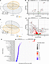 Figure 1
Differences in plasma proteins and metabolome between PWH enrolled in the United States and Mexico. (A) Principal component analysis of plasma protein concentrations, colored by country of enrollment and tested by PERMANOVA. (B) Volcano plot of cytokines and gut barrier integrity markers differentially associated with country of enrollment. (C) Principal component analysis of plasma metabolomic features, colored by country of enrollment and tested by PERMANOVA. (D) Volcano plot of metabolomic features differentially associated with country of enrollment. (E) GSEA of metabolomic data comparing KEGG pathway enrichment between PWH in the United States and Mexico; asterisks denote FDR-adjusted q ≤ 0.25.
We next evaluated the plasma metabolome of PWH from the United States or Mexico using a comprehensive targeted liquid chromatography–tandem mass spectrometry panel of 1,006 metabolites, including 751 lipids, 255 small molecules, and 273 sums or ratios, for a total of 1,279 metabolomic features after filtering. Once again, PCA showed that country was the largest and only significant effect on the overall metabolomic profile composition (R2 = 0.077, q = 0.004, Figure 1C), followed by age. Differential analysis revealed 357 metabolomic features that were significantly different between PWH in the United States and Mexico at an adjusted P value threshold of 0.25 or less, and 236 at a threshold of 0.05 or less, most of which were elevated in PWH from the United States (Figure 1D and Supplemental Data 1). When looking at the classes of significantly different metabolites (q ≤ 0.25, Supplemental Figure 1), phospholipids were especially higher in PWH from the United States, including phosphatidylethanolamines (PEs), phosphatidylcholines, phosphatidylglycerols (PGs), and phosphatidylserines, which represented 39.6% of the significantly different metabolites. The lyso-forms of these lipid classes were also elevated in PWH from the United States including lyso-PEs and lysophosphatidylcholines. Sphingomyelins were also exclusively elevated in PWH from the United States. The primary metabolite class that was most represented by PWH in Mexico was acylcarnitines. When looking at individual metabolites, taurine, nicotinamide, oxaloacetic acid, spermidine, and acetylneuraminic acid were some of the top US-associated metabolites (P < 0.001 and q < 0.001 for all metabolites), and sarcosine (P < 0.001, q < 0.001), biotin (P < 0.001, q < 0.001), glycolic acid (P < 0.001, q = 0.001), octanoylcarnitine (P < 0.001, q = 0.012), and decanoylcarnitine (P < 0.001, q = 0.012) were some of the top Mexico-associated metabolites. Gene set enrichment analysis (GSEA) of the metabolite data showed that the Kyoto Encyclopedia of Genes and Genomes (KEGG) pathways for primary bile acid biosynthesis, taurine/hypotaurine metabolism, and nicotinate/nicotinamide metabolism were all enriched in PWH from the United States (q < 0.25, Figure 1E).
At the phylum level, ileal and rectal biopsies were primarily composed of Bacillota (formerly Firmicutes) and Bacteroidota (Supplemental Figure 2A). We did not observe any difference in alpha diversity metrics for rectal or ileal biopsies between the United States and Mexico (Figure 2, A and B, respectively). Overall microbial composition of the rectal biopsies (Figure 2C), but not ileal biopsies (Figure 2D), was significantly associated with country of enrollment (PERMANOVA: R2 = 0.076, q = 0.02). Differential abundance analysis performed at the genus and species level showed increased abundances of Ruminococcus, Eubacterium eligens group, Enterococcus, Veillonella, Escherichia-Shigella, Blautia stercoris, and Gemmiger formicilis in the rectal biopsies of PWH from Mexico and increased abundances of Agathobaculum butyriciproducens, Flavonifractor, Flavonifractor plautii, Enterocloster, Parabacteroides distasonis, and Parabacteroides in PWH from the United States (q < 0.25, Figure 2E). When comparing the ileal biopsies, we observed increased abundance of Eubacterium eligens group, Escherichia-Shigella, Oscillospiraceae NK4A214 group, and Haemophilus in PWH from Mexico, whereas Akkermansia, Akkermansia muciniphila, Desulfovibrio, Phascolarctobacterium faecium, Bacteroides uniformis, Parabacteroides distasonis, and Phascolarctobacterium were all increased in PWH from the United States (q < 0.25, Figure 2F). Overall, some of the most striking differences between countries were the consistent increased abundance of Escherichia-Shigella (Figure 2G) and decreased abundance of Parabacteroides distasonis (Figure 2H) in PWH from Mexico versus the United States in both the ileal and rectal biopsies. We did not see any significant correlations between Escherichia-Shigella abundance in PWH from Mexico with mucosal cytokines or markers of gut barrier damage (Supplemental Figure 3).
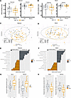 Figure 2
Differences in the ileal and rectal microbiomes of PWH enrolled from the United States and Mexico. Alpha diversity metrics for rectum (A) and ileum (B) biopsies between PWH from the United States and Mexico. Beta diversity of rectum (C) and ileum (D) biopsies, tested by PERMANOVA using the robust Aitchison distance of genus-level taxa. Differential abundance of genus- and species-level taxa between the United States and Mexico, tested by MaAsLin2 for rectum (E) and ileum (F) biopsies, showing the top 15 differentially abundant taxa; asterisks denote FDR-adjusted q ≤ 0.25. Representative box plots of Escherichia-Shigella (G) and Parabacteroides distasonis (H) in both the ileum and rectum.
We next examined whether these multi-omic differences between geographic locations were also present in PWoH. No significant differences in cytokine concentrations were observed between PWoH from the United States and Mexico, though this may reflect the limited sample size of the HIV-negative cohort given that several cytokines, including IL-17A, showed nonsignificant but concordant effect directions (Supplemental Figure 4A). For rectal and ileal biopsy taxa, none of the significant differences identified in PWoH overlapped with those seen in PWH, although a number of PWH-significant taxa again showed nonsignificant concordant effects in PWoH (e.g., ileal Escherichia-Shigella and Bacteroides uniformis; Supplemental Figure 4B). In contrast, 98 of the metabolites that differed significantly between PWH cohorts were also significantly different (q < 0.25) and directionally concordant in the PWoH comparison (Supplemental Figure 4C), suggesting that many of these metabolic differences are a geographic effect not unique to PWH.
Systemic inflammation and metabolomic disruptions in PWH at high risk of SNAEs. Given the geographic differences between participants from the United States and Mexico, we next examined whether there were biomarkers distinguishing PWH at low versus high risk for SNAEs, defined by CD4+ T cell counts and the CD4/CD8 ratio, that were independent of geographic effects. Such biomarkers could provide important insights into the processes driving SNAE risk. We first compared concentrations of plasma cytokines and markers of gut epithelial dysfunction. In contrast to country of enrollment, SNAE risk group did not have a significant effect on the overall plasma protein profile when tested by PERMANOVA (R2 = 0.031, q = 0.102, Figure 3A). When looking at individual markers, IL-6 was the only plasma protein we measured that was significantly increased in PWH at high risk for SNAEs (P = 0.021, q = 0.159, Figure 3B). Although not statistically significant after P value adjustment, IL-10 was also elevated in the high-risk group (P = 0.039, q = 0.264). Of note, when stratified by country, IL-6 was nonsignificantly (q > 0.25) increased in PWH at high-risk of SNAEs (Supplemental Figure 5), and only reached statistical significance when the cohorts were combined.
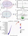 Figure 3
Differences in plasma proteins and metabolome between PWH at low or high risk of SNAEs. (A) Principal component analysis of plasma protein concentrations, colored by SNAE risk group, tested by PERMANOVA. (B) Volcano plot of cytokines and gut barrier integrity markers differentially associated with SNAE risk group. (C) Principal component analysis of plasma metabolomic features, colored by SNAE risk group and tested by PERMANOVA. (D) Volcano plot of metabolomic features differentially associated with SNAE risk group. (E) GSEA of metabolomic data comparing KEGG pathway enrichment between low and high SNAE risk groups; asterisks denote FDR-adjusted q ≤ 0.25.
We next compared the plasma metabolome of individuals at low and high risk of SNAE development. Risk group did not have a significant effect on the overall profile of metabolomic features when tested by PERMANOVA (R2 = 0.011, q = 0.782, Figure 3C). Overall, we found 13 metabolomic features that were significantly different between SNAE risk groups (q < 0.25, Figure 3D). Individuals with high risk of SNAEs had increased plasma concentrations of dicarboxylic acids (adipic acid and suberic acid), bile acids (cholic acid and 12-ketodeoxycholic acid), triglycerides (TG 20:1 34:3), polyamines (N1,N8-Di-Ac-spermidine), monoglycerides (MG 20:1), and lysophosphatidic acids (LPA 15:0), whereas those at low risk had higher concentrations of specific phosphatidic acids (PAs) (PA 14:0 14:1), PGs (PG 16:0 19:1), and PEs (PE P-18:0/20:3). Individuals at high risk also had a reduced ratio of chenodeoxycholic acid to cholic acid, and higher activity of imidazole propionic acid synthesis as measured by the ratio of imidazole propionic acid to histidine. GSEA showed enrichment of KEGG pathways for aminoacyl-tRNA biosynthesis, biosynthesis of amino acids, glycine/serine/threonine metabolism, and sphingolipid metabolism in patients with low risk, and enrichment of biosynthesis of unsaturated fatty acids in patients at high SNAE risk (q < 0.25, Figure 3E).
Reduced commensal bacteria in gut biopsies of PWH at high risk of SNAEs. We did not see any significant differences in alpha or beta diversity metrics in both the rectum and ileum of PWH with low versus high risk for SNAEs (Figure 4, A–D). However, differential abundance analysis revealed several genus and species level bacterial taxa that were associated with SNAE risk group. In the rectal biopsies, the Ruminococcus gauvreauii group was increased in PWH at high risk for SNAEs, while Ruminococcus and Bacteroides uniformis were both decreased (q < 0.25, Figure 4, E and G). In the ileal biopsies, Intestinibacter, Intestinibacter bartlettii, Oscillospiraceae NK4A214 group, and Collinsella bouchesdurhonensis were increased in PWH at high risk of SNAEs, while Akkermansia and A. muciniphila were decreased (q < 0.25, Figure 4, F and H). Notably, Segatella (formerly part of the Prevotella genus) was increased in the ileal biopsies of the high-risk SNAE group, though it did not reach statistical significance after adjusting for multiple comparisons (P = 0.016, q = 0.289, Figure 4F).
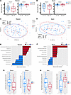 Figure 4
Differences in the ileal and rectal microbiomes of PWH at low or high risk of SNAEs. Alpha diversity metrics for rectum (A) and ileum (B) biopsies between SNAE risk groups. Beta diversity of rectum (C) and ileum (D) biopsies, tested by PERMANOVA using the robust Aitchison distance of genus-level taxa. Differential abundance of genus- and species-level taxa between SNAE risk groups, tested by MaAsLin2 for rectum (E) and ileum (F) biopsies, showing the top 10 differentially abundant taxa; asterisks denote FDR-adjusted q values ≤ 0.25. Representative box plots of differentially abundant taxa in the rectum (G) and ileum (H) biopsies.
Multi-omic machine learning discriminates between SNAE risk groups. We next applied supervised machine learning methods from the mixOmics R package to evaluate how well each ome (i.e., proteins, metabolome, ileum, rectum) classified SNAE risk, to identify key discriminative variables, and to examine cross-omic relationships. For single-ome models, we used partial least squares discriminant analysis (PLS-DA) or sparse PLS-DA when there were more variables than samples (Figure 5A). To integrate all omes, we used Data Integration Analysis for Biomarker discovery using Latent variable approaches for Omics studies (DIABLO, Figure 5B).
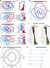 Figure 5
Supervised machine learning of SNAE risk groups. (A) Scores plot and top loading vectors on component 1 for the protein block PLS-DA, metabolome block sparse PLS-DA (sPLS-DA), rectum block sPLS-DA, and ileum block sPLS-DA. Loading vectors are colored by the group with the highest mean value. (B) Scores plot of the integrated DIABLO model, combining all 4 data blocks and showing the weighted average of components 1 and 2 according to their correlation with SNAE risk group. (C) Top loading scores on components 1 and 2 for each block in the DIABLO model; bars are colored by the corresponding block the variable belongs to. (D) Correlation circle plot of the DIABLO model, showing variables with correlations (>0.5) on components 1 and 2. (E) A variable’s location depicts both its correlation on each component and its correlation with other variables in proximity. Performance of each model in predicting SNAE risk based on AUROC, determined by 5-fold cross-validation repeated 50 times.
Many of the top discriminatory features for each model were also identified in our differential analysis. For example, IL-6 and N1,N8-Di-Ac-spermidine were the top features for component 1 in the protein and metabolome models, both discriminatory for the high-risk SNAE group. Similarly, A. muciniphila and Intestinibacter bartlettii were the top features in the ileum model, in the direction of low- and high-risk SNAE groups, respectively. In the integrated DIABLO model, top features in the direction of PWH at high risk included rectum abundance of Coprococcus and Coprococcus comes as well as I-FAPB, IL-12p70, and MDH1 activity as measured by the ratio of malic to oxaloacetic acid (Figure 5, C and D). Top features in the direction of PWH at low risk included LPE 18:0, rectum Enterocloster abundance, MIP1-β, and ileum abundances of Akkermansia and A. muciniphila. On component 2, top features in the direction of PWH at high risk included IL-6 and both ileum and rectum abundances of Eggerthella and Eggerthella lenta, whereas features in the direction of PWH at low risk included the lipids PE P-18:0/18:2, PA 18:0/18:2, and PA P-16:0/18:2.
The ability of each model to discriminate between low- and high-risk groups was estimated by the mean area under the receiver operator curve (AUROC) from 5-fold cross-validation repeated 50 times. Individually, blocks performed modestly, with the rectal microbiome having the relative best performance (AUROC = 0.649, Figure 5E). In contrast, the integrated DIABLO model combining all omes achieved the top performance (AUROC = 0.872), indicating a strong ability to discriminate between low- and high-risk SNAE groups.
Individual associations between omic features and T cell criteria. Because our high-risk SNAE group included criteria for both CD4+ T cell counts and the CD4/CD8 ratio, we next sought to understand how each biomarker was correlated with individual T cell criteria. We performed partial Spearman’s correlations between each biomarker and CD4+ T cell counts, CD8+ T cell counts, and the CD4/CD8 ratio, adjusting for the effect of country, age, and years living with HIV (Figure 6A). To reduce the number of comparisons, we focused only on biomarkers that were significantly different between our low- and high-risk groups. Notably, CD4+ T cell counts were negatively correlated with the ileum abundance of both Intestinibacter bartlettii (r = –0.405, P = 0.001, q = 0.011) and the NK4A214 group (r = –0.41, P < 0.001, q = 0.011). These taxa were similarly negatively correlated with the CD4/CD8 ratio (r = –0.312, P = 0.014, q = 0.15 and r = –0.305, P = 0.016, q = 0.15 respectively). In the opposite direction, ileum abundance of A. muciniphila and rectum abundance of Bacteroides uniformis were positively correlated with the CD4/CD8 ratio (r = 0.264, P = 0.038, q = 0.164 and r = 0.248, P = 0.043, q = 0.164, respectively). In the metabolomic data, CD4+ T cell counts were negatively correlated with TG 20:1 34:4 (r = –0.359, P = 0.002, q = 0.012), imidazole propionic acid synthesis (r = –0.269, P = 0.021, q = 0.089), and cholic acid (r = –0.253, P = 0.031, q = 0.107), while positively correlated with PA 14:0 14:1 (r = 0.274, P = 0.019, q = 0.089). The CD4/CD8 ratio was positively correlated with PE P-18:0/20:3 (r = 0.269, P = 0.021, q = 0.15). We did not observe any significant correlations with CD8+ T cell counts.
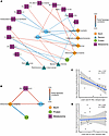 Figure 6
Partial correlations between variables of interest, CD4 counts, and the CD4/CD8 ratio. (A) Network of partial Spearman’s correlations between variables that were significantly different between SNAE risk groups, adjusted for country, age, and years living with HIV. (B) Selected correlations between bacterial taxa and the CD4/CD8 ratio with all connected nodes, highlighting potential mechanisms involved in driving immune activation. All correlations with an FDR-adjusted q ≤ 0.25 and nominal P ≤ 0.05 are shown. (C and D) Spearman’s correlations between ileum A. muciniphila abundance and HIV RNA+ (C) and DNA+ (D) cells/gram in lymph node tissue.
Correlation analysis highlights the potential role of A. muciniphila in reducing immune activation via IL-6 and the active HIV reservoir. We next performed partial correlations between these omic layers to understand how the relationships between them may be driving SNAE risk (Figure 6, A and B). Interestingly, we observed a negative correlation between ileum A. muciniphila abundance and plasma IL-6 concentrations (r = –0.388, P = 0.002, q = 0.007), with the same pattern observed when stratified by country (Supplemental Figure 6). IL-6 was further positively correlated with the secondary bile acid 12-ketodeoxycholic acid (r = 0.247, P = 0.035, q = 0.23) and negatively correlated with PG 16:0 19:1 (r = –0.302, P = 0.009, q = 0.121). Rectum Bacteroides uniformis abundance was similarly positively correlated with PG 16:0 19:1 (r = 0.269, P = 0.028, q = 0.242) and negatively correlated with 12-ketodeoxycholic acid (r = –0.415, P < 0.001, q = 0.019). Other notable correlations included a positive correlation between ileum Collinsella bouchesdurhonensis abundance and N1, N8-Di-Ac-spermidine (r = 0.375, P = 0.003, q = 0.118), and a positive correlation between A. muciniphila and PE P-18:0/20:3 (r = 0.354, P = 0.005, q = 0.112).
Given the negative correlation we observed between A. muciniphila and plasma IL-6, we next asked whether ileal A. muciniphila abundance was associated with the HIV reservoir. For a subset (n = 20) of the US participants, we were able to obtain lymph node tissue and measure the quantity of HIV RNA-producing cells and proviral DNA+ cells, as we have done previously (13, 18). Interestingly, ileal A. muciniphila abundance was negatively correlated with the number of HIV RNA+ cells/gram of lymph node tissue (Spearman’s r = –0.57, P = 0.014, Figure 6C), though was not correlated with the number of HIV DNA+ cells/gram (Spearman’s r = 0.05, P = 0.842, Figure 6D). Further, we did not observe a significant correlation between either IL-6 or IL-10 plasma concentrations and lymph node HIV reservoir measures (Supplemental Figure 7). Collectively, these findings suggest that A. muciniphila may reduce SNAE risk through a potential mechanism involving suppression of IL-6 and HIV RNA production in latently infected cells.
Multi-omic SNAE risk signatures contextualized in PWoH. To assess whether the observed differences between SNAE risk groups reflected true biological signals rather than confounding between groups, we compared the significantly different variables to a group of PWoH (n = 10) from both the United States and Mexico (n = 5 per country) using linear regression adjusted for age and country. Although limited statistical power likely hindered significance in many comparisons with PWoH, nearly all variables followed the expected ordinal trend, whereby features elevated in the high-risk relative to low-risk group were also higher than in PWoH and vice versa. For example, IL-6 was nonsignificantly elevated in the high-risk group relative to PWoH (β = 0.85, P = 0.272) and nonsignificantly reduced in the low-risk group (β = –0.31, P = 0.657, Figure 7A). Among metabolomic features, N1,N8-Di-Ac-spermidine, TG 20:1 34:3, and imidazole propionic acid synthesis were each significantly higher in the high-risk group compared with PWoH (P < 0.05, Figure 7B). Microbiome trends were similarly conserved, with Ruminococcus and Bacteroides uniformis significantly reduced in the high-risk group relative to PWoH in the rectum (P < 0.05, Figure 7C). In the ileum, Akkermansia and A. muciniphila were likewise reduced in the high-risk group, while the NK4A214 group, Intestinibacter, Intestinibacter bartlettii, and Collinsella bouchesdurhonensis were all significantly increased compared with PWoH (P < 0.05, Figure 7D). The only exception to these patterns was 12-ketodeoxycholic acid, which was lowest in the low-risk group, followed by the high-risk group and PWoH. Collectively, the significant differences observed between the high-risk group and PWoH in key metabolomic features and microbiome taxa, combined with the directional consistency of nearly all remaining variables, supports the biological plausibility of these signatures as indicators of SNAE risk.
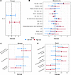 Figure 7
Comparison of SNAE risk groups to people without HIV. Variables that were significantly different between SNAE risk groups (q < 0.25) were compared against a PWoH reference group (n = 10, n = 5 per country) using linear regression adjusted for age and country. Panels display the linear model estimate and 95% CI for each SNAE risk group relative to PWoH for log2-transformed plasma IL-6 (A), log2-transformed metabolomic features (B), and CLR-transformed rectal (C) and ileal (D) microbiome taxa abundances, with microbiome estimates derived from MaAsLin2. Asterisks denote nominally significant differences from PWoH (P < 0.05).


