
Research ArticleCell biologyOncology
Open Access |  10.1172/jci.insight.202853
Derivation and characterization of ubiquitin-specific protease 18 inhibitors
Blessing O. Ogunlade,1 Kevin N. Dalby,2,3 Samuel C. Okpechi,1 Eun Jeong Cho,2,3 Liliya Tyutyunyk-Massey,1 Zibo Chen,1 Xiuxia Liu,1 Joseph Ivanic,4 Brian Luke,4 Shyamal D. Desai,5 Yair Alfaro,1,6 Ashwini K. Devkota,2,3 Rae M. Sammons,2,3 Gilbert G. Privé,7,8,9 Xi Liu,1 and Ethan Dmitrovsky1
1Molecular Pharmacology Program, Cancer Research Technology Program, Frederick National Laboratory for Cancer Research, Frederick, Maryland, USA.
2Targeted Therapeutic Drug Discovery and Development Program and
3Division of Chemical Biology and Medicinal Chemistry, The University of Texas at Austin, Austin, Texas, USA.
4Advanced Biomedical Computational Science, Frederick National Laboratory for Cancer Research, Frederick, Maryland, USA.
5Department of Biochemistry and Molecular Biology, Louisiana State University Health Science Center-School of Medicine, New Orleans, Louisiana, USA.
6School of Medicine and Health Sciences, Tecnologico de Monterrey, Monterrey, Mexico.
7Division of Cancer Genomics and Proteomics, Ontario Cancer Institute,
8Department of Medical Biophysics, and
9Department of Biochemistry, University of Toronto, Toronto, Ontario, Canada.
Address correspondence to: Ethan Dmitrovsky, Frederick National Laboratory for Cancer Research, PO Box B, Frederick, Maryland, 21701, USA. Phone: 301.846.1154; Email: ethan.dmitrovsky@gmail.com.
Find articles by Ogunlade, B. in: PubMed | Google Scholar
1Molecular Pharmacology Program, Cancer Research Technology Program, Frederick National Laboratory for Cancer Research, Frederick, Maryland, USA.
2Targeted Therapeutic Drug Discovery and Development Program and
3Division of Chemical Biology and Medicinal Chemistry, The University of Texas at Austin, Austin, Texas, USA.
4Advanced Biomedical Computational Science, Frederick National Laboratory for Cancer Research, Frederick, Maryland, USA.
5Department of Biochemistry and Molecular Biology, Louisiana State University Health Science Center-School of Medicine, New Orleans, Louisiana, USA.
6School of Medicine and Health Sciences, Tecnologico de Monterrey, Monterrey, Mexico.
7Division of Cancer Genomics and Proteomics, Ontario Cancer Institute,
8Department of Medical Biophysics, and
9Department of Biochemistry, University of Toronto, Toronto, Ontario, Canada.
Address correspondence to: Ethan Dmitrovsky, Frederick National Laboratory for Cancer Research, PO Box B, Frederick, Maryland, 21701, USA. Phone: 301.846.1154; Email: ethan.dmitrovsky@gmail.com.
Find articles by Dalby, K. in: PubMed | Google Scholar
1Molecular Pharmacology Program, Cancer Research Technology Program, Frederick National Laboratory for Cancer Research, Frederick, Maryland, USA.
2Targeted Therapeutic Drug Discovery and Development Program and
3Division of Chemical Biology and Medicinal Chemistry, The University of Texas at Austin, Austin, Texas, USA.
4Advanced Biomedical Computational Science, Frederick National Laboratory for Cancer Research, Frederick, Maryland, USA.
5Department of Biochemistry and Molecular Biology, Louisiana State University Health Science Center-School of Medicine, New Orleans, Louisiana, USA.
6School of Medicine and Health Sciences, Tecnologico de Monterrey, Monterrey, Mexico.
7Division of Cancer Genomics and Proteomics, Ontario Cancer Institute,
8Department of Medical Biophysics, and
9Department of Biochemistry, University of Toronto, Toronto, Ontario, Canada.
Address correspondence to: Ethan Dmitrovsky, Frederick National Laboratory for Cancer Research, PO Box B, Frederick, Maryland, 21701, USA. Phone: 301.846.1154; Email: ethan.dmitrovsky@gmail.com.
Find articles by Okpechi, S. in: PubMed | Google Scholar
1Molecular Pharmacology Program, Cancer Research Technology Program, Frederick National Laboratory for Cancer Research, Frederick, Maryland, USA.
2Targeted Therapeutic Drug Discovery and Development Program and
3Division of Chemical Biology and Medicinal Chemistry, The University of Texas at Austin, Austin, Texas, USA.
4Advanced Biomedical Computational Science, Frederick National Laboratory for Cancer Research, Frederick, Maryland, USA.
5Department of Biochemistry and Molecular Biology, Louisiana State University Health Science Center-School of Medicine, New Orleans, Louisiana, USA.
6School of Medicine and Health Sciences, Tecnologico de Monterrey, Monterrey, Mexico.
7Division of Cancer Genomics and Proteomics, Ontario Cancer Institute,
8Department of Medical Biophysics, and
9Department of Biochemistry, University of Toronto, Toronto, Ontario, Canada.
Address correspondence to: Ethan Dmitrovsky, Frederick National Laboratory for Cancer Research, PO Box B, Frederick, Maryland, 21701, USA. Phone: 301.846.1154; Email: ethan.dmitrovsky@gmail.com.
Find articles by Cho, E. in: PubMed | Google Scholar
1Molecular Pharmacology Program, Cancer Research Technology Program, Frederick National Laboratory for Cancer Research, Frederick, Maryland, USA.
2Targeted Therapeutic Drug Discovery and Development Program and
3Division of Chemical Biology and Medicinal Chemistry, The University of Texas at Austin, Austin, Texas, USA.
4Advanced Biomedical Computational Science, Frederick National Laboratory for Cancer Research, Frederick, Maryland, USA.
5Department of Biochemistry and Molecular Biology, Louisiana State University Health Science Center-School of Medicine, New Orleans, Louisiana, USA.
6School of Medicine and Health Sciences, Tecnologico de Monterrey, Monterrey, Mexico.
7Division of Cancer Genomics and Proteomics, Ontario Cancer Institute,
8Department of Medical Biophysics, and
9Department of Biochemistry, University of Toronto, Toronto, Ontario, Canada.
Address correspondence to: Ethan Dmitrovsky, Frederick National Laboratory for Cancer Research, PO Box B, Frederick, Maryland, 21701, USA. Phone: 301.846.1154; Email: ethan.dmitrovsky@gmail.com.
Find articles by Tyutyunyk-Massey, L. in: PubMed | Google Scholar
1Molecular Pharmacology Program, Cancer Research Technology Program, Frederick National Laboratory for Cancer Research, Frederick, Maryland, USA.
2Targeted Therapeutic Drug Discovery and Development Program and
3Division of Chemical Biology and Medicinal Chemistry, The University of Texas at Austin, Austin, Texas, USA.
4Advanced Biomedical Computational Science, Frederick National Laboratory for Cancer Research, Frederick, Maryland, USA.
5Department of Biochemistry and Molecular Biology, Louisiana State University Health Science Center-School of Medicine, New Orleans, Louisiana, USA.
6School of Medicine and Health Sciences, Tecnologico de Monterrey, Monterrey, Mexico.
7Division of Cancer Genomics and Proteomics, Ontario Cancer Institute,
8Department of Medical Biophysics, and
9Department of Biochemistry, University of Toronto, Toronto, Ontario, Canada.
Address correspondence to: Ethan Dmitrovsky, Frederick National Laboratory for Cancer Research, PO Box B, Frederick, Maryland, 21701, USA. Phone: 301.846.1154; Email: ethan.dmitrovsky@gmail.com.
Find articles by Chen, Z. in: PubMed | Google Scholar
1Molecular Pharmacology Program, Cancer Research Technology Program, Frederick National Laboratory for Cancer Research, Frederick, Maryland, USA.
2Targeted Therapeutic Drug Discovery and Development Program and
3Division of Chemical Biology and Medicinal Chemistry, The University of Texas at Austin, Austin, Texas, USA.
4Advanced Biomedical Computational Science, Frederick National Laboratory for Cancer Research, Frederick, Maryland, USA.
5Department of Biochemistry and Molecular Biology, Louisiana State University Health Science Center-School of Medicine, New Orleans, Louisiana, USA.
6School of Medicine and Health Sciences, Tecnologico de Monterrey, Monterrey, Mexico.
7Division of Cancer Genomics and Proteomics, Ontario Cancer Institute,
8Department of Medical Biophysics, and
9Department of Biochemistry, University of Toronto, Toronto, Ontario, Canada.
Address correspondence to: Ethan Dmitrovsky, Frederick National Laboratory for Cancer Research, PO Box B, Frederick, Maryland, 21701, USA. Phone: 301.846.1154; Email: ethan.dmitrovsky@gmail.com.
Find articles by Liu, X. in: PubMed | Google Scholar
1Molecular Pharmacology Program, Cancer Research Technology Program, Frederick National Laboratory for Cancer Research, Frederick, Maryland, USA.
2Targeted Therapeutic Drug Discovery and Development Program and
3Division of Chemical Biology and Medicinal Chemistry, The University of Texas at Austin, Austin, Texas, USA.
4Advanced Biomedical Computational Science, Frederick National Laboratory for Cancer Research, Frederick, Maryland, USA.
5Department of Biochemistry and Molecular Biology, Louisiana State University Health Science Center-School of Medicine, New Orleans, Louisiana, USA.
6School of Medicine and Health Sciences, Tecnologico de Monterrey, Monterrey, Mexico.
7Division of Cancer Genomics and Proteomics, Ontario Cancer Institute,
8Department of Medical Biophysics, and
9Department of Biochemistry, University of Toronto, Toronto, Ontario, Canada.
Address correspondence to: Ethan Dmitrovsky, Frederick National Laboratory for Cancer Research, PO Box B, Frederick, Maryland, 21701, USA. Phone: 301.846.1154; Email: ethan.dmitrovsky@gmail.com.
Find articles by Ivanic, J. in: PubMed | Google Scholar
1Molecular Pharmacology Program, Cancer Research Technology Program, Frederick National Laboratory for Cancer Research, Frederick, Maryland, USA.
2Targeted Therapeutic Drug Discovery and Development Program and
3Division of Chemical Biology and Medicinal Chemistry, The University of Texas at Austin, Austin, Texas, USA.
4Advanced Biomedical Computational Science, Frederick National Laboratory for Cancer Research, Frederick, Maryland, USA.
5Department of Biochemistry and Molecular Biology, Louisiana State University Health Science Center-School of Medicine, New Orleans, Louisiana, USA.
6School of Medicine and Health Sciences, Tecnologico de Monterrey, Monterrey, Mexico.
7Division of Cancer Genomics and Proteomics, Ontario Cancer Institute,
8Department of Medical Biophysics, and
9Department of Biochemistry, University of Toronto, Toronto, Ontario, Canada.
Address correspondence to: Ethan Dmitrovsky, Frederick National Laboratory for Cancer Research, PO Box B, Frederick, Maryland, 21701, USA. Phone: 301.846.1154; Email: ethan.dmitrovsky@gmail.com.
Find articles by
Luke, B.
in:
PubMed
|
Google Scholar
|

1Molecular Pharmacology Program, Cancer Research Technology Program, Frederick National Laboratory for Cancer Research, Frederick, Maryland, USA.
2Targeted Therapeutic Drug Discovery and Development Program and
3Division of Chemical Biology and Medicinal Chemistry, The University of Texas at Austin, Austin, Texas, USA.
4Advanced Biomedical Computational Science, Frederick National Laboratory for Cancer Research, Frederick, Maryland, USA.
5Department of Biochemistry and Molecular Biology, Louisiana State University Health Science Center-School of Medicine, New Orleans, Louisiana, USA.
6School of Medicine and Health Sciences, Tecnologico de Monterrey, Monterrey, Mexico.
7Division of Cancer Genomics and Proteomics, Ontario Cancer Institute,
8Department of Medical Biophysics, and
9Department of Biochemistry, University of Toronto, Toronto, Ontario, Canada.
Address correspondence to: Ethan Dmitrovsky, Frederick National Laboratory for Cancer Research, PO Box B, Frederick, Maryland, 21701, USA. Phone: 301.846.1154; Email: ethan.dmitrovsky@gmail.com.
Find articles by Desai, S. in: PubMed | Google Scholar
1Molecular Pharmacology Program, Cancer Research Technology Program, Frederick National Laboratory for Cancer Research, Frederick, Maryland, USA.
2Targeted Therapeutic Drug Discovery and Development Program and
3Division of Chemical Biology and Medicinal Chemistry, The University of Texas at Austin, Austin, Texas, USA.
4Advanced Biomedical Computational Science, Frederick National Laboratory for Cancer Research, Frederick, Maryland, USA.
5Department of Biochemistry and Molecular Biology, Louisiana State University Health Science Center-School of Medicine, New Orleans, Louisiana, USA.
6School of Medicine and Health Sciences, Tecnologico de Monterrey, Monterrey, Mexico.
7Division of Cancer Genomics and Proteomics, Ontario Cancer Institute,
8Department of Medical Biophysics, and
9Department of Biochemistry, University of Toronto, Toronto, Ontario, Canada.
Address correspondence to: Ethan Dmitrovsky, Frederick National Laboratory for Cancer Research, PO Box B, Frederick, Maryland, 21701, USA. Phone: 301.846.1154; Email: ethan.dmitrovsky@gmail.com.
Find articles by Alfaro, Y. in: PubMed | Google Scholar
1Molecular Pharmacology Program, Cancer Research Technology Program, Frederick National Laboratory for Cancer Research, Frederick, Maryland, USA.
2Targeted Therapeutic Drug Discovery and Development Program and
3Division of Chemical Biology and Medicinal Chemistry, The University of Texas at Austin, Austin, Texas, USA.
4Advanced Biomedical Computational Science, Frederick National Laboratory for Cancer Research, Frederick, Maryland, USA.
5Department of Biochemistry and Molecular Biology, Louisiana State University Health Science Center-School of Medicine, New Orleans, Louisiana, USA.
6School of Medicine and Health Sciences, Tecnologico de Monterrey, Monterrey, Mexico.
7Division of Cancer Genomics and Proteomics, Ontario Cancer Institute,
8Department of Medical Biophysics, and
9Department of Biochemistry, University of Toronto, Toronto, Ontario, Canada.
Address correspondence to: Ethan Dmitrovsky, Frederick National Laboratory for Cancer Research, PO Box B, Frederick, Maryland, 21701, USA. Phone: 301.846.1154; Email: ethan.dmitrovsky@gmail.com.
Find articles by Devkota, A. in: PubMed | Google Scholar
1Molecular Pharmacology Program, Cancer Research Technology Program, Frederick National Laboratory for Cancer Research, Frederick, Maryland, USA.
2Targeted Therapeutic Drug Discovery and Development Program and
3Division of Chemical Biology and Medicinal Chemistry, The University of Texas at Austin, Austin, Texas, USA.
4Advanced Biomedical Computational Science, Frederick National Laboratory for Cancer Research, Frederick, Maryland, USA.
5Department of Biochemistry and Molecular Biology, Louisiana State University Health Science Center-School of Medicine, New Orleans, Louisiana, USA.
6School of Medicine and Health Sciences, Tecnologico de Monterrey, Monterrey, Mexico.
7Division of Cancer Genomics and Proteomics, Ontario Cancer Institute,
8Department of Medical Biophysics, and
9Department of Biochemistry, University of Toronto, Toronto, Ontario, Canada.
Address correspondence to: Ethan Dmitrovsky, Frederick National Laboratory for Cancer Research, PO Box B, Frederick, Maryland, 21701, USA. Phone: 301.846.1154; Email: ethan.dmitrovsky@gmail.com.
Find articles by Sammons, R. in: PubMed | Google Scholar
1Molecular Pharmacology Program, Cancer Research Technology Program, Frederick National Laboratory for Cancer Research, Frederick, Maryland, USA.
2Targeted Therapeutic Drug Discovery and Development Program and
3Division of Chemical Biology and Medicinal Chemistry, The University of Texas at Austin, Austin, Texas, USA.
4Advanced Biomedical Computational Science, Frederick National Laboratory for Cancer Research, Frederick, Maryland, USA.
5Department of Biochemistry and Molecular Biology, Louisiana State University Health Science Center-School of Medicine, New Orleans, Louisiana, USA.
6School of Medicine and Health Sciences, Tecnologico de Monterrey, Monterrey, Mexico.
7Division of Cancer Genomics and Proteomics, Ontario Cancer Institute,
8Department of Medical Biophysics, and
9Department of Biochemistry, University of Toronto, Toronto, Ontario, Canada.
Address correspondence to: Ethan Dmitrovsky, Frederick National Laboratory for Cancer Research, PO Box B, Frederick, Maryland, 21701, USA. Phone: 301.846.1154; Email: ethan.dmitrovsky@gmail.com.
Find articles by Privé, G. in: PubMed | Google Scholar
1Molecular Pharmacology Program, Cancer Research Technology Program, Frederick National Laboratory for Cancer Research, Frederick, Maryland, USA.
2Targeted Therapeutic Drug Discovery and Development Program and
3Division of Chemical Biology and Medicinal Chemistry, The University of Texas at Austin, Austin, Texas, USA.
4Advanced Biomedical Computational Science, Frederick National Laboratory for Cancer Research, Frederick, Maryland, USA.
5Department of Biochemistry and Molecular Biology, Louisiana State University Health Science Center-School of Medicine, New Orleans, Louisiana, USA.
6School of Medicine and Health Sciences, Tecnologico de Monterrey, Monterrey, Mexico.
7Division of Cancer Genomics and Proteomics, Ontario Cancer Institute,
8Department of Medical Biophysics, and
9Department of Biochemistry, University of Toronto, Toronto, Ontario, Canada.
Address correspondence to: Ethan Dmitrovsky, Frederick National Laboratory for Cancer Research, PO Box B, Frederick, Maryland, 21701, USA. Phone: 301.846.1154; Email: ethan.dmitrovsky@gmail.com.
Find articles by Liu, X. in: PubMed | Google Scholar
1Molecular Pharmacology Program, Cancer Research Technology Program, Frederick National Laboratory for Cancer Research, Frederick, Maryland, USA.
2Targeted Therapeutic Drug Discovery and Development Program and
3Division of Chemical Biology and Medicinal Chemistry, The University of Texas at Austin, Austin, Texas, USA.
4Advanced Biomedical Computational Science, Frederick National Laboratory for Cancer Research, Frederick, Maryland, USA.
5Department of Biochemistry and Molecular Biology, Louisiana State University Health Science Center-School of Medicine, New Orleans, Louisiana, USA.
6School of Medicine and Health Sciences, Tecnologico de Monterrey, Monterrey, Mexico.
7Division of Cancer Genomics and Proteomics, Ontario Cancer Institute,
8Department of Medical Biophysics, and
9Department of Biochemistry, University of Toronto, Toronto, Ontario, Canada.
Address correspondence to: Ethan Dmitrovsky, Frederick National Laboratory for Cancer Research, PO Box B, Frederick, Maryland, 21701, USA. Phone: 301.846.1154; Email: ethan.dmitrovsky@gmail.com.
Find articles by Dmitrovsky, E. in: PubMed | Google Scholar
Published May 19, 2026 - More info
JCI Insight. 2026;11(14):e202853. https://doi.org/10.1172/jci.insight.202853.
© 2026 Ogunlade et al. This work is licensed under the Creative Commons Attribution 4.0 International License. To view a copy of this license, visit http://creativecommons.org/licenses/by/4.0/.
Received: December 1, 2025; Accepted: May 13, 2026

-
Results
High-throughput screen identifies candidate USP18 inhibitors. To identify small-molecule inhibitors of USP18, high-throughput screening was performed using a diverse compound library containing 79,493 small molecules, as shown in Figure 1A. A fluorescence-based enzymatic assay was established using recombinant, enzymatically active USP18 protein as displayed in Figure 1, B and C. Purified USP18 displayed deISGylating activity, which was independently confirmed by immunoblot assays in Figure 1D. Primary screening, followed by counter screening, identified 13 candidate inhibitors that were subjected to dose–response assays at various treatment durations (0 hour, 0.5 hour, 1 hour, and 4 hours).
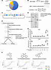 Figure 1
High-throughput screen identifies candidate USP18 small molecule inhibitors. (A) The features of the 79,493-compound library used for high-throughput screening are displayed, showing the proportions of fragment, kinase, diversity, biologically annotated, and academic compound sets that were interrogated. (B) The diagram of the fluorogenic enzymatic assay used to identify small-molecule USP18 inhibitors is shown. Recombinant, enzymatically active recombinant USP18 deconjugated ISG15-AMC substrate, and fluorescence intensity was used to calculate the percentage inhibition of each tested compound. (C) Schematic representation of the GFP-tagged USP18 expression vector used to generate enzymatically active USP18 protein. (D) Immunoblot analysis of USP18 species before and after 3C protease cleavage confirmed the production of the enzymatically active USP18 used for the biochemical assays. (E) Workflow of the screening strategy indicated the progression from primary screening to hit validation, including the selection of 13 primary hits, 3 reproducible USP18-inhibitory compounds, and identification of β-lapachone as the lead compound for further evaluation. (F) Dose-response inhibition curves of β-lapachone, 9,10-phenanthrenequinone, and 2-nitrophenanthrene-9,10-dione are displayed using the same fluorogenic enzymatic assay described in B. (G) The percent of ROS production was measured in human A549 and H1299 lung cancer cell lines following β-lapachone treatment at the indicated concentrations with or without cotreatment with the ROS scavenger N-acetylcysteine (NAC, 10 mM). Two-tailed Student’s t tests compared differences between arms with a P < 0.05 deemed statistically significant. Data are shown as mean ± SD with the symbols indicating *P < 0.05 and **P < 0.01, respectively.
The strategy deployed in this screen is summarized in Figure 1E. This approach progressed from high-throughput enzymatic assays through multiple biochemical, transcriptional, and cell-based assays. Five compounds displayed reproducible enzymatic inhibition against USP18 in concentration- and time-dependent manners. The increased inhibition observed with prolonged pretreatment suggests that these compounds may act via a noninstantaneous or potentially covalent mechanism, and the detailed mechanism of action is under investigation. Among the 5 candidate inhibitors, 3 compounds (β-lapachone; 9,10-phenanthrenequinone; and 2-nitrophenanthrene-9,10-dione) shared similar structural scaffolds and physicochemical properties, prompting further interest in their mode of action, as shown in Figure 1F. Two of these 5 compounds did not appreciably suppress lung cancer cell proliferation and were not moved forward for further screening, as shown in Supplemental Figure 1 (supplemental material available online with this article; https://doi.org/10.1172/jci.insight.202853DS1). The observation of similar scaffolds in the 3 most active USP18 inhibitors motivated the subsequent molecular modeling studies to investigate how these compounds, particularly β-lapachone, engaged USP18 at functional and structural levels.
β-Lapachone is reported to confer cytotoxic effects through NQO1-mediated redox cycling, generating ROS that cause DNA damage and necrosis (32–34). However, the data displayed in Figure 1G found that β-lapachone treatment generated negligible ROS at 1 μM and 2.5 μM concentrations. A marked increase in ROS was only observed at the higher dosage of β-lapachone (4 μM), as measured by flow cytometry. Notably, combined treatment with the ROS scavenger N-acetylcysteine (NAC) substantially attenuated this ROS elevation, at the β-lapachone 4 μM dosage, but hardly at the lower dosages as shown in Figure 1G. These findings indicated that β-lapachone was activating anticancer mechanisms at concentrations lower than 4 μM that did not engage ROS. Studies were next conducted to elucidate these mechanisms.
β-Lapachone treatment suppresses lung cancer cell proliferation. Functional consequences of β-lapachone treatment were explored. β-Lapachone statistically significantly repressed proliferation of human lung cancer cell lines A549 and H1299 (Figure 2A) in a dose-dependent manner. β-Lapachone treatment augmented apoptosis in A549 and H1299 cells, as shown in Figure 2B. This growth inhibition was not reversed by cotreatment with the ROS scavenger NAC, as seen in Figure 2C. The β-lapachone treatment at 2.5 μM concentrations also increased apoptosis in these cancer cells, but NAC cotreatment did not appreciably attenuate this response, as displayed in Figure 2C. These results suggest that the antiproliferative and proapoptotic activities of β-lapachone occur at lower concentrations largely through an ROS-independent mechanism. In addition, β-lapachone suppressed proliferation and induced apoptosis of murine lung cancer 344SQ cells in a similar dose-dependent pattern as in human lung cancer cells, as shown in Supplemental Figure 2.
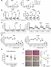 Figure 2
β-Lapachone reduces lung cancer cell proliferation and increases apoptosis. (A and B) Proliferation and apoptosis assays of human lung cancer A549 and H1299 lung cancer cell lines following β-lapachone treatment at the indicated concentrations are displayed. β-Lapachone treatment statistically significantly reduced proliferation and augmented apoptosis in a dose-dependent manner as compared with vehicle controls. (C) Apoptosis and proliferation were displayed for A549 lung cancer cells following β-lapachone treatment (versus vehicle treatment) at the indicated concentrations with or without cotreatment with the ROS scavenger N-acetylcysteine (NAC, 10 mM). β-Lapachone increased apoptosis significantly at the 4 μM dosage but not at lower concentrations. NAC cotreatment statistically significantly antagonized this effect only at the 4 μM dosage. (D) Growth suppression by β-lapachone treatment was also antagonized by engineered gain of USP18 expression. β-Lapachone treatment repressed proliferation of A549 and H1299 lung cancer cells transfected with an empty vector (EV). In contrast, USP18 overexpression partially reversed this growth inhibition at the 2 μM and 4 μM dosages. (E) In vivo antitumor activity of β-lapachone treatment is displayed at the left panel (12.5 mg/kg or 25 mg/kg body weight) as evaluated using the H1299 human lung cancer xenograft model. The 25 mg/kg body weight of β-lapachone statistically significantly reduced tumor growth as compared with vehicle-treated controls after 29 days of treatment. (F) Representative H&E staining, Ki-67 immunostaining, and apoptosis scoring are displayed for different treatment groups. Two-tailed Student’s t tests compared differences between study groups with a P value below 0.05 deemed statistically significant. Data are shown as mean ± SD. *P < 0.05, **P < 0.01, and ***P < 0.001.
To explore if these effects are mediated through USP18, USP18 rescue experiments were performed. Independently engineered gain of USP18 expression in human A549 and H1299 lung cancer cells antagonized β-lapachone–induced growth inhibition (Figure 2D). This indicates that reduction of USP18 activity contributed to the antiproliferative signals of β-lapachone.
To confirm and extend these in vitro findings to the in vivo setting, human lung cancer xenograft models were interrogated using the H1299 lung cancer cell line. Oral administration of β-lapachone at the 12.5 mg/kg and 25 mg/kg of mouse body weight decreased lung tumor growth as compared with controls. The tumor growth decline at the 25 mg/kg of mouse body weight reached statistical significance versus vehicle controls (Figure 2E). Notably, this decline in lung cancer growth was associated with a statistically significant reduction in proliferation as measured by Ki-67 staining and an increase in apoptosis TUNEL scoring, as displayed in Figure 2F. These findings demonstrate that β-lapachone treatments (as compared with vehicle control treatments) confer anticancer effects in immunodeficient mice harboring human lung cancer cells. Given these antineoplastic effects, it was next studied whether these actions were mediated through USP18 inhibition.
β-Lapachone antagonizes USP18-dependent activities. To confirm target specificity, it was explored whether β-lapachone treatment affected USP18-dependent signaling in A549 lung cancer cells. Consistent with an antagonist of USP18 function, β-lapachone treatment enhanced activity in a dose-dependent manner of the transfected ISRE reporter assay, as depicted in Figure 3A. In contrast, engineered gain of human USP18 expression repressed this ISRE activity. This inhibited the increase in reported activity that was conferred by β-lapachone treatment. Consistent with these findings, immunoblot analysis demonstrated that β-lapachone treatment also increased global ISGylation in A549 human lung cells, as seen in Figure 3B. Notably, β-lapachone treatment preferentially affected ISGylation, given that this treatment had little effect on ubiquitin, SUMO-2/3, or NEDD8 conjugation other than a minor effect on SUMO-2/3 conjugation observed only at the highest β-lapachone examined treatment concentrations (Figure 3, C and D).
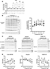 Figure 3
β-Lapachone antagonizes USP18-dependent activity. (A) IFN-stimulated response element (ISRE) reporter assays were performed to evaluate the effect of β-lapachone treatment on IFN pathway activation. A549 lung cancer cells were transiently transfected with an insertless vector (EV) as a control or a USP18 expression vector and treated with the indicated concentrations of β-lapachone with or without IFN (200 units) cotreatment. β-Lapachone treatment statistically significantly increased ISRE reporter activity in a concentration-dependent manner, whereas USP18 overexpression antagonized this activation. (B) Immunoblot analyses were performed to assess the effects of β-lapachone treatment on global ISGylation in A549 cells. Representative immunoblot images and the quantification of conjugated ISG15 species within the indicated bracketed region of 4 independent experiments are displayed. A statistically significant increase in global ISGylation was evident after dose-dependent β-lapachone treatments. (C) Independent immunoblot analyses of NEDD8, ubiquitin, and SUMO-2/3 conjugation after β-lapachone treatment as compared with vehicle control were performed to evaluate pathway selectivity. β-Lapachone treatment did not confer detectable changes in ubiquitin or NEDD8 conjugation. Only a modest change in SUMO-2/3 conjugation was observed at the highest β-lapachone concentration. The vinculin signals present in the middle and right panels were from the same immunoblot and lysate that were independently probed for ubiquitin and SUMO-2/3. The NEDD8 immunoblot was from a different lysate. These immunoblots present representative signals for each of these species. (D) Quantification of the NEDD8, ubiquitin, and SUMO-2/3 conjugates shown in C was performed using 3 independent replicate experiments. Two-tailed Student’s t tests compared differences between study groups with a P value below 0.05 deemed statistically significant. Data are shown as mean ± SD. *P < 0.05, **P < 0.01, ***P < 0.001 and ****P < 0.0001.
Molecular modeling of β-lapachone binding to a USP18. To elucidate the molecular basis of USP18 inhibition by individual β-lapachone, 9,10-phenanthrenequinone, and 2-nitrophenanthrene-9,10-dione treatments, molecular modeling of the USP18–ISG15 interaction interface was performed. Intriguingly, structural analyses identified a hydrophobic pocket needed for substrate engagement. Within this pocket, the phenylalanine residue F259 was found to be a critical determinant of ligand binding. As shown in Figure 4A, the conformation of F259 differs between ISG15-bound (interacting) and unbound (noninteracting) USP18, indicating its dynamic role in mediating substrate recognition. Additional modeling highlighted 3 residues — D139, F259, and T266 — as key interaction points through which β-lapachone engages USP18 within this hydrophobic pocket (Figure 4B).
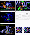 Figure 4
Molecular modeling of β-lapachone binding to USP18. (A) Structural modeling of USP18 and ISG15 complex formation identified a hydrophobic pocket in USP18 required for substrate engagement. Within this pocket, 3 key amino acid residues — D139, F259, and T266 were found to mediate ISG15 binding. (B) Molecular docking analysis revealed that β-lapachone (green) interacts with USP18 at the identified hydrophobic pocket, with the critical residues D139, F259, and T266 highlighted in brown. (C) Docking simulations of β-lapachone; 9,10-phenanthrenequinone; and 2-nitrophenanthrene-9,10-dione showed that all 3 compounds occupy this same hydrophobic pocket of USP18 in a similar spatial configuration. (D) Calculated binding energies for β-lapachone; 9,10-phenanthrenequinone; and 2-nitrophenanthrene-9,10-dione are individually displayed, supporting comparable affinities for binding to USP18 protein.
Consistent with these findings, docking analyses revealed the compounds having USP18 enzymatic inhibitory properties (β-lapachone, 9,10-phenanthrenequinone, and 2-nitrophenanthrene-9,10-dione) bound the identified USP18 pocket in a similar manner, as displayed in Figure 4C. Binding energy calculations supported these findings, revealing favorable energetics for ligand engagement, with β-lapachone exhibiting a calculated binding energy of approximately –6.1 kcal/mol, comparable with those of 9,10-phenanthrenequinone and 2-nitrophenanthrene-9,10-dione, as shown in Figure 4D. These results provide a mechanistic basis through which β-lapachone and its analogs inhibit USP18 enzymatic activity through occupancy of a conserved hydrophobic binding pocket defined by residues D139, F259, and T266.
To independently validate these molecular modeling predictions, site-directed mutagenesis of the 3 critical amino acid residues (D139, F259, and T266) in USP18 was next performed. In silico energy calculations revealed that mutation of any of these residues would markedly reduce the predicted binding affinity between β-lapachone and USP18, with the greatest loss of interaction observed in examination of the triple mutant (Figure 5A and Supplemental Figure 3). Structural visualization confirmed that β-lapachone bound tightly to WT USP18 but did not similarly bind mutant USP18 proteins, consistent with loss of the hydrophobic-pocket interaction displayed in Figure 5B.
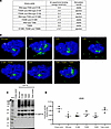 Figure 5
Mutational analyses validate the USP18 residues critical for β-lapachone binding and activity. (A) Binding energy calculations and 1,000 nanosecond molecular dynamics simulations were performed to evaluate the effects of single (D139K, F259K, T266K), double, and triple amino acid substitutions on β-lapachone–USP18 interactions. Mutation of any of the 3 key residues markedly reduced predicted binding affinity, with the triple mutant (D139K/F259K/T266K) abolishing stable binding. (B) Molecular dynamics visualization of β-lapachone (green) bound to WT and mutant USP18 proteins (blue) after 1,000 nanosecond simulations showed stable binding to WT USP18 but with ejection of β-lapachone from mutant USP18 species. (C) Immunoblot analysis confirmed comparable expression of WT and mutant USP18 proteins following transfection of each construct, including single and triple mutants. (D) The A549 lung cancer cell line was transfected with an ISRE luciferase reporter plasmid together with either empty vector, WT USP18, or USP18 constructs harboring individual (D139K, F259K, T266K) or triple mutations at the indicated residues. Luciferase activity was measured 48 hours after transfection and normalized to Renilla luciferase. Two-tailed Student’s t tests compared differences between study groups with a P value below 0.05 deemed statistically significant. Data are shown as mean ± SD. *P < 0.05 and **P < 0.01.
To ensure proper protein expression, immunoblot analyses verified comparable expression levels of WT and mutant USP18 constructs in Figure 5C. Functional testing using the ISRE reporter assay showed that IFN treatment elevated the luciferase activity, while transfection of WT USP18 species into A549 lung cancer cells opposed this activation. In marked contrast, transfection of the triple mutant USP18 species did not appreciably affect this activation and indicated that the triple mutant USP18 species was no longer functional, as displayed in Figure 5D. Together, these findings provide a mechanistic basis through which β-lapachone disrupts USP18 enzymatic activity by binding the hydrophobic pocket needed for ISG15 recognition (44).
Among the 3 highlighted USP18 candidate inhibitors, β-lapachone was prioritized for further pharmacodynamic studies because it exhibited the most robust inhibitory activity in enzymatic, biochemical, cellular, and tumorigenicity assays. β-Lapachone is a natural product with known antineoplastic properties (35–37). It is a tool compound to further assess the consequences of USP18 inhibition.
Transcriptomic profiling and USP18-dependent β-lapachone response. RNAseq was next performed to determine transcriptional pathways engaged by β-lapachone treatment. This treatment of A549 lung cancer cells as compared with vehicle controls caused a distinct pattern of differentially expressed genes, including both upregulated and downregulated transcripts (Figure 6A and Supplemental Figure 4A). To determine whether these responses were mediated by USP18 inhibition, transcriptomes of USP18-overexpressing A549 lung cancer cells were compared with that of empty-vector controls treated with β-lapachone. Exogenous USP18 expression antagonized a subset of β-lapachone–induced transcriptional changes, indicating that the effects displayed in Figure 6B and Supplemental Figure 4, B and C, are likely USP18 dependent.
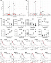 Figure 6
Transcriptomic profiling identifies USP18-dependent gene expression changes conferred by β-lapachone treatment relative to vehicle control. (A) Volcano plot displayed the differentially expressed genes in A549 human lung cancer cells treated with β-lapachone (2.5 μM) as compared with vehicle-treated controls. β-Lapachone treatment caused distinct patterns of gene upregulation and downregulation. (B) Volcano plot of A549 lung cancer cells engineered to overexpress USP18 versus empty vector controls is shown following β-lapachone treatment (2.5 μM) as compared with vehicle control. Genes whose expression changes were opposed by USP18 overexpression represent β-lapachone regulated transcripts that are USP18 dependent. (C) Quantitative real-time PCR assays validated representative β-lapachone–regulated genes identified in B, including those decreased (NR4A2, ENTPD1, DCC, and USP41) and those increased (FGF13, IL31RA, OTUD7A, and KLK2) after β-lapachone treatment. (D) Representative β-lapachone treatment repressed expression of USP18-dependent genes, including LINC00319, NCCRP1, and ENTPD1. Differential expression profiles of many of species showed statistically significant associations with overall survival in TCGA lung adenocarcinoma cohort. (E) Representative β-lapachone treatment increased expression of USP18-regulated genes, including FGF13, IL31RA, OTUD7A, and KLK2, exhibited statistically significant associations with improved overall survival in the same TCGA cohort. Two-tailed Student’s t tests compared differences between study groups with a P value below 0.05 deemed statistically significant. Data are shown as mean ± SD. *P < 0.05, **P < 0.01 and ***P < 0.001. Survival analyses for D and E were performed using the Kaplan-Meier method using the log-rank test with a P value below 0.05 deemed statistically significant.
Real-time PCR assays independently validated representative expressed genes that were either reduced (NR4A2, ENTPD1, DCC, and USP41) or increased (FGF13, IL31RA, OTUD7A, and KLK2) following β-lapachone treatment (Figure 6C). To assess the translational relevance of these β-lapachone–regulated and USP18-dependent genes, transcriptomic and survival data were interrogated using The Cancer Genome Atlas (TCGA) lung adenocarcinoma cohort. High mRNA expression levels of USP18 or ISG15 were each associated with poorer overall lung cancer survival in Figure 6D. The prognostic value of USP18 and ISG15 in lung squamous cell carcinoma is shown in Supplemental Figure 4D. Among β-lapachone–suppressed genes whose expression was rescued by USP18 overexpression, including LINC00319, NCCRP1, and ENTPD1, higher expression in human lung adenocarcinomas was associated with unfavorable survival outcomes in Figure 6D.
In contrast, β-lapachone–expressed genes that were antagonized by engineered USP18 overexpression, including FGF13, IL31RA, OTUD7A, and KLK2, showed the opposite survival patterns with higher expression levels in lung adenocarcinomas that were associated with an improved survival (Figure 6E). These findings serve to identify USP18-dependent gene expression pathways that are engaged by β-lapachone treatment. To delineate species that could mediate the antineoplastic effects of β-lapachone, proteomic analyses were next done.
Proteomic analyses reveal β-lapachone–regulated ISG15-related proteins. To further define the consequences of USP18 inhibition, we performed ISG15 immunoprecipitation–mass spectrometry analyses in A549 lung cancer cells expressing an empty vector or a USP18-expressing vector. These transfectants were each treated with vehicle or β-lapachone at the 2.5 μM concentration. Under stringent elution conditions designed to retain preferentially the covalently bound ISG15 conjugates, β-lapachone treatment substantially affected the ISG15-conjugated proteome. Comparison between β-lapachone–treated control cells and USP18-overexpressing A549 lung cancer transfectants identified proteins whose ISGylation were rescued by USP18 expression, as expected for effects that were USP18-dependent. Among the ISG15-conjugated proteins identified, 86 matched previously reported ISG15 targets, while 70 represented new candidate ISG15-conjugated proteins, as displayed in Figure 7A and Supplemental Figure 5. Pathway enrichment analysis revealed that these β-lapachone–regulated ISG15 substrates were distributed across multiple cellular processes, including signal transduction, RNA and protein modification, mitochondrial transport, vesicle trafficking, and chromatin regulation, as shown in Figure 7B.
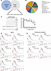 Figure 7
Proteomic analyses reveal β-lapachone treatment affected ISG15-related proteins. (A) ISG15 immunoprecipitation followed by mass spectrometry proteomics analysis was performed in A549 lung cancer cells that expressed an empty vector or a USP18 expression construct, and these transfectants were treated with β-lapachone (2.5 μM) or vehicle treatments. Comparative proteomic analyses identified potential ISG15-conjugated proteins whose levels were increased by β-lapachone and rescued by engineered USP18 overexpression. Among the ISG15-conjugated targets detected, 86 were previously reported ISG15 substrates and 70 were found as new ISG15-conjugated proteins. (B) Functional categorization of the β-lapachone treatment arm that regulated ISG15 conjugates revealed broad distribution across multiple cellular processes. (C) Immunoblot validation of the representative ISG15-conjugated protein, DYNLL1, confirmed that its expression was regulated by β-lapachone–treatment relative to vehicle controls (quantification is shown). Data are shown as mean ± SD. *P < 0.05, **P < 0.01, and ***P < 0.001. (D) TCGA lung adenocarcinoma cohort analyses demonstrated that several β-lapachone upregulated ISG15-conjugated proteins, including DYNLL1, HAUS6, LAMB3, BANF1, EIF5A, and ANXA2P2, were statistically significantly associated with lung cancer survival outcomes. (E) Four previously unrecognized ISG15-conjugated proteins, PCBP2, NME1, TUBA1C, and TUBA4A, were confirmed as β-lapachone regulated ISG15 targets whose elevated expression predicted an unfavorable survival in TCGA lung adenocarcinoma cases. Survival analysis for D and E highlighted species was performed using the Kaplan-Meier method, using the log-rank test. A P value below 0.05 was deemed statistically significant.
The expression of a representative potentially novel ISGylated protein, DYNLL1, was interrogated by immunoblot analyses following β-lapachone as compared with vehicle treatment. These findings are shown in Figure 7C and reveal a β-lapachone dose-dependent repression of DYNLL1 immunoblot expression. To establish the translational relevance of this observation, TCGA lung adenocarcinoma cohort was examined for the effect on survival of differential expression of DYNLL1. Intriguingly, a favorable survival outcome was observed in lung cancer cases having low as compared with high levels of DYNLL1 levels within the lung cancers. Other novel and previously recognized ISGylated species were examined for their associations for lung cancer survival in Figure 7, D and E. Differential expression profiles of several identified ISG15-conjugated proteins were explored for their prognostic effect across different cancer types, as shown in Supplemental Figure 6.
Several β-lapachone–upregulated ISG15 substrates, including DYNLL1, HAUS6, LAMB3, BANF1, EIF5A, and ANXA2P2 showed statistically significant associations with survival of patients with cancer, as seen in Figure 7D. Four previously unrecognized ISG15 targets (PCBP2, NME1, TUBA1C, and TUBA4A) were confirmed as β-lapachone–regulated conjugates whose high expression profiles were associated with unfavorable survival outcomes in Figure 7E. These observations broaden an understanding of β-lapachone treatment effects on the ISGylated proteome.


