
Research ArticleImmunologyInflammationNeuroscience
Open Access |  10.1172/jci.insight.200419
Systemic immune dysregulation and neutrophil activation define prognostic inflammatory signatures in drug-resistant epilepsy
Coraly Simoës Da Gama,1 Aurélie Hanin,2,3,4 Gwen Goudard,5 Véronique Masson,5 Aurore Besnard,5 Karim Dorgham,6 Guy Gorochov,6 Guillaume Dorothée,1 Valerio Frazzini,2,5 Vincent Navarro,2,5,7 and Mélanie Morin-Brureau1
1Sorbonne Université, Inserm, Centre de Recherche Saint-Antoine (CRSA), Hôpital Saint-Antoine, Paris, France.
2Sorbonne Université, Paris Brain Institute, ICM, INSERM, CNRS, AP-HP, Pitié-Salpêtrière Hospital, Paris, France.
3AP-HP, Sorbonne Université, Pitié-Salpêtrière - Charles Foix University Hospital, Department of Metabolic Biochemistry, DMU BioGeMH, Paris, France.
4Université Paris Cité, Faculté de Pharmacie, Paris, France.
5AP-HP, Epilepsy Unit, Pitié-Salpêtrière Hospital, Paris, France.
6Sorbonne Université, Inserm, Centre d’Immunologie et des Maladies Infectieuses, AP-HP, Département d’Immunologie, Hôpital de la Pitié-Salpêtrière, Paris, France.
7Center of Reference for Rare Epilepsies, ERN EPICARE, AP-HP, Pitié-Salpêtrière Hospital, Paris, France.
Address correspondence to: Mélanie Morin-Brureau - INSERM UMRS938 - Bâtiment Kourilsky-34 rue Crozatier-75012, Paris, France. Phone: 33149286673; Email: melanie.morin-brureau@sorbonne-universite.fr.
Find articles by Simoës Da Gama, C. in: PubMed | Google Scholar
1Sorbonne Université, Inserm, Centre de Recherche Saint-Antoine (CRSA), Hôpital Saint-Antoine, Paris, France.
2Sorbonne Université, Paris Brain Institute, ICM, INSERM, CNRS, AP-HP, Pitié-Salpêtrière Hospital, Paris, France.
3AP-HP, Sorbonne Université, Pitié-Salpêtrière - Charles Foix University Hospital, Department of Metabolic Biochemistry, DMU BioGeMH, Paris, France.
4Université Paris Cité, Faculté de Pharmacie, Paris, France.
5AP-HP, Epilepsy Unit, Pitié-Salpêtrière Hospital, Paris, France.
6Sorbonne Université, Inserm, Centre d’Immunologie et des Maladies Infectieuses, AP-HP, Département d’Immunologie, Hôpital de la Pitié-Salpêtrière, Paris, France.
7Center of Reference for Rare Epilepsies, ERN EPICARE, AP-HP, Pitié-Salpêtrière Hospital, Paris, France.
Address correspondence to: Mélanie Morin-Brureau - INSERM UMRS938 - Bâtiment Kourilsky-34 rue Crozatier-75012, Paris, France. Phone: 33149286673; Email: melanie.morin-brureau@sorbonne-universite.fr.
Find articles by Hanin, A. in: PubMed | Google Scholar
1Sorbonne Université, Inserm, Centre de Recherche Saint-Antoine (CRSA), Hôpital Saint-Antoine, Paris, France.
2Sorbonne Université, Paris Brain Institute, ICM, INSERM, CNRS, AP-HP, Pitié-Salpêtrière Hospital, Paris, France.
3AP-HP, Sorbonne Université, Pitié-Salpêtrière - Charles Foix University Hospital, Department of Metabolic Biochemistry, DMU BioGeMH, Paris, France.
4Université Paris Cité, Faculté de Pharmacie, Paris, France.
5AP-HP, Epilepsy Unit, Pitié-Salpêtrière Hospital, Paris, France.
6Sorbonne Université, Inserm, Centre d’Immunologie et des Maladies Infectieuses, AP-HP, Département d’Immunologie, Hôpital de la Pitié-Salpêtrière, Paris, France.
7Center of Reference for Rare Epilepsies, ERN EPICARE, AP-HP, Pitié-Salpêtrière Hospital, Paris, France.
Address correspondence to: Mélanie Morin-Brureau - INSERM UMRS938 - Bâtiment Kourilsky-34 rue Crozatier-75012, Paris, France. Phone: 33149286673; Email: melanie.morin-brureau@sorbonne-universite.fr.
Find articles by Goudard, G. in: PubMed | Google Scholar
1Sorbonne Université, Inserm, Centre de Recherche Saint-Antoine (CRSA), Hôpital Saint-Antoine, Paris, France.
2Sorbonne Université, Paris Brain Institute, ICM, INSERM, CNRS, AP-HP, Pitié-Salpêtrière Hospital, Paris, France.
3AP-HP, Sorbonne Université, Pitié-Salpêtrière - Charles Foix University Hospital, Department of Metabolic Biochemistry, DMU BioGeMH, Paris, France.
4Université Paris Cité, Faculté de Pharmacie, Paris, France.
5AP-HP, Epilepsy Unit, Pitié-Salpêtrière Hospital, Paris, France.
6Sorbonne Université, Inserm, Centre d’Immunologie et des Maladies Infectieuses, AP-HP, Département d’Immunologie, Hôpital de la Pitié-Salpêtrière, Paris, France.
7Center of Reference for Rare Epilepsies, ERN EPICARE, AP-HP, Pitié-Salpêtrière Hospital, Paris, France.
Address correspondence to: Mélanie Morin-Brureau - INSERM UMRS938 - Bâtiment Kourilsky-34 rue Crozatier-75012, Paris, France. Phone: 33149286673; Email: melanie.morin-brureau@sorbonne-universite.fr.
Find articles by Masson, V. in: PubMed | Google Scholar
1Sorbonne Université, Inserm, Centre de Recherche Saint-Antoine (CRSA), Hôpital Saint-Antoine, Paris, France.
2Sorbonne Université, Paris Brain Institute, ICM, INSERM, CNRS, AP-HP, Pitié-Salpêtrière Hospital, Paris, France.
3AP-HP, Sorbonne Université, Pitié-Salpêtrière - Charles Foix University Hospital, Department of Metabolic Biochemistry, DMU BioGeMH, Paris, France.
4Université Paris Cité, Faculté de Pharmacie, Paris, France.
5AP-HP, Epilepsy Unit, Pitié-Salpêtrière Hospital, Paris, France.
6Sorbonne Université, Inserm, Centre d’Immunologie et des Maladies Infectieuses, AP-HP, Département d’Immunologie, Hôpital de la Pitié-Salpêtrière, Paris, France.
7Center of Reference for Rare Epilepsies, ERN EPICARE, AP-HP, Pitié-Salpêtrière Hospital, Paris, France.
Address correspondence to: Mélanie Morin-Brureau - INSERM UMRS938 - Bâtiment Kourilsky-34 rue Crozatier-75012, Paris, France. Phone: 33149286673; Email: melanie.morin-brureau@sorbonne-universite.fr.
Find articles by Besnard, A. in: PubMed | Google Scholar
1Sorbonne Université, Inserm, Centre de Recherche Saint-Antoine (CRSA), Hôpital Saint-Antoine, Paris, France.
2Sorbonne Université, Paris Brain Institute, ICM, INSERM, CNRS, AP-HP, Pitié-Salpêtrière Hospital, Paris, France.
3AP-HP, Sorbonne Université, Pitié-Salpêtrière - Charles Foix University Hospital, Department of Metabolic Biochemistry, DMU BioGeMH, Paris, France.
4Université Paris Cité, Faculté de Pharmacie, Paris, France.
5AP-HP, Epilepsy Unit, Pitié-Salpêtrière Hospital, Paris, France.
6Sorbonne Université, Inserm, Centre d’Immunologie et des Maladies Infectieuses, AP-HP, Département d’Immunologie, Hôpital de la Pitié-Salpêtrière, Paris, France.
7Center of Reference for Rare Epilepsies, ERN EPICARE, AP-HP, Pitié-Salpêtrière Hospital, Paris, France.
Address correspondence to: Mélanie Morin-Brureau - INSERM UMRS938 - Bâtiment Kourilsky-34 rue Crozatier-75012, Paris, France. Phone: 33149286673; Email: melanie.morin-brureau@sorbonne-universite.fr.
Find articles by
Dorgham, K.
in:
PubMed
|
Google Scholar
|

1Sorbonne Université, Inserm, Centre de Recherche Saint-Antoine (CRSA), Hôpital Saint-Antoine, Paris, France.
2Sorbonne Université, Paris Brain Institute, ICM, INSERM, CNRS, AP-HP, Pitié-Salpêtrière Hospital, Paris, France.
3AP-HP, Sorbonne Université, Pitié-Salpêtrière - Charles Foix University Hospital, Department of Metabolic Biochemistry, DMU BioGeMH, Paris, France.
4Université Paris Cité, Faculté de Pharmacie, Paris, France.
5AP-HP, Epilepsy Unit, Pitié-Salpêtrière Hospital, Paris, France.
6Sorbonne Université, Inserm, Centre d’Immunologie et des Maladies Infectieuses, AP-HP, Département d’Immunologie, Hôpital de la Pitié-Salpêtrière, Paris, France.
7Center of Reference for Rare Epilepsies, ERN EPICARE, AP-HP, Pitié-Salpêtrière Hospital, Paris, France.
Address correspondence to: Mélanie Morin-Brureau - INSERM UMRS938 - Bâtiment Kourilsky-34 rue Crozatier-75012, Paris, France. Phone: 33149286673; Email: melanie.morin-brureau@sorbonne-universite.fr.
Find articles by
Gorochov, G.
in:
PubMed
|
Google Scholar
|
1Sorbonne Université, Inserm, Centre de Recherche Saint-Antoine (CRSA), Hôpital Saint-Antoine, Paris, France.
2Sorbonne Université, Paris Brain Institute, ICM, INSERM, CNRS, AP-HP, Pitié-Salpêtrière Hospital, Paris, France.
3AP-HP, Sorbonne Université, Pitié-Salpêtrière - Charles Foix University Hospital, Department of Metabolic Biochemistry, DMU BioGeMH, Paris, France.
4Université Paris Cité, Faculté de Pharmacie, Paris, France.
5AP-HP, Epilepsy Unit, Pitié-Salpêtrière Hospital, Paris, France.
6Sorbonne Université, Inserm, Centre d’Immunologie et des Maladies Infectieuses, AP-HP, Département d’Immunologie, Hôpital de la Pitié-Salpêtrière, Paris, France.
7Center of Reference for Rare Epilepsies, ERN EPICARE, AP-HP, Pitié-Salpêtrière Hospital, Paris, France.
Address correspondence to: Mélanie Morin-Brureau - INSERM UMRS938 - Bâtiment Kourilsky-34 rue Crozatier-75012, Paris, France. Phone: 33149286673; Email: melanie.morin-brureau@sorbonne-universite.fr.
Find articles by Dorothée, G. in: PubMed | Google Scholar
1Sorbonne Université, Inserm, Centre de Recherche Saint-Antoine (CRSA), Hôpital Saint-Antoine, Paris, France.
2Sorbonne Université, Paris Brain Institute, ICM, INSERM, CNRS, AP-HP, Pitié-Salpêtrière Hospital, Paris, France.
3AP-HP, Sorbonne Université, Pitié-Salpêtrière - Charles Foix University Hospital, Department of Metabolic Biochemistry, DMU BioGeMH, Paris, France.
4Université Paris Cité, Faculté de Pharmacie, Paris, France.
5AP-HP, Epilepsy Unit, Pitié-Salpêtrière Hospital, Paris, France.
6Sorbonne Université, Inserm, Centre d’Immunologie et des Maladies Infectieuses, AP-HP, Département d’Immunologie, Hôpital de la Pitié-Salpêtrière, Paris, France.
7Center of Reference for Rare Epilepsies, ERN EPICARE, AP-HP, Pitié-Salpêtrière Hospital, Paris, France.
Address correspondence to: Mélanie Morin-Brureau - INSERM UMRS938 - Bâtiment Kourilsky-34 rue Crozatier-75012, Paris, France. Phone: 33149286673; Email: melanie.morin-brureau@sorbonne-universite.fr.
Find articles by Frazzini, V. in: PubMed | Google Scholar
1Sorbonne Université, Inserm, Centre de Recherche Saint-Antoine (CRSA), Hôpital Saint-Antoine, Paris, France.
2Sorbonne Université, Paris Brain Institute, ICM, INSERM, CNRS, AP-HP, Pitié-Salpêtrière Hospital, Paris, France.
3AP-HP, Sorbonne Université, Pitié-Salpêtrière - Charles Foix University Hospital, Department of Metabolic Biochemistry, DMU BioGeMH, Paris, France.
4Université Paris Cité, Faculté de Pharmacie, Paris, France.
5AP-HP, Epilepsy Unit, Pitié-Salpêtrière Hospital, Paris, France.
6Sorbonne Université, Inserm, Centre d’Immunologie et des Maladies Infectieuses, AP-HP, Département d’Immunologie, Hôpital de la Pitié-Salpêtrière, Paris, France.
7Center of Reference for Rare Epilepsies, ERN EPICARE, AP-HP, Pitié-Salpêtrière Hospital, Paris, France.
Address correspondence to: Mélanie Morin-Brureau - INSERM UMRS938 - Bâtiment Kourilsky-34 rue Crozatier-75012, Paris, France. Phone: 33149286673; Email: melanie.morin-brureau@sorbonne-universite.fr.
Find articles by Navarro, V. in: PubMed | Google Scholar
1Sorbonne Université, Inserm, Centre de Recherche Saint-Antoine (CRSA), Hôpital Saint-Antoine, Paris, France.
2Sorbonne Université, Paris Brain Institute, ICM, INSERM, CNRS, AP-HP, Pitié-Salpêtrière Hospital, Paris, France.
3AP-HP, Sorbonne Université, Pitié-Salpêtrière - Charles Foix University Hospital, Department of Metabolic Biochemistry, DMU BioGeMH, Paris, France.
4Université Paris Cité, Faculté de Pharmacie, Paris, France.
5AP-HP, Epilepsy Unit, Pitié-Salpêtrière Hospital, Paris, France.
6Sorbonne Université, Inserm, Centre d’Immunologie et des Maladies Infectieuses, AP-HP, Département d’Immunologie, Hôpital de la Pitié-Salpêtrière, Paris, France.
7Center of Reference for Rare Epilepsies, ERN EPICARE, AP-HP, Pitié-Salpêtrière Hospital, Paris, France.
Address correspondence to: Mélanie Morin-Brureau - INSERM UMRS938 - Bâtiment Kourilsky-34 rue Crozatier-75012, Paris, France. Phone: 33149286673; Email: melanie.morin-brureau@sorbonne-universite.fr.
Find articles by Morin-Brureau, M. in: PubMed | Google Scholar
Published April 14, 2026 - More info
JCI Insight. 2026;11(10):e200419. https://doi.org/10.1172/jci.insight.200419.
© 2026 Da Gama et al. This work is licensed under the Creative Commons Attribution 4.0 International License. To view a copy of this license, visit http://creativecommons.org/licenses/by/4.0/.
Received: September 23, 2025; Accepted: March 24, 2026


-
Results
Population of patients with DRE. Serum samples were collected from 67 patients with DRE (median age 35 years [IQR 28–47]) and 35 age-matched healthy controls. Clinical timing variables were defined using standardized criteria. Disease duration corresponded to the interval between first seizure and blood sampling. Seizure frequency and latency were estimated from patient reports, and seizure frequency was corroborated by video electroencephalogram (EEG) when available. Thirty-two patients had seizures within 48 hours before sampling (“post seizure,” median 0.66 hours [0.33–1]), and 31 patients had samples collected more than 48 hours after the last seizure (“baseline,” median 7 [4–10]). Most patients had TLE (n = 54, 81%). Regarding seizure frequency, patients experienced multiple seizures daily (n = 15), weekly (n = 32), monthly (n = 15), and annually (n = 4). Median disease duration was 17 years [IQR 8–28]. Among the 67 patients, 49 (73%) underwent epilepsy surgery, with neuropathology revealing hippocampal sclerosis (n = 25, 51%); tumors (n = 13, 26%); and other lesions such as dysplasia, gliosis, or cavernoma (n = 11, 22%). Seizures were mostly focal with impaired awareness (n = 40, 60%), and about half of the patients experienced multi-weekly seizures (n = 32, 48%). Information regarding post-surgery outcomes was available for 48 patients and revealed seizure recurrence for 15 of them (47%) at 1 year after surgery. All clinical features are detailed in Table 1. Information regarding antiseizure medications was collected for all patients. Patients receiving systemic glucocorticoids or immunotherapy at the time of blood sampling were excluded to avoid confounding effects on leukocyte activation. Among the 110 patients, 17 different antiseizure medication molecules were used, with most patients receiving polytherapy (> 100 distinct combinations). Comparison between patients receiving antiseizure medications with reported antiinflammatory effects (e.g., valproate, levetiracetam) and those not receiving them revealed no significant differences in neutrophil activation or inflammatory markers. Similarly, grouping antiseizure medications by mechanism of action did not reveal significant associations (data not shown).
Peripheral immune dysregulation reflected by altered blood cell counts and neutrophil activation states. Compared with healthy controls, patients with DRE exhibited a significant increase in neutrophil/lymphocyte ratios (1.365 vs. 1,940, P = 0.02), and neutrophil counts (2.78 × 10⁹/L vs. 3.42 × 10⁹/L, P = 0.013; normal range: 2.5–7 × 10⁹/L), with no significant change in lymphocyte and monocyte counts (Figure 1, A–D).
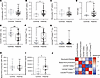 Figure 1
Peripheral immune profiling and neutrophil activation states in healthy controls and patients with epilepsy. (A–D) Quantification of lymphocytes, neutrophils, monocytes, and neutrophil-to-lymphocyte (NLR) ratio in whole blood from healthy controls (gray, n = 10) and patients with epilepsy (black, n = 57). (E–G) Percentage of CXCR4+CD62+ (E), CXCR4+CD62Llo (F), and CXCR4hiCD62Llo (G) neutrophils in control (gray, n = 42) and patients with epilepsy (black, n = 59). (H) Basal ROS production capacity in neutrophils from controls (gray, n = 27) and patients with epilepsy (black, n = 35). (I) Serum neutrophil elastase concentration in controls (gray, n = 12) and patients with epilepsy (black, n = 26). (J) Correlation table showing Spearman’s correlation coefficients between basal ROS production, serum neutrophil elastase concentration, and the 3 neutrophil activation states: CXCR4+CD62+, CXCR4+CD62Llo, and CXCR4hiCD62Llo. Normality was assessed using the Shapiro-Wilk test; comparisons were performed using 2-tailed Student’s t test or Mann-Whitney U test as appropriate. #P < 0.05, ###P < 0.001. Correlations were analyzed using Spearman’s rank correlation test.
To further characterize neutrophil involvement, we assessed their activation state using flow cytometry based on CXCR4 and CD62L expression. This approach allowed us to distinguish 3 activation subsets, steady state (CXCR4+CD62L+), activated (CXCR4+CD62Llo), and hyperactivated (CXCR4hiCD62Llo), according to our gating strategy (33). Briefly, after removing doublets in the neutrophil population, CXCR4hi and CD62Llo were determined in the control group using a mean intensity fluorescence (MIF) histogram and then applied in all patients (Supplemental Figure 1; supplemental material available online with this article; https://doi.org/10.1172/jci.insight.200419DS1). Patients with DRE showed a distinct activation pattern compared with healthy controls, with a reduced frequency of steady-state CXCR4+CD62L+ neutrophils (62.7% vs. 40.4%; P < 0.0001) and a higher percentage of activated CXCR4+CD62Llo neutrophils (12.8% vs. 35.7%; P = 0.0001), while the proportion of hyperactivated CXCR4hiCD62Llo neutrophils (2.75% vs. 2.18%) remained unchanged (Figure 1, E–G).
To further investigate basal neutrophil activation in patients with DRE, we assessed their degranulation capacity and ROS production. No differences were observed in basal ROS output between patients and healthy controls (Figure 1H). In contrast, neutrophil elastase levels were significantly elevated in the serum of patients with DRE compared with controls (Figure 1I). We then examined correlations between these functional markers and neutrophil activation phenotypes. Interestingly, the proportion of CXCR4hiCD62Llo neutrophils correlated positively with ROS levels but negatively with serum neutrophil elastase levels (Figure 1J). Altogether, these data support a higher basal activation of neutrophils in patients with DRE compared with healthy controls with a higher degranulation capacity.
Chronic peripheral low-grade inflammation in patients with DRE. To further investigate the inflammatory environment, we measured the levels of 10 cytokines in nonepileptic and epileptic sera (Figure 2, A–D, and Supplemental Figure 2). Serum analysis revealed elevated levels of proinflammatory cytokines in patients with DRE compared with healthy controls: IL-6 (0.83 vs. 0.41 pg/mL; P = 0.005), CXCL8/IL-8 (10.96 vs. 6.92 pg/mL; P = 0.03, I, and TNF-α (2.23 vs. 0.82 pg/mL; P = 0.006); antiinflammatory cytokine IL-22 levels were reduced (0.70 vs. 2.66 pg/mL; P < 0.0001) (Figure 2, A–D). Cluster analysis revealed significant positive correlations among most cytokines (Figure 2G, pink square outline).
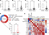 Figure 2
Chronic peripheral low-grade inflammation in patients with epilepsy. (A–D) Graphics displaying cytokine concentrations in sera of control individuals (gray, n = 12) and patients with epilepsy (black, n = 48) and expressed in pg/mL. (E) Distribution of inflammation assessed by CRP in patients with epilepsy: normal inflammation (CRP < 1, blue), slight inflammation (1 < CRP < 3, light red), significant (CRP > 3, red) and (F) histogram showing the distribution of CRP levels in patients (n = 36), categorized as moderate (1 < CRP ≤ 3 mg/L) and high (CRP > 3 mg/L). (G) Heatmap showing Spearman’s correlation coefficients (r) between serum cytokine levels in patients with epilepsy (pink square outline) and neutrophil activation states in blood (green square outline). Color intensity reflects the strength and direction of correlation, with red indicating positive correlations and blue indicating negative correlations. Normality was assessed using the Shapiro-Wilk test; comparisons were performed using 2-tailed Student’s t test or Mann-Whitney U test as appropriate. #P < 0.05, ###P < 0.001. Statistical comparisons between patients and controls are denoted by #, whereas comparisons among different patient groups are denoted by *.
Given the proinflammatory profile observed, we next assessed C-reactive protein (CRP) levels as a general marker of systemic inflammation. Based on established thresholds (CRP < 1 mg/L: no inflammation; 1–3 mg/L: slight inflammation; > 3 mg/L: significant inflammation) (34, 35), we found that 22% of patients with DRE presented slightly elevated CRP (1.685 mg/mL), and 46% had CRP levels above 3 mg/L (6.9 mg/mL) (Figure 2, E and F).
To investigate a potential association between inflammatory cytokines and neutrophil phenotypes, we performed correlation analyses. We observed that steady-state CXCR4+CD62L+ neutrophils were positively correlated with IL-6 levels (r = 0.437, P = 0.001). In contrast, activated CXCR4+CD62Llo neutrophils were negatively correlated with IL-6 (r = –0.472, P = 0.0004), TNF-α (r = –0.405, P = 0.008), and IL-10 (r = –0.358, P = 0.02) but positively correlated with IL-12 (r = 0.273, P = 0.05) (Figure 2G, green square outline). CXCR4+CD62Llo neutrophils were negatively correlated with IL-6 (r = –0.472, P = 0.0004), TNF-α (r = –0.405, P = 0.008), and IL-10 (r = –0.358, P = 0.02) but positively correlated with IL-12 (r = 0.273, P = 0.05). Finally, hyperactivated CXCR4hiCD62Llo neutrophils were positively correlated with IL-8 and TNF-α (IL-8_ r = 0.349, P = 0.01; TNF-α_ r = 0.347, P = 0.04).
Taken together, these findings indicate a peripheral low-grade chronic inflammation characterized by elevated levels of proinflammatory cytokines (CXCL8/IL-8, IL-6, TNF-α) and CRP and associated with an altered neutrophil activation in patients with DRE.
Peripheral low-grade chronic inflammation is independent of a recent seizure. Given that epileptic seizures are associated with increased inflammatory processes (7), we evaluated whether this low-grade inflammation was related to a recent seizure. When considering the delay since the last seizure as a continuous variable, no significant correlations were observed between seizure latency and neutrophil phenotypes or circulating cytokine levels (Table 2). We stratified our patients with DRE into 2 groups: patients who experienced a seizure within 48 hours before blood collection (< 48 hours, n = 32) and those who had been seizure-free for more than 48 hours (> 48 hours, n = 31). This 48-hour window was chosen based on previous studies showing that inflammatory cytokines, such as IL-6, return to baseline within approximately 48 hours (31, 36–38). Moreover, neutrophils exhibit rapid turnover in the circulation, with a residence time typically of several hours to less than 1–2 days, particularly under inflammatory conditions (39, 40). Patients were classified as baseline or “post seizure,” with samples collected a median of 7 and 0.5 days, respectively, after the last seizure.
When comparing these 2 subgroups, no significant differences were found in the absolute counts of lymphocytes, neutrophils, or monocytes. However, the neutrophil/lymphocyte ratio was significantly higher in patients who had a seizure within the last 48 hours (2.26 vs. 1.67; P = 0.03) (Figure 3, A–D). Neutrophil counts were not elevated, and analysis of their activation states showed no significant differences between patients with recent seizures and those without across the 3 defined subpopulations (Figure 3, E–G). Among the cytokines analyzed, IL-22 was the only one found to be significantly elevated in patients who had experienced a seizure within the previous 48 hours, relative to those who had been seizure-free for more than 48 hours (0.8 vs. 0.5 pg/mL; P = 0.05) (Figure 4, A–J). Finally, we observed no significant difference in the CRP levels between these 2 subgroups of patients (Figure 4K). These findings suggest that although recent seizure activity may acutely influence systemic immune responses, the chronic low-grade inflammation observed in patients with DRE appears to persist independently of seizure timing.
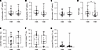 Figure 3
Neutrophil activation is independent of seizure latency. (A–D) Measurement of lymphocyte (A), neutrophil (B), and monocyte (C) counts, as well as the neutrophil-to-lymphocyte ratio (NLR) (D), in the whole blood of patients with epilepsy with a seizure within the last 48 hours (< 48 hours, n = 32) and those seizure-free for more than 48 hours (> 48 hours, n = 31). (E–G) Graphical representation of the distribution of CXCR4+CD62L+ (E), CXCR4+CD62Llo (F), and CXCR4hiCD62Llo (G) neutrophils in patients with epilepsy with a seizure within the last 48 hours (< 48 hours, n = 32) and those seizure-free for more than 48 hours (> 48 hours, n = 31). Normality was assessed using the Shapiro-Wilk test; comparisons were performed using 2-tailed Student’s t test or Mann-Whitney U test as appropriate. *P < 0.05.
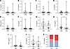 Figure 4
Chronic peripheral low-grade inflammation independent of seizure latency. (A–J) Graphics displaying cytokine concentrations between patients with epilepsy with a seizure within the last 48 hours (< 48 hours, n = 27) and those seizure-free for more than 48 hours (> 48 hours, n = 28). (K) Distribution of inflammation assessed by CRP in patients with epilepsy with a seizure within the last 48 hours and those seizure-free for more than 48 hours. No inflammation (CRP < 1, blue), slight inflammation (1 < CRP < 3, light red), significant (CRP > 3, red). Normality was assessed using the Shapiro-Wilk test; comparisons were performed using 2-tailed Student’s t test or Mann-Whitney U test as appropriate. For distribution comparisons, the χ2 test was used. *P < 0.05.
Two distinct inflammatory phenotypes identified in patients with DRE. The clinical phenotype of patients with DRE is heterogeneous, primarily due to the diversity of clinical parameters in our cohort. We investigated the impact of the clinical parameters of lesion localization (i.e., temporal vs. others) and the presence of hippocampal sclerosis or other lesions, including tumors, on systemic inflammation. Despite a significant increase in lymphocyte counts and variability in CRP levels, these alterations did not seem to affect cytokine profiles or neutrophil phenotypic distribution (Supplemental Figures 3 and 4).
Then, we investigated the impact of monthly seizure frequency, disease duration, and latency of the most recent seizures on systemic inflammation. We performed a principal component analysis (PCA) integrating neutrophil activation states (steady, activated, hyperactivated) with 3 clinical parameters: seizure latency, epilepsy duration, and seizure frequency per month. This multivariate analysis revealed 2 distinct patient clusters (Figure 5, A and B).
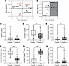 Figure 5
Patient stratification according to seizure frequency, duration of epilepsy, and neutrophil activation. (A-B) The PCA of 3 neutrophil phenotypes and 3 clinical variables for patients with epilepsy. Variable contribution in building the PCA is represented by the length of each arrow. Variable correlation plot illustrating the relationship between clinical data and activation state data. Graph depicting the distribution of individuals following PCA and the clustering of patients into 2 groups (n = 55). (C–E) Graphics displaying clinical data: latency (C), duration (D), and seizure frequency per month (E) among the 2 clusters (n = 29, cluster 1; n = 26, cluster 2). (F–H) Percentage of CXCR4+CD62+ (F), CXCR4+CD62Llo (G), and CXCR4hiCD62Llo (H) in each patients’ clusters (n = 29, cluster 1; n = 26, cluster 2). Normality was assessed using the Shapiro-Wilk test; comparisons were performed using 2-tailed Student’s t test or Mann-Whitney U test as appropriate. *P < 0.05, ***P < 0.001.
An initial comparison of clinical characteristics between the 2 clusters revealed no significant difference in seizure latency, with both groups exhibiting a median of around 5 days (Figure 5C). Conversely, patients in cluster 1 had a significantly longer disease duration (23 vs. 14 years, P = 0.02), whereas those in cluster 2 experienced a substantially higher monthly seizure frequency (11.6 vs. 4 seizures/month, P = 0.01) (Figure 5, D and E). Regarding neutrophil activation, patients in cluster 1 exhibited a significantly higher proportion of steady-state neutrophils (CXCR4+CD62L+) compared with those in cluster 2 (61.3% vs. 23.7%, P < 0.0001) (Figure 5F). In contrast, cluster 2 showed increased proportions of both activated (CXCR4+CD62Llo) and hyperactivated (CXCR4hiCD62Llo) neutrophils (68.3% vs. 21.4%, P < 0.0001 and 2.6% vs. 1.5%, P = 0.03, respectively) (Figure 5, G and H).
Regarding blood count, no significant differences in lymphocyte, neutrophil, or monocyte counts were found between the 2 clusters (Figure 6, A–D). Cytokine analysis revealed that cluster 1 had significantly higher levels of IL-6 and IL-10 compared with cluster 2 (IL-6: 1.3 vs. 0.7 pg/mL, P = 0.02; IL-10: 0.5 vs. 0.1 pg/mL, P = 0.03). Conversely, cluster 2 displayed significantly elevated levels of IL-12 (0.06 vs. 0.01 pg/mL, P = 0.02), a cytokine with well-established proinflammatory and immune-activating properties (Figure 6, E–G). Higher proportions of patients with DRE with CRP greater than 1 mg/mL or greater than 3 mg/mL were present in cluster 1 (χ2 = 43.01, P < 0.0001) (Figure 6H).
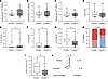 Figure 6
Characterization of peripheral inflammation in patients’ clusters. (A–D) Histogram representing lymphocyte (A), neutrophil (B), and monocyte (C) counts, as well as the neutrophil-to-lymphocyte ratio (NLR) (D), in the whole blood of patients with epilepsy of each cluster (n = 29, cluster 1; n = 26 cluster2). (E–G) Graphics displaying IL-12 (E), IL-6 (F), and IL-10 (G) concentrations in sera of patients with epilepsy in each cluster (n = 7–14, cluster 1; n = 26, cluster 2). (H) Distribution of inflammation assessed by CRP in patients with epilepsy in each cluster according to seizure frequency: No inflammation (CRP < 1, blue), slight inflammation (1 < CRP < 3, light red), significant (CRP > 3, red). (I) Histogram representing the percentage of immature neutrophils in cluster 1 (n = 10) and cluster 2 (n = 9) patients with epilepsy. (J) Radar chart illustrating the immune profiles of cluster 1 and cluster 2 based on IL-6, IL-12, immature neutrophils, and activation state of neutrophils. Each axis represents 1 immune parameter, and values correspond to the ratio of expression or frequency between cluster 1 and cluster 2, allowing visual comparison of immune characteristics across clusters. Normality was assessed using the Shapiro-Wilk test; comparisons were performed using 2-tailed Student’s t test or Mann-Whitney U test as appropriate. For distribution comparisons, the χ2 test was used. *P < 0.05, ***P < 0.001.
Given that IL-6 is a pleiotropic cytokine associated with emergency hematopoiesis (41), and that CXCR4+CD62L+ neutrophils are typically associated with steady-state or immature phenotypes (42), we further investigated the presence of immature neutrophils defined as CD15+CD10–. We found that patients in cluster 1 presented a significantly higher proportion of immature neutrophils in peripheral blood compared with cluster 2 (11.65 vs. 3.12 P = 0.02) (Figure 6I).
Taken together, these findings reveal the existence of 2 chronic low-grade inflammatory profiles among patients with DRE. Profile 1, associated with longer disease duration and lower seizure frequency, is characterized by a low-grade chronic inflammation enriched in immature neutrophils and elevated levels of pleiotropic (IL-6) and antiinflammatory (IL-10) cytokines. In contrast, profile 2, defined by higher seizure frequency, is marked by increased proportions of activated and hyperactivated neutrophils and elevated concentrations of the proinflammatory cytokine IL-12 (Figure 6J).
Peripheral inflammation as a predictor of postsurgical epilepsy prognosis. Seizure recurrence after epilepsy surgery remains a major clinical challenge. In our cohort, 35% of patients experienced seizure recurrence within 1 year after surgery (Figure 7A). Several risk factors have been identified, such as seizure frequency, disease duration, and defined lesion (43). In our cohort, patients who presented with recurrent seizures did not exhibit a higher monthly seizure frequency or longer disease duration (Figure 7, B and C). Given the known role of inflammation in epileptogenesis, we hypothesized that specific immune and inflammatory parameters might serve as predictive biomarkers for seizure recurrence after surgery. We explored CRP levels as a marker of systemic inflammation. Patients who relapsed had significantly higher CRP values compared with seizure-free patients (χ2 = 30.02, P < 0,0001) (Figure 7E). However, the proportion of patients who experienced seizure recurrence 1 year after surgery was similar in cluster 1 and cluster 2 (Figure 7D).
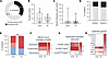 Figure 7
Peripheral inflammation as a predictor of postsurgical epilepsy prognosis. (A) Proportion of patients with recurrence (black) or non-recurrence (gray) of seizure within the year after surgery. (B and C) Graphics displaying clinical data disease duration (B), and seizure frequency per month (C) among patients with no seizure recurrence (N.R; gray, n = 33) or with seizure recurrence (R; black, n = 18). (D) Proportion of patients with recurrence (black) and non-recurrence (gray) of seizures among cluster 1 and cluster 2 patients with epilepsy. (E) Distribution of inflammation assessed by CRP in patients with epilepsy without (N.R) or with (R) recurrence: No inflammation (CRP < 1, blue), slight inflammation (1 < CRP < 3, light red), significant (CRP > 3, red). (F–H) Heatmap showing the leucocyte blood count (F), percentage of CXCR4+CD62L+, CXCR4+CD62Llo, or CXCR4hiCD62Llo (G), and cytokine concentration (H) in blood of patients with epilepsy with or without seizure recurrence. Normality was assessed using the Shapiro-Wilk test; comparisons were performed using 2-tailed Student’s t test or Mann-Whitney U test as appropriate. For distribution comparisons, the χ2 test was used. ***P < 0.001.
Analysis of complete blood count (CBC) revealed no significant differences in neutrophil, lymphocyte, and monocyte counts between the 2 groups (Figure 7F). However, analysis of serum cytokines showed that patients with postoperative seizure recurrence had significantly higher levels of IL-6 and TNF-α before the surgery (IL-6: 0.71 vs. 1.4; P = 0.02; TNF-α: 1.9 vs. 3.9; P = 0.007) (Figure 7H). Although the steady and activated phenotypes were comparable between groups, patients with seizure recurrence exhibited a significantly higher proportion of hyperactivated neutrophils (CXCR4hiCD62Llo) (6.4 vs. 2.2; P = 0.04) (Figure 7G). Normalization of z scores revealed a significant difference in preoperative IL-6 and TNF-α and CXCR4hiCD62Llo levels in patients with seizure recurrence after surgery, supporting an association between the 3 inflammatory components and unfavorable postsurgical outcome (Figure 8, A–C).
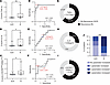 Figure 8
Peripheral inflammation as a predictor of postsurgical epilepsy prognosis. We calculated the z score for TNF-α, IL-6, and CXCR4hiCD62Llo neutrophils (A–C) in blood from patients with DRE at preoperative evaluation comparing patients with (n = 8) and without (n = 24) postsurgery seizure recurrence at 1 year after surgery. (D–F) ROC curves illustrating the predictive performance of preoperative biomarkers for seizure recurrence 1 year after surgery. Each curve represents a distinct biomarker, and AUC reflects the overall discriminative ability. AUC values and corresponding P values are indicated for each marker. Youden index is plotted in red. (G–I) Proportion of patients with a TNF-α, IL-6, and percentage of CXCR4hiCD62Llo neutrophils above the Youden index cutoff, stratified by seizure recurrence status 1 year after surgery. (J) Distribution of patients according to the number of elevated preoperative biomarkers (TNF-α, IL-6, and CXCR4hiCD62Llo neutrophils) in relation to seizure recurrence 1 year after surgery. Normality was assessed using the Shapiro-Wilk test; comparisons were performed using 2-tailed Student’s t test or Mann-Whitney U test as appropriate. *P < 0.05, **P < 0.01, ***P < 0.001.
Using ROC curve analysis, we assessed the predictive value of the 3 markers (i.e., TNF-α IL-6, CXCR4hiCD62Llo) for postoperative seizure recurrence. TNF-α showed the highest discriminative ability, with an AUC of 0.83 (95% CI, 0.5749–1.000; P = 0.01). IL-6 demonstrated moderate predictive accuracy, with an AUC of 0.71 (95% CI, 0.5472–0.8872; P = 0.04), and CXCR4hiCD62Llo neutrophils showed a higher than IL-6 significant discriminative capacity, with an AUC of 0.74 (95% CI, 0.5388–0.9404; P = 0.04) (Figure 8, D–F). To further evaluate the clinical utility of each biomarker, the optimal cutoff values were determined using the Youden index. For TNF-α, the optimal threshold was 2.417 pg/mL, yielding a sensitivity of 0.86 and a specificity of 0.86 (Figure 8D). Among patients with concentrations above this cutoff, 63% experienced seizure recurrence within 1 year after surgery (Figure 8G). For IL-6, the optimal cutoff was 0.82 pg/mL (sensitivity 0.78, specificity 0.57), and 27% of patients above this threshold developed recurrent seizures (Figure 8, E–H). CXCR4hiCD62Llo neutrophils had a lower sensitivity (0.63) but a higher specificity (0.87), and 63% of patients with values above the identified threshold experienced postoperative seizures (Figure 8, F–I). Each parameter was initially evaluated individually using the Youden index to determine optimal thresholds, and then these parameters were combined to improve the overall diagnostic accuracy. We evaluated the proportion of patients, with or without recurrence, who presented at least 1 of the 3 biomarkers above their respective predefined thresholds. This analysis was performed in the subgroup of patients with available values for all 3 parameters (n = 24). We found that 84% of patients with seizure recurrence had at least 1 elevated biomarker compared with 47% of patients without recurrence. Notably, 50% of patients with recurrence had all 3 biomarkers elevated, whereas none of the patients without recurrence showed this pattern (χ2 = 95.56, P < 0.0001) (Figure 8J). These findings suggest that the combined evaluation of these preoperative biomarkers may improve the identification of patients at higher risk of postoperative seizure recurrence.


