
Research ArticleImmunologyInflammation
Open Access |  10.1172/jci.insight.199040
Hypomorphic Lig4 gene mutation in mice predisposes to Th1-skewed intestinal inflammation
Yusuke Yamashita,1 Hideki Kosako,1 Takashi Kato,2,3 Izumi Sasaki,2 Sadahiro Iwabuchi,4 Yuri Fukuda-Ohta,2 Tadashi Okamura,1 Misato Tane,1 Shotaro Tabata,1 Kazutaka Nakashima,5 Ken Tanaka,1,5 Kazunori Shiraishi,6 Yuki Uchihara,7 Daisuke Okuzaki,8 Kyoichi Isono,9 Atsushi Shibata,7 Tsunehiro Mizushima,10 Hiroaki Hemmi,2,11 Nobuo Kanazawa,12 Seiji Kodama,6 Hiroaki Miyoshi,5 Koichi Ohshima,5 Shinichi Hashimoto,4 Yoshio Fujitani,13 Takashi Sonoki,1 Shinobu Tamura,1,14 and Tsuneyasu Kaisho2,15
1Department of Hematology/Oncology,
2Department of Immunology, Institute of Advanced Medicine,
3Department of Rheumatology and Clinical Immunology, and
4Department of Molecular Pathophysiology, Institute of Advanced Medicine, Wakayama Medical University, Wakayama, Japan.
5Department of Pathology, Kurume University School of Medicine, Fukuoka, Japan.
6Laboratory of Radiation Biology, Department of Biological Chemistry, Graduate School of Science, Osaka Metropolitan University, Osaka, Japan.
7Division of Molecular Oncological Pharmacy, Faculty of Pharmacy, Keio University, Tokyo, Japan.
8Laboratory of Human Immunology (Single Cell Genomics), WPI Immunology Frontier Research Center, University of Osaka, Osaka, Japan.
9Laboratory Animal Center, School of Medicine, Wakayama Medical University, Wakayama, Japan.
10Graduate School of Science, University of Hyogo, Hyogo, Japan.
11Laboratory of Immunology, Faculty of Veterinary Medicine, Okayama University of Science, Ehime, Japan.
12Department of Dermatology, Hyogo Medical University, Hyogo, Japan.
13Laboratory of Developmental Biology and Metabolism, Institute for Molecular and Cellular Regulation, Gunma University, Gunma, Japan.
14First Department of Internal Medicine and
15Industry-Government-Academia Collaboration Promotion Headquarters, Wakayama Medical University, Wakayama, Japan.
Address correspondence to: Shinobu Tamura, First Department of Internal Medicine, Wakayama Medical University, 811-1 Kimiidera, Wakayama 641-8509, Japan. Email: stamura@wakayama-med.ac.jp. Or to: Tsuneyasu Kaisho, Department of Immunology, Institute of Advanced Medicine, Wakayama Medical University, 811-1 Kimiidera, Wakayama 641-8509, Japan. Email: tkaisho@wakayama-med.ac.jp.
Authorship note: YY and HK are co–first authors and contributed equally to this work.
Find articles by Yamashita, Y. in: PubMed | Google Scholar
1Department of Hematology/Oncology,
2Department of Immunology, Institute of Advanced Medicine,
3Department of Rheumatology and Clinical Immunology, and
4Department of Molecular Pathophysiology, Institute of Advanced Medicine, Wakayama Medical University, Wakayama, Japan.
5Department of Pathology, Kurume University School of Medicine, Fukuoka, Japan.
6Laboratory of Radiation Biology, Department of Biological Chemistry, Graduate School of Science, Osaka Metropolitan University, Osaka, Japan.
7Division of Molecular Oncological Pharmacy, Faculty of Pharmacy, Keio University, Tokyo, Japan.
8Laboratory of Human Immunology (Single Cell Genomics), WPI Immunology Frontier Research Center, University of Osaka, Osaka, Japan.
9Laboratory Animal Center, School of Medicine, Wakayama Medical University, Wakayama, Japan.
10Graduate School of Science, University of Hyogo, Hyogo, Japan.
11Laboratory of Immunology, Faculty of Veterinary Medicine, Okayama University of Science, Ehime, Japan.
12Department of Dermatology, Hyogo Medical University, Hyogo, Japan.
13Laboratory of Developmental Biology and Metabolism, Institute for Molecular and Cellular Regulation, Gunma University, Gunma, Japan.
14First Department of Internal Medicine and
15Industry-Government-Academia Collaboration Promotion Headquarters, Wakayama Medical University, Wakayama, Japan.
Address correspondence to: Shinobu Tamura, First Department of Internal Medicine, Wakayama Medical University, 811-1 Kimiidera, Wakayama 641-8509, Japan. Email: stamura@wakayama-med.ac.jp. Or to: Tsuneyasu Kaisho, Department of Immunology, Institute of Advanced Medicine, Wakayama Medical University, 811-1 Kimiidera, Wakayama 641-8509, Japan. Email: tkaisho@wakayama-med.ac.jp.
Authorship note: YY and HK are co–first authors and contributed equally to this work.
Find articles by Kosako, H. in: PubMed | Google Scholar
1Department of Hematology/Oncology,
2Department of Immunology, Institute of Advanced Medicine,
3Department of Rheumatology and Clinical Immunology, and
4Department of Molecular Pathophysiology, Institute of Advanced Medicine, Wakayama Medical University, Wakayama, Japan.
5Department of Pathology, Kurume University School of Medicine, Fukuoka, Japan.
6Laboratory of Radiation Biology, Department of Biological Chemistry, Graduate School of Science, Osaka Metropolitan University, Osaka, Japan.
7Division of Molecular Oncological Pharmacy, Faculty of Pharmacy, Keio University, Tokyo, Japan.
8Laboratory of Human Immunology (Single Cell Genomics), WPI Immunology Frontier Research Center, University of Osaka, Osaka, Japan.
9Laboratory Animal Center, School of Medicine, Wakayama Medical University, Wakayama, Japan.
10Graduate School of Science, University of Hyogo, Hyogo, Japan.
11Laboratory of Immunology, Faculty of Veterinary Medicine, Okayama University of Science, Ehime, Japan.
12Department of Dermatology, Hyogo Medical University, Hyogo, Japan.
13Laboratory of Developmental Biology and Metabolism, Institute for Molecular and Cellular Regulation, Gunma University, Gunma, Japan.
14First Department of Internal Medicine and
15Industry-Government-Academia Collaboration Promotion Headquarters, Wakayama Medical University, Wakayama, Japan.
Address correspondence to: Shinobu Tamura, First Department of Internal Medicine, Wakayama Medical University, 811-1 Kimiidera, Wakayama 641-8509, Japan. Email: stamura@wakayama-med.ac.jp. Or to: Tsuneyasu Kaisho, Department of Immunology, Institute of Advanced Medicine, Wakayama Medical University, 811-1 Kimiidera, Wakayama 641-8509, Japan. Email: tkaisho@wakayama-med.ac.jp.
Authorship note: YY and HK are co–first authors and contributed equally to this work.
Find articles by Kato, T. in: PubMed | Google Scholar
1Department of Hematology/Oncology,
2Department of Immunology, Institute of Advanced Medicine,
3Department of Rheumatology and Clinical Immunology, and
4Department of Molecular Pathophysiology, Institute of Advanced Medicine, Wakayama Medical University, Wakayama, Japan.
5Department of Pathology, Kurume University School of Medicine, Fukuoka, Japan.
6Laboratory of Radiation Biology, Department of Biological Chemistry, Graduate School of Science, Osaka Metropolitan University, Osaka, Japan.
7Division of Molecular Oncological Pharmacy, Faculty of Pharmacy, Keio University, Tokyo, Japan.
8Laboratory of Human Immunology (Single Cell Genomics), WPI Immunology Frontier Research Center, University of Osaka, Osaka, Japan.
9Laboratory Animal Center, School of Medicine, Wakayama Medical University, Wakayama, Japan.
10Graduate School of Science, University of Hyogo, Hyogo, Japan.
11Laboratory of Immunology, Faculty of Veterinary Medicine, Okayama University of Science, Ehime, Japan.
12Department of Dermatology, Hyogo Medical University, Hyogo, Japan.
13Laboratory of Developmental Biology and Metabolism, Institute for Molecular and Cellular Regulation, Gunma University, Gunma, Japan.
14First Department of Internal Medicine and
15Industry-Government-Academia Collaboration Promotion Headquarters, Wakayama Medical University, Wakayama, Japan.
Address correspondence to: Shinobu Tamura, First Department of Internal Medicine, Wakayama Medical University, 811-1 Kimiidera, Wakayama 641-8509, Japan. Email: stamura@wakayama-med.ac.jp. Or to: Tsuneyasu Kaisho, Department of Immunology, Institute of Advanced Medicine, Wakayama Medical University, 811-1 Kimiidera, Wakayama 641-8509, Japan. Email: tkaisho@wakayama-med.ac.jp.
Authorship note: YY and HK are co–first authors and contributed equally to this work.
Find articles by Sasaki, I. in: PubMed | Google Scholar
1Department of Hematology/Oncology,
2Department of Immunology, Institute of Advanced Medicine,
3Department of Rheumatology and Clinical Immunology, and
4Department of Molecular Pathophysiology, Institute of Advanced Medicine, Wakayama Medical University, Wakayama, Japan.
5Department of Pathology, Kurume University School of Medicine, Fukuoka, Japan.
6Laboratory of Radiation Biology, Department of Biological Chemistry, Graduate School of Science, Osaka Metropolitan University, Osaka, Japan.
7Division of Molecular Oncological Pharmacy, Faculty of Pharmacy, Keio University, Tokyo, Japan.
8Laboratory of Human Immunology (Single Cell Genomics), WPI Immunology Frontier Research Center, University of Osaka, Osaka, Japan.
9Laboratory Animal Center, School of Medicine, Wakayama Medical University, Wakayama, Japan.
10Graduate School of Science, University of Hyogo, Hyogo, Japan.
11Laboratory of Immunology, Faculty of Veterinary Medicine, Okayama University of Science, Ehime, Japan.
12Department of Dermatology, Hyogo Medical University, Hyogo, Japan.
13Laboratory of Developmental Biology and Metabolism, Institute for Molecular and Cellular Regulation, Gunma University, Gunma, Japan.
14First Department of Internal Medicine and
15Industry-Government-Academia Collaboration Promotion Headquarters, Wakayama Medical University, Wakayama, Japan.
Address correspondence to: Shinobu Tamura, First Department of Internal Medicine, Wakayama Medical University, 811-1 Kimiidera, Wakayama 641-8509, Japan. Email: stamura@wakayama-med.ac.jp. Or to: Tsuneyasu Kaisho, Department of Immunology, Institute of Advanced Medicine, Wakayama Medical University, 811-1 Kimiidera, Wakayama 641-8509, Japan. Email: tkaisho@wakayama-med.ac.jp.
Authorship note: YY and HK are co–first authors and contributed equally to this work.
Find articles by
Iwabuchi, S.
in:
PubMed
|
Google Scholar
|

1Department of Hematology/Oncology,
2Department of Immunology, Institute of Advanced Medicine,
3Department of Rheumatology and Clinical Immunology, and
4Department of Molecular Pathophysiology, Institute of Advanced Medicine, Wakayama Medical University, Wakayama, Japan.
5Department of Pathology, Kurume University School of Medicine, Fukuoka, Japan.
6Laboratory of Radiation Biology, Department of Biological Chemistry, Graduate School of Science, Osaka Metropolitan University, Osaka, Japan.
7Division of Molecular Oncological Pharmacy, Faculty of Pharmacy, Keio University, Tokyo, Japan.
8Laboratory of Human Immunology (Single Cell Genomics), WPI Immunology Frontier Research Center, University of Osaka, Osaka, Japan.
9Laboratory Animal Center, School of Medicine, Wakayama Medical University, Wakayama, Japan.
10Graduate School of Science, University of Hyogo, Hyogo, Japan.
11Laboratory of Immunology, Faculty of Veterinary Medicine, Okayama University of Science, Ehime, Japan.
12Department of Dermatology, Hyogo Medical University, Hyogo, Japan.
13Laboratory of Developmental Biology and Metabolism, Institute for Molecular and Cellular Regulation, Gunma University, Gunma, Japan.
14First Department of Internal Medicine and
15Industry-Government-Academia Collaboration Promotion Headquarters, Wakayama Medical University, Wakayama, Japan.
Address correspondence to: Shinobu Tamura, First Department of Internal Medicine, Wakayama Medical University, 811-1 Kimiidera, Wakayama 641-8509, Japan. Email: stamura@wakayama-med.ac.jp. Or to: Tsuneyasu Kaisho, Department of Immunology, Institute of Advanced Medicine, Wakayama Medical University, 811-1 Kimiidera, Wakayama 641-8509, Japan. Email: tkaisho@wakayama-med.ac.jp.
Authorship note: YY and HK are co–first authors and contributed equally to this work.
Find articles by Fukuda-Ohta, Y. in: PubMed | Google Scholar
1Department of Hematology/Oncology,
2Department of Immunology, Institute of Advanced Medicine,
3Department of Rheumatology and Clinical Immunology, and
4Department of Molecular Pathophysiology, Institute of Advanced Medicine, Wakayama Medical University, Wakayama, Japan.
5Department of Pathology, Kurume University School of Medicine, Fukuoka, Japan.
6Laboratory of Radiation Biology, Department of Biological Chemistry, Graduate School of Science, Osaka Metropolitan University, Osaka, Japan.
7Division of Molecular Oncological Pharmacy, Faculty of Pharmacy, Keio University, Tokyo, Japan.
8Laboratory of Human Immunology (Single Cell Genomics), WPI Immunology Frontier Research Center, University of Osaka, Osaka, Japan.
9Laboratory Animal Center, School of Medicine, Wakayama Medical University, Wakayama, Japan.
10Graduate School of Science, University of Hyogo, Hyogo, Japan.
11Laboratory of Immunology, Faculty of Veterinary Medicine, Okayama University of Science, Ehime, Japan.
12Department of Dermatology, Hyogo Medical University, Hyogo, Japan.
13Laboratory of Developmental Biology and Metabolism, Institute for Molecular and Cellular Regulation, Gunma University, Gunma, Japan.
14First Department of Internal Medicine and
15Industry-Government-Academia Collaboration Promotion Headquarters, Wakayama Medical University, Wakayama, Japan.
Address correspondence to: Shinobu Tamura, First Department of Internal Medicine, Wakayama Medical University, 811-1 Kimiidera, Wakayama 641-8509, Japan. Email: stamura@wakayama-med.ac.jp. Or to: Tsuneyasu Kaisho, Department of Immunology, Institute of Advanced Medicine, Wakayama Medical University, 811-1 Kimiidera, Wakayama 641-8509, Japan. Email: tkaisho@wakayama-med.ac.jp.
Authorship note: YY and HK are co–first authors and contributed equally to this work.
Find articles by Okamura, T. in: PubMed | Google Scholar
1Department of Hematology/Oncology,
2Department of Immunology, Institute of Advanced Medicine,
3Department of Rheumatology and Clinical Immunology, and
4Department of Molecular Pathophysiology, Institute of Advanced Medicine, Wakayama Medical University, Wakayama, Japan.
5Department of Pathology, Kurume University School of Medicine, Fukuoka, Japan.
6Laboratory of Radiation Biology, Department of Biological Chemistry, Graduate School of Science, Osaka Metropolitan University, Osaka, Japan.
7Division of Molecular Oncological Pharmacy, Faculty of Pharmacy, Keio University, Tokyo, Japan.
8Laboratory of Human Immunology (Single Cell Genomics), WPI Immunology Frontier Research Center, University of Osaka, Osaka, Japan.
9Laboratory Animal Center, School of Medicine, Wakayama Medical University, Wakayama, Japan.
10Graduate School of Science, University of Hyogo, Hyogo, Japan.
11Laboratory of Immunology, Faculty of Veterinary Medicine, Okayama University of Science, Ehime, Japan.
12Department of Dermatology, Hyogo Medical University, Hyogo, Japan.
13Laboratory of Developmental Biology and Metabolism, Institute for Molecular and Cellular Regulation, Gunma University, Gunma, Japan.
14First Department of Internal Medicine and
15Industry-Government-Academia Collaboration Promotion Headquarters, Wakayama Medical University, Wakayama, Japan.
Address correspondence to: Shinobu Tamura, First Department of Internal Medicine, Wakayama Medical University, 811-1 Kimiidera, Wakayama 641-8509, Japan. Email: stamura@wakayama-med.ac.jp. Or to: Tsuneyasu Kaisho, Department of Immunology, Institute of Advanced Medicine, Wakayama Medical University, 811-1 Kimiidera, Wakayama 641-8509, Japan. Email: tkaisho@wakayama-med.ac.jp.
Authorship note: YY and HK are co–first authors and contributed equally to this work.
Find articles by Tane, M. in: PubMed | Google Scholar
1Department of Hematology/Oncology,
2Department of Immunology, Institute of Advanced Medicine,
3Department of Rheumatology and Clinical Immunology, and
4Department of Molecular Pathophysiology, Institute of Advanced Medicine, Wakayama Medical University, Wakayama, Japan.
5Department of Pathology, Kurume University School of Medicine, Fukuoka, Japan.
6Laboratory of Radiation Biology, Department of Biological Chemistry, Graduate School of Science, Osaka Metropolitan University, Osaka, Japan.
7Division of Molecular Oncological Pharmacy, Faculty of Pharmacy, Keio University, Tokyo, Japan.
8Laboratory of Human Immunology (Single Cell Genomics), WPI Immunology Frontier Research Center, University of Osaka, Osaka, Japan.
9Laboratory Animal Center, School of Medicine, Wakayama Medical University, Wakayama, Japan.
10Graduate School of Science, University of Hyogo, Hyogo, Japan.
11Laboratory of Immunology, Faculty of Veterinary Medicine, Okayama University of Science, Ehime, Japan.
12Department of Dermatology, Hyogo Medical University, Hyogo, Japan.
13Laboratory of Developmental Biology and Metabolism, Institute for Molecular and Cellular Regulation, Gunma University, Gunma, Japan.
14First Department of Internal Medicine and
15Industry-Government-Academia Collaboration Promotion Headquarters, Wakayama Medical University, Wakayama, Japan.
Address correspondence to: Shinobu Tamura, First Department of Internal Medicine, Wakayama Medical University, 811-1 Kimiidera, Wakayama 641-8509, Japan. Email: stamura@wakayama-med.ac.jp. Or to: Tsuneyasu Kaisho, Department of Immunology, Institute of Advanced Medicine, Wakayama Medical University, 811-1 Kimiidera, Wakayama 641-8509, Japan. Email: tkaisho@wakayama-med.ac.jp.
Authorship note: YY and HK are co–first authors and contributed equally to this work.
Find articles by Tabata, S. in: PubMed | Google Scholar
1Department of Hematology/Oncology,
2Department of Immunology, Institute of Advanced Medicine,
3Department of Rheumatology and Clinical Immunology, and
4Department of Molecular Pathophysiology, Institute of Advanced Medicine, Wakayama Medical University, Wakayama, Japan.
5Department of Pathology, Kurume University School of Medicine, Fukuoka, Japan.
6Laboratory of Radiation Biology, Department of Biological Chemistry, Graduate School of Science, Osaka Metropolitan University, Osaka, Japan.
7Division of Molecular Oncological Pharmacy, Faculty of Pharmacy, Keio University, Tokyo, Japan.
8Laboratory of Human Immunology (Single Cell Genomics), WPI Immunology Frontier Research Center, University of Osaka, Osaka, Japan.
9Laboratory Animal Center, School of Medicine, Wakayama Medical University, Wakayama, Japan.
10Graduate School of Science, University of Hyogo, Hyogo, Japan.
11Laboratory of Immunology, Faculty of Veterinary Medicine, Okayama University of Science, Ehime, Japan.
12Department of Dermatology, Hyogo Medical University, Hyogo, Japan.
13Laboratory of Developmental Biology and Metabolism, Institute for Molecular and Cellular Regulation, Gunma University, Gunma, Japan.
14First Department of Internal Medicine and
15Industry-Government-Academia Collaboration Promotion Headquarters, Wakayama Medical University, Wakayama, Japan.
Address correspondence to: Shinobu Tamura, First Department of Internal Medicine, Wakayama Medical University, 811-1 Kimiidera, Wakayama 641-8509, Japan. Email: stamura@wakayama-med.ac.jp. Or to: Tsuneyasu Kaisho, Department of Immunology, Institute of Advanced Medicine, Wakayama Medical University, 811-1 Kimiidera, Wakayama 641-8509, Japan. Email: tkaisho@wakayama-med.ac.jp.
Authorship note: YY and HK are co–first authors and contributed equally to this work.
Find articles by Nakashima, K. in: PubMed | Google Scholar
1Department of Hematology/Oncology,
2Department of Immunology, Institute of Advanced Medicine,
3Department of Rheumatology and Clinical Immunology, and
4Department of Molecular Pathophysiology, Institute of Advanced Medicine, Wakayama Medical University, Wakayama, Japan.
5Department of Pathology, Kurume University School of Medicine, Fukuoka, Japan.
6Laboratory of Radiation Biology, Department of Biological Chemistry, Graduate School of Science, Osaka Metropolitan University, Osaka, Japan.
7Division of Molecular Oncological Pharmacy, Faculty of Pharmacy, Keio University, Tokyo, Japan.
8Laboratory of Human Immunology (Single Cell Genomics), WPI Immunology Frontier Research Center, University of Osaka, Osaka, Japan.
9Laboratory Animal Center, School of Medicine, Wakayama Medical University, Wakayama, Japan.
10Graduate School of Science, University of Hyogo, Hyogo, Japan.
11Laboratory of Immunology, Faculty of Veterinary Medicine, Okayama University of Science, Ehime, Japan.
12Department of Dermatology, Hyogo Medical University, Hyogo, Japan.
13Laboratory of Developmental Biology and Metabolism, Institute for Molecular and Cellular Regulation, Gunma University, Gunma, Japan.
14First Department of Internal Medicine and
15Industry-Government-Academia Collaboration Promotion Headquarters, Wakayama Medical University, Wakayama, Japan.
Address correspondence to: Shinobu Tamura, First Department of Internal Medicine, Wakayama Medical University, 811-1 Kimiidera, Wakayama 641-8509, Japan. Email: stamura@wakayama-med.ac.jp. Or to: Tsuneyasu Kaisho, Department of Immunology, Institute of Advanced Medicine, Wakayama Medical University, 811-1 Kimiidera, Wakayama 641-8509, Japan. Email: tkaisho@wakayama-med.ac.jp.
Authorship note: YY and HK are co–first authors and contributed equally to this work.
Find articles by Tanaka, K. in: PubMed | Google Scholar
1Department of Hematology/Oncology,
2Department of Immunology, Institute of Advanced Medicine,
3Department of Rheumatology and Clinical Immunology, and
4Department of Molecular Pathophysiology, Institute of Advanced Medicine, Wakayama Medical University, Wakayama, Japan.
5Department of Pathology, Kurume University School of Medicine, Fukuoka, Japan.
6Laboratory of Radiation Biology, Department of Biological Chemistry, Graduate School of Science, Osaka Metropolitan University, Osaka, Japan.
7Division of Molecular Oncological Pharmacy, Faculty of Pharmacy, Keio University, Tokyo, Japan.
8Laboratory of Human Immunology (Single Cell Genomics), WPI Immunology Frontier Research Center, University of Osaka, Osaka, Japan.
9Laboratory Animal Center, School of Medicine, Wakayama Medical University, Wakayama, Japan.
10Graduate School of Science, University of Hyogo, Hyogo, Japan.
11Laboratory of Immunology, Faculty of Veterinary Medicine, Okayama University of Science, Ehime, Japan.
12Department of Dermatology, Hyogo Medical University, Hyogo, Japan.
13Laboratory of Developmental Biology and Metabolism, Institute for Molecular and Cellular Regulation, Gunma University, Gunma, Japan.
14First Department of Internal Medicine and
15Industry-Government-Academia Collaboration Promotion Headquarters, Wakayama Medical University, Wakayama, Japan.
Address correspondence to: Shinobu Tamura, First Department of Internal Medicine, Wakayama Medical University, 811-1 Kimiidera, Wakayama 641-8509, Japan. Email: stamura@wakayama-med.ac.jp. Or to: Tsuneyasu Kaisho, Department of Immunology, Institute of Advanced Medicine, Wakayama Medical University, 811-1 Kimiidera, Wakayama 641-8509, Japan. Email: tkaisho@wakayama-med.ac.jp.
Authorship note: YY and HK are co–first authors and contributed equally to this work.
Find articles by Shiraishi, K. in: PubMed | Google Scholar
1Department of Hematology/Oncology,
2Department of Immunology, Institute of Advanced Medicine,
3Department of Rheumatology and Clinical Immunology, and
4Department of Molecular Pathophysiology, Institute of Advanced Medicine, Wakayama Medical University, Wakayama, Japan.
5Department of Pathology, Kurume University School of Medicine, Fukuoka, Japan.
6Laboratory of Radiation Biology, Department of Biological Chemistry, Graduate School of Science, Osaka Metropolitan University, Osaka, Japan.
7Division of Molecular Oncological Pharmacy, Faculty of Pharmacy, Keio University, Tokyo, Japan.
8Laboratory of Human Immunology (Single Cell Genomics), WPI Immunology Frontier Research Center, University of Osaka, Osaka, Japan.
9Laboratory Animal Center, School of Medicine, Wakayama Medical University, Wakayama, Japan.
10Graduate School of Science, University of Hyogo, Hyogo, Japan.
11Laboratory of Immunology, Faculty of Veterinary Medicine, Okayama University of Science, Ehime, Japan.
12Department of Dermatology, Hyogo Medical University, Hyogo, Japan.
13Laboratory of Developmental Biology and Metabolism, Institute for Molecular and Cellular Regulation, Gunma University, Gunma, Japan.
14First Department of Internal Medicine and
15Industry-Government-Academia Collaboration Promotion Headquarters, Wakayama Medical University, Wakayama, Japan.
Address correspondence to: Shinobu Tamura, First Department of Internal Medicine, Wakayama Medical University, 811-1 Kimiidera, Wakayama 641-8509, Japan. Email: stamura@wakayama-med.ac.jp. Or to: Tsuneyasu Kaisho, Department of Immunology, Institute of Advanced Medicine, Wakayama Medical University, 811-1 Kimiidera, Wakayama 641-8509, Japan. Email: tkaisho@wakayama-med.ac.jp.
Authorship note: YY and HK are co–first authors and contributed equally to this work.
Find articles by Uchihara, Y. in: PubMed | Google Scholar
1Department of Hematology/Oncology,
2Department of Immunology, Institute of Advanced Medicine,
3Department of Rheumatology and Clinical Immunology, and
4Department of Molecular Pathophysiology, Institute of Advanced Medicine, Wakayama Medical University, Wakayama, Japan.
5Department of Pathology, Kurume University School of Medicine, Fukuoka, Japan.
6Laboratory of Radiation Biology, Department of Biological Chemistry, Graduate School of Science, Osaka Metropolitan University, Osaka, Japan.
7Division of Molecular Oncological Pharmacy, Faculty of Pharmacy, Keio University, Tokyo, Japan.
8Laboratory of Human Immunology (Single Cell Genomics), WPI Immunology Frontier Research Center, University of Osaka, Osaka, Japan.
9Laboratory Animal Center, School of Medicine, Wakayama Medical University, Wakayama, Japan.
10Graduate School of Science, University of Hyogo, Hyogo, Japan.
11Laboratory of Immunology, Faculty of Veterinary Medicine, Okayama University of Science, Ehime, Japan.
12Department of Dermatology, Hyogo Medical University, Hyogo, Japan.
13Laboratory of Developmental Biology and Metabolism, Institute for Molecular and Cellular Regulation, Gunma University, Gunma, Japan.
14First Department of Internal Medicine and
15Industry-Government-Academia Collaboration Promotion Headquarters, Wakayama Medical University, Wakayama, Japan.
Address correspondence to: Shinobu Tamura, First Department of Internal Medicine, Wakayama Medical University, 811-1 Kimiidera, Wakayama 641-8509, Japan. Email: stamura@wakayama-med.ac.jp. Or to: Tsuneyasu Kaisho, Department of Immunology, Institute of Advanced Medicine, Wakayama Medical University, 811-1 Kimiidera, Wakayama 641-8509, Japan. Email: tkaisho@wakayama-med.ac.jp.
Authorship note: YY and HK are co–first authors and contributed equally to this work.
Find articles by
Okuzaki, D.
in:
PubMed
|
Google Scholar
|
1Department of Hematology/Oncology,
2Department of Immunology, Institute of Advanced Medicine,
3Department of Rheumatology and Clinical Immunology, and
4Department of Molecular Pathophysiology, Institute of Advanced Medicine, Wakayama Medical University, Wakayama, Japan.
5Department of Pathology, Kurume University School of Medicine, Fukuoka, Japan.
6Laboratory of Radiation Biology, Department of Biological Chemistry, Graduate School of Science, Osaka Metropolitan University, Osaka, Japan.
7Division of Molecular Oncological Pharmacy, Faculty of Pharmacy, Keio University, Tokyo, Japan.
8Laboratory of Human Immunology (Single Cell Genomics), WPI Immunology Frontier Research Center, University of Osaka, Osaka, Japan.
9Laboratory Animal Center, School of Medicine, Wakayama Medical University, Wakayama, Japan.
10Graduate School of Science, University of Hyogo, Hyogo, Japan.
11Laboratory of Immunology, Faculty of Veterinary Medicine, Okayama University of Science, Ehime, Japan.
12Department of Dermatology, Hyogo Medical University, Hyogo, Japan.
13Laboratory of Developmental Biology and Metabolism, Institute for Molecular and Cellular Regulation, Gunma University, Gunma, Japan.
14First Department of Internal Medicine and
15Industry-Government-Academia Collaboration Promotion Headquarters, Wakayama Medical University, Wakayama, Japan.
Address correspondence to: Shinobu Tamura, First Department of Internal Medicine, Wakayama Medical University, 811-1 Kimiidera, Wakayama 641-8509, Japan. Email: stamura@wakayama-med.ac.jp. Or to: Tsuneyasu Kaisho, Department of Immunology, Institute of Advanced Medicine, Wakayama Medical University, 811-1 Kimiidera, Wakayama 641-8509, Japan. Email: tkaisho@wakayama-med.ac.jp.
Authorship note: YY and HK are co–first authors and contributed equally to this work.
Find articles by Isono, K. in: PubMed | Google Scholar
1Department of Hematology/Oncology,
2Department of Immunology, Institute of Advanced Medicine,
3Department of Rheumatology and Clinical Immunology, and
4Department of Molecular Pathophysiology, Institute of Advanced Medicine, Wakayama Medical University, Wakayama, Japan.
5Department of Pathology, Kurume University School of Medicine, Fukuoka, Japan.
6Laboratory of Radiation Biology, Department of Biological Chemistry, Graduate School of Science, Osaka Metropolitan University, Osaka, Japan.
7Division of Molecular Oncological Pharmacy, Faculty of Pharmacy, Keio University, Tokyo, Japan.
8Laboratory of Human Immunology (Single Cell Genomics), WPI Immunology Frontier Research Center, University of Osaka, Osaka, Japan.
9Laboratory Animal Center, School of Medicine, Wakayama Medical University, Wakayama, Japan.
10Graduate School of Science, University of Hyogo, Hyogo, Japan.
11Laboratory of Immunology, Faculty of Veterinary Medicine, Okayama University of Science, Ehime, Japan.
12Department of Dermatology, Hyogo Medical University, Hyogo, Japan.
13Laboratory of Developmental Biology and Metabolism, Institute for Molecular and Cellular Regulation, Gunma University, Gunma, Japan.
14First Department of Internal Medicine and
15Industry-Government-Academia Collaboration Promotion Headquarters, Wakayama Medical University, Wakayama, Japan.
Address correspondence to: Shinobu Tamura, First Department of Internal Medicine, Wakayama Medical University, 811-1 Kimiidera, Wakayama 641-8509, Japan. Email: stamura@wakayama-med.ac.jp. Or to: Tsuneyasu Kaisho, Department of Immunology, Institute of Advanced Medicine, Wakayama Medical University, 811-1 Kimiidera, Wakayama 641-8509, Japan. Email: tkaisho@wakayama-med.ac.jp.
Authorship note: YY and HK are co–first authors and contributed equally to this work.
Find articles by Shibata, A. in: PubMed | Google Scholar
1Department of Hematology/Oncology,
2Department of Immunology, Institute of Advanced Medicine,
3Department of Rheumatology and Clinical Immunology, and
4Department of Molecular Pathophysiology, Institute of Advanced Medicine, Wakayama Medical University, Wakayama, Japan.
5Department of Pathology, Kurume University School of Medicine, Fukuoka, Japan.
6Laboratory of Radiation Biology, Department of Biological Chemistry, Graduate School of Science, Osaka Metropolitan University, Osaka, Japan.
7Division of Molecular Oncological Pharmacy, Faculty of Pharmacy, Keio University, Tokyo, Japan.
8Laboratory of Human Immunology (Single Cell Genomics), WPI Immunology Frontier Research Center, University of Osaka, Osaka, Japan.
9Laboratory Animal Center, School of Medicine, Wakayama Medical University, Wakayama, Japan.
10Graduate School of Science, University of Hyogo, Hyogo, Japan.
11Laboratory of Immunology, Faculty of Veterinary Medicine, Okayama University of Science, Ehime, Japan.
12Department of Dermatology, Hyogo Medical University, Hyogo, Japan.
13Laboratory of Developmental Biology and Metabolism, Institute for Molecular and Cellular Regulation, Gunma University, Gunma, Japan.
14First Department of Internal Medicine and
15Industry-Government-Academia Collaboration Promotion Headquarters, Wakayama Medical University, Wakayama, Japan.
Address correspondence to: Shinobu Tamura, First Department of Internal Medicine, Wakayama Medical University, 811-1 Kimiidera, Wakayama 641-8509, Japan. Email: stamura@wakayama-med.ac.jp. Or to: Tsuneyasu Kaisho, Department of Immunology, Institute of Advanced Medicine, Wakayama Medical University, 811-1 Kimiidera, Wakayama 641-8509, Japan. Email: tkaisho@wakayama-med.ac.jp.
Authorship note: YY and HK are co–first authors and contributed equally to this work.
Find articles by Mizushima, T. in: PubMed | Google Scholar
1Department of Hematology/Oncology,
2Department of Immunology, Institute of Advanced Medicine,
3Department of Rheumatology and Clinical Immunology, and
4Department of Molecular Pathophysiology, Institute of Advanced Medicine, Wakayama Medical University, Wakayama, Japan.
5Department of Pathology, Kurume University School of Medicine, Fukuoka, Japan.
6Laboratory of Radiation Biology, Department of Biological Chemistry, Graduate School of Science, Osaka Metropolitan University, Osaka, Japan.
7Division of Molecular Oncological Pharmacy, Faculty of Pharmacy, Keio University, Tokyo, Japan.
8Laboratory of Human Immunology (Single Cell Genomics), WPI Immunology Frontier Research Center, University of Osaka, Osaka, Japan.
9Laboratory Animal Center, School of Medicine, Wakayama Medical University, Wakayama, Japan.
10Graduate School of Science, University of Hyogo, Hyogo, Japan.
11Laboratory of Immunology, Faculty of Veterinary Medicine, Okayama University of Science, Ehime, Japan.
12Department of Dermatology, Hyogo Medical University, Hyogo, Japan.
13Laboratory of Developmental Biology and Metabolism, Institute for Molecular and Cellular Regulation, Gunma University, Gunma, Japan.
14First Department of Internal Medicine and
15Industry-Government-Academia Collaboration Promotion Headquarters, Wakayama Medical University, Wakayama, Japan.
Address correspondence to: Shinobu Tamura, First Department of Internal Medicine, Wakayama Medical University, 811-1 Kimiidera, Wakayama 641-8509, Japan. Email: stamura@wakayama-med.ac.jp. Or to: Tsuneyasu Kaisho, Department of Immunology, Institute of Advanced Medicine, Wakayama Medical University, 811-1 Kimiidera, Wakayama 641-8509, Japan. Email: tkaisho@wakayama-med.ac.jp.
Authorship note: YY and HK are co–first authors and contributed equally to this work.
Find articles by Hemmi, H. in: PubMed | Google Scholar
1Department of Hematology/Oncology,
2Department of Immunology, Institute of Advanced Medicine,
3Department of Rheumatology and Clinical Immunology, and
4Department of Molecular Pathophysiology, Institute of Advanced Medicine, Wakayama Medical University, Wakayama, Japan.
5Department of Pathology, Kurume University School of Medicine, Fukuoka, Japan.
6Laboratory of Radiation Biology, Department of Biological Chemistry, Graduate School of Science, Osaka Metropolitan University, Osaka, Japan.
7Division of Molecular Oncological Pharmacy, Faculty of Pharmacy, Keio University, Tokyo, Japan.
8Laboratory of Human Immunology (Single Cell Genomics), WPI Immunology Frontier Research Center, University of Osaka, Osaka, Japan.
9Laboratory Animal Center, School of Medicine, Wakayama Medical University, Wakayama, Japan.
10Graduate School of Science, University of Hyogo, Hyogo, Japan.
11Laboratory of Immunology, Faculty of Veterinary Medicine, Okayama University of Science, Ehime, Japan.
12Department of Dermatology, Hyogo Medical University, Hyogo, Japan.
13Laboratory of Developmental Biology and Metabolism, Institute for Molecular and Cellular Regulation, Gunma University, Gunma, Japan.
14First Department of Internal Medicine and
15Industry-Government-Academia Collaboration Promotion Headquarters, Wakayama Medical University, Wakayama, Japan.
Address correspondence to: Shinobu Tamura, First Department of Internal Medicine, Wakayama Medical University, 811-1 Kimiidera, Wakayama 641-8509, Japan. Email: stamura@wakayama-med.ac.jp. Or to: Tsuneyasu Kaisho, Department of Immunology, Institute of Advanced Medicine, Wakayama Medical University, 811-1 Kimiidera, Wakayama 641-8509, Japan. Email: tkaisho@wakayama-med.ac.jp.
Authorship note: YY and HK are co–first authors and contributed equally to this work.
Find articles by Kanazawa, N. in: PubMed | Google Scholar
1Department of Hematology/Oncology,
2Department of Immunology, Institute of Advanced Medicine,
3Department of Rheumatology and Clinical Immunology, and
4Department of Molecular Pathophysiology, Institute of Advanced Medicine, Wakayama Medical University, Wakayama, Japan.
5Department of Pathology, Kurume University School of Medicine, Fukuoka, Japan.
6Laboratory of Radiation Biology, Department of Biological Chemistry, Graduate School of Science, Osaka Metropolitan University, Osaka, Japan.
7Division of Molecular Oncological Pharmacy, Faculty of Pharmacy, Keio University, Tokyo, Japan.
8Laboratory of Human Immunology (Single Cell Genomics), WPI Immunology Frontier Research Center, University of Osaka, Osaka, Japan.
9Laboratory Animal Center, School of Medicine, Wakayama Medical University, Wakayama, Japan.
10Graduate School of Science, University of Hyogo, Hyogo, Japan.
11Laboratory of Immunology, Faculty of Veterinary Medicine, Okayama University of Science, Ehime, Japan.
12Department of Dermatology, Hyogo Medical University, Hyogo, Japan.
13Laboratory of Developmental Biology and Metabolism, Institute for Molecular and Cellular Regulation, Gunma University, Gunma, Japan.
14First Department of Internal Medicine and
15Industry-Government-Academia Collaboration Promotion Headquarters, Wakayama Medical University, Wakayama, Japan.
Address correspondence to: Shinobu Tamura, First Department of Internal Medicine, Wakayama Medical University, 811-1 Kimiidera, Wakayama 641-8509, Japan. Email: stamura@wakayama-med.ac.jp. Or to: Tsuneyasu Kaisho, Department of Immunology, Institute of Advanced Medicine, Wakayama Medical University, 811-1 Kimiidera, Wakayama 641-8509, Japan. Email: tkaisho@wakayama-med.ac.jp.
Authorship note: YY and HK are co–first authors and contributed equally to this work.
Find articles by Kodama, S. in: PubMed | Google Scholar
1Department of Hematology/Oncology,
2Department of Immunology, Institute of Advanced Medicine,
3Department of Rheumatology and Clinical Immunology, and
4Department of Molecular Pathophysiology, Institute of Advanced Medicine, Wakayama Medical University, Wakayama, Japan.
5Department of Pathology, Kurume University School of Medicine, Fukuoka, Japan.
6Laboratory of Radiation Biology, Department of Biological Chemistry, Graduate School of Science, Osaka Metropolitan University, Osaka, Japan.
7Division of Molecular Oncological Pharmacy, Faculty of Pharmacy, Keio University, Tokyo, Japan.
8Laboratory of Human Immunology (Single Cell Genomics), WPI Immunology Frontier Research Center, University of Osaka, Osaka, Japan.
9Laboratory Animal Center, School of Medicine, Wakayama Medical University, Wakayama, Japan.
10Graduate School of Science, University of Hyogo, Hyogo, Japan.
11Laboratory of Immunology, Faculty of Veterinary Medicine, Okayama University of Science, Ehime, Japan.
12Department of Dermatology, Hyogo Medical University, Hyogo, Japan.
13Laboratory of Developmental Biology and Metabolism, Institute for Molecular and Cellular Regulation, Gunma University, Gunma, Japan.
14First Department of Internal Medicine and
15Industry-Government-Academia Collaboration Promotion Headquarters, Wakayama Medical University, Wakayama, Japan.
Address correspondence to: Shinobu Tamura, First Department of Internal Medicine, Wakayama Medical University, 811-1 Kimiidera, Wakayama 641-8509, Japan. Email: stamura@wakayama-med.ac.jp. Or to: Tsuneyasu Kaisho, Department of Immunology, Institute of Advanced Medicine, Wakayama Medical University, 811-1 Kimiidera, Wakayama 641-8509, Japan. Email: tkaisho@wakayama-med.ac.jp.
Authorship note: YY and HK are co–first authors and contributed equally to this work.
Find articles by Miyoshi, H. in: PubMed | Google Scholar
1Department of Hematology/Oncology,
2Department of Immunology, Institute of Advanced Medicine,
3Department of Rheumatology and Clinical Immunology, and
4Department of Molecular Pathophysiology, Institute of Advanced Medicine, Wakayama Medical University, Wakayama, Japan.
5Department of Pathology, Kurume University School of Medicine, Fukuoka, Japan.
6Laboratory of Radiation Biology, Department of Biological Chemistry, Graduate School of Science, Osaka Metropolitan University, Osaka, Japan.
7Division of Molecular Oncological Pharmacy, Faculty of Pharmacy, Keio University, Tokyo, Japan.
8Laboratory of Human Immunology (Single Cell Genomics), WPI Immunology Frontier Research Center, University of Osaka, Osaka, Japan.
9Laboratory Animal Center, School of Medicine, Wakayama Medical University, Wakayama, Japan.
10Graduate School of Science, University of Hyogo, Hyogo, Japan.
11Laboratory of Immunology, Faculty of Veterinary Medicine, Okayama University of Science, Ehime, Japan.
12Department of Dermatology, Hyogo Medical University, Hyogo, Japan.
13Laboratory of Developmental Biology and Metabolism, Institute for Molecular and Cellular Regulation, Gunma University, Gunma, Japan.
14First Department of Internal Medicine and
15Industry-Government-Academia Collaboration Promotion Headquarters, Wakayama Medical University, Wakayama, Japan.
Address correspondence to: Shinobu Tamura, First Department of Internal Medicine, Wakayama Medical University, 811-1 Kimiidera, Wakayama 641-8509, Japan. Email: stamura@wakayama-med.ac.jp. Or to: Tsuneyasu Kaisho, Department of Immunology, Institute of Advanced Medicine, Wakayama Medical University, 811-1 Kimiidera, Wakayama 641-8509, Japan. Email: tkaisho@wakayama-med.ac.jp.
Authorship note: YY and HK are co–first authors and contributed equally to this work.
Find articles by Ohshima, K. in: PubMed | Google Scholar
1Department of Hematology/Oncology,
2Department of Immunology, Institute of Advanced Medicine,
3Department of Rheumatology and Clinical Immunology, and
4Department of Molecular Pathophysiology, Institute of Advanced Medicine, Wakayama Medical University, Wakayama, Japan.
5Department of Pathology, Kurume University School of Medicine, Fukuoka, Japan.
6Laboratory of Radiation Biology, Department of Biological Chemistry, Graduate School of Science, Osaka Metropolitan University, Osaka, Japan.
7Division of Molecular Oncological Pharmacy, Faculty of Pharmacy, Keio University, Tokyo, Japan.
8Laboratory of Human Immunology (Single Cell Genomics), WPI Immunology Frontier Research Center, University of Osaka, Osaka, Japan.
9Laboratory Animal Center, School of Medicine, Wakayama Medical University, Wakayama, Japan.
10Graduate School of Science, University of Hyogo, Hyogo, Japan.
11Laboratory of Immunology, Faculty of Veterinary Medicine, Okayama University of Science, Ehime, Japan.
12Department of Dermatology, Hyogo Medical University, Hyogo, Japan.
13Laboratory of Developmental Biology and Metabolism, Institute for Molecular and Cellular Regulation, Gunma University, Gunma, Japan.
14First Department of Internal Medicine and
15Industry-Government-Academia Collaboration Promotion Headquarters, Wakayama Medical University, Wakayama, Japan.
Address correspondence to: Shinobu Tamura, First Department of Internal Medicine, Wakayama Medical University, 811-1 Kimiidera, Wakayama 641-8509, Japan. Email: stamura@wakayama-med.ac.jp. Or to: Tsuneyasu Kaisho, Department of Immunology, Institute of Advanced Medicine, Wakayama Medical University, 811-1 Kimiidera, Wakayama 641-8509, Japan. Email: tkaisho@wakayama-med.ac.jp.
Authorship note: YY and HK are co–first authors and contributed equally to this work.
Find articles by Hashimoto, S. in: PubMed | Google Scholar
1Department of Hematology/Oncology,
2Department of Immunology, Institute of Advanced Medicine,
3Department of Rheumatology and Clinical Immunology, and
4Department of Molecular Pathophysiology, Institute of Advanced Medicine, Wakayama Medical University, Wakayama, Japan.
5Department of Pathology, Kurume University School of Medicine, Fukuoka, Japan.
6Laboratory of Radiation Biology, Department of Biological Chemistry, Graduate School of Science, Osaka Metropolitan University, Osaka, Japan.
7Division of Molecular Oncological Pharmacy, Faculty of Pharmacy, Keio University, Tokyo, Japan.
8Laboratory of Human Immunology (Single Cell Genomics), WPI Immunology Frontier Research Center, University of Osaka, Osaka, Japan.
9Laboratory Animal Center, School of Medicine, Wakayama Medical University, Wakayama, Japan.
10Graduate School of Science, University of Hyogo, Hyogo, Japan.
11Laboratory of Immunology, Faculty of Veterinary Medicine, Okayama University of Science, Ehime, Japan.
12Department of Dermatology, Hyogo Medical University, Hyogo, Japan.
13Laboratory of Developmental Biology and Metabolism, Institute for Molecular and Cellular Regulation, Gunma University, Gunma, Japan.
14First Department of Internal Medicine and
15Industry-Government-Academia Collaboration Promotion Headquarters, Wakayama Medical University, Wakayama, Japan.
Address correspondence to: Shinobu Tamura, First Department of Internal Medicine, Wakayama Medical University, 811-1 Kimiidera, Wakayama 641-8509, Japan. Email: stamura@wakayama-med.ac.jp. Or to: Tsuneyasu Kaisho, Department of Immunology, Institute of Advanced Medicine, Wakayama Medical University, 811-1 Kimiidera, Wakayama 641-8509, Japan. Email: tkaisho@wakayama-med.ac.jp.
Authorship note: YY and HK are co–first authors and contributed equally to this work.
Find articles by Fujitani, Y. in: PubMed | Google Scholar
1Department of Hematology/Oncology,
2Department of Immunology, Institute of Advanced Medicine,
3Department of Rheumatology and Clinical Immunology, and
4Department of Molecular Pathophysiology, Institute of Advanced Medicine, Wakayama Medical University, Wakayama, Japan.
5Department of Pathology, Kurume University School of Medicine, Fukuoka, Japan.
6Laboratory of Radiation Biology, Department of Biological Chemistry, Graduate School of Science, Osaka Metropolitan University, Osaka, Japan.
7Division of Molecular Oncological Pharmacy, Faculty of Pharmacy, Keio University, Tokyo, Japan.
8Laboratory of Human Immunology (Single Cell Genomics), WPI Immunology Frontier Research Center, University of Osaka, Osaka, Japan.
9Laboratory Animal Center, School of Medicine, Wakayama Medical University, Wakayama, Japan.
10Graduate School of Science, University of Hyogo, Hyogo, Japan.
11Laboratory of Immunology, Faculty of Veterinary Medicine, Okayama University of Science, Ehime, Japan.
12Department of Dermatology, Hyogo Medical University, Hyogo, Japan.
13Laboratory of Developmental Biology and Metabolism, Institute for Molecular and Cellular Regulation, Gunma University, Gunma, Japan.
14First Department of Internal Medicine and
15Industry-Government-Academia Collaboration Promotion Headquarters, Wakayama Medical University, Wakayama, Japan.
Address correspondence to: Shinobu Tamura, First Department of Internal Medicine, Wakayama Medical University, 811-1 Kimiidera, Wakayama 641-8509, Japan. Email: stamura@wakayama-med.ac.jp. Or to: Tsuneyasu Kaisho, Department of Immunology, Institute of Advanced Medicine, Wakayama Medical University, 811-1 Kimiidera, Wakayama 641-8509, Japan. Email: tkaisho@wakayama-med.ac.jp.
Authorship note: YY and HK are co–first authors and contributed equally to this work.
Find articles by Sonoki, T. in: PubMed | Google Scholar
1Department of Hematology/Oncology,
2Department of Immunology, Institute of Advanced Medicine,
3Department of Rheumatology and Clinical Immunology, and
4Department of Molecular Pathophysiology, Institute of Advanced Medicine, Wakayama Medical University, Wakayama, Japan.
5Department of Pathology, Kurume University School of Medicine, Fukuoka, Japan.
6Laboratory of Radiation Biology, Department of Biological Chemistry, Graduate School of Science, Osaka Metropolitan University, Osaka, Japan.
7Division of Molecular Oncological Pharmacy, Faculty of Pharmacy, Keio University, Tokyo, Japan.
8Laboratory of Human Immunology (Single Cell Genomics), WPI Immunology Frontier Research Center, University of Osaka, Osaka, Japan.
9Laboratory Animal Center, School of Medicine, Wakayama Medical University, Wakayama, Japan.
10Graduate School of Science, University of Hyogo, Hyogo, Japan.
11Laboratory of Immunology, Faculty of Veterinary Medicine, Okayama University of Science, Ehime, Japan.
12Department of Dermatology, Hyogo Medical University, Hyogo, Japan.
13Laboratory of Developmental Biology and Metabolism, Institute for Molecular and Cellular Regulation, Gunma University, Gunma, Japan.
14First Department of Internal Medicine and
15Industry-Government-Academia Collaboration Promotion Headquarters, Wakayama Medical University, Wakayama, Japan.
Address correspondence to: Shinobu Tamura, First Department of Internal Medicine, Wakayama Medical University, 811-1 Kimiidera, Wakayama 641-8509, Japan. Email: stamura@wakayama-med.ac.jp. Or to: Tsuneyasu Kaisho, Department of Immunology, Institute of Advanced Medicine, Wakayama Medical University, 811-1 Kimiidera, Wakayama 641-8509, Japan. Email: tkaisho@wakayama-med.ac.jp.
Authorship note: YY and HK are co–first authors and contributed equally to this work.
Find articles by Tamura, S. in: PubMed | Google Scholar
1Department of Hematology/Oncology,
2Department of Immunology, Institute of Advanced Medicine,
3Department of Rheumatology and Clinical Immunology, and
4Department of Molecular Pathophysiology, Institute of Advanced Medicine, Wakayama Medical University, Wakayama, Japan.
5Department of Pathology, Kurume University School of Medicine, Fukuoka, Japan.
6Laboratory of Radiation Biology, Department of Biological Chemistry, Graduate School of Science, Osaka Metropolitan University, Osaka, Japan.
7Division of Molecular Oncological Pharmacy, Faculty of Pharmacy, Keio University, Tokyo, Japan.
8Laboratory of Human Immunology (Single Cell Genomics), WPI Immunology Frontier Research Center, University of Osaka, Osaka, Japan.
9Laboratory Animal Center, School of Medicine, Wakayama Medical University, Wakayama, Japan.
10Graduate School of Science, University of Hyogo, Hyogo, Japan.
11Laboratory of Immunology, Faculty of Veterinary Medicine, Okayama University of Science, Ehime, Japan.
12Department of Dermatology, Hyogo Medical University, Hyogo, Japan.
13Laboratory of Developmental Biology and Metabolism, Institute for Molecular and Cellular Regulation, Gunma University, Gunma, Japan.
14First Department of Internal Medicine and
15Industry-Government-Academia Collaboration Promotion Headquarters, Wakayama Medical University, Wakayama, Japan.
Address correspondence to: Shinobu Tamura, First Department of Internal Medicine, Wakayama Medical University, 811-1 Kimiidera, Wakayama 641-8509, Japan. Email: stamura@wakayama-med.ac.jp. Or to: Tsuneyasu Kaisho, Department of Immunology, Institute of Advanced Medicine, Wakayama Medical University, 811-1 Kimiidera, Wakayama 641-8509, Japan. Email: tkaisho@wakayama-med.ac.jp.
Authorship note: YY and HK are co–first authors and contributed equally to this work.
Find articles by
Kaisho, T.
in:
PubMed
|
Google Scholar
|
Published September 1, 2026 - More info
JCI Insight. 2026;11(20):e199040. https://doi.org/10.1172/jci.insight.199040.
© 2026 Yamashita et al. This work is licensed under the Creative Commons Attribution 4.0 International License. To view a copy of this license, visit http://creativecommons.org/licenses/by/4.0/.
Received: August 13, 2025; Accepted: August 28, 2026


-
Results
Lig4W447C/W447C mice show microcephaly, growth retardation, and radiosensitivity. To clarify the pathological roles of a missense variant (p.W447C), we introduced the variant in mice by the CRISPR/Cas9 system. Lig4W447C/+ mice were born and appeared healthy. After crossing between Lig4W447C/+ mice, Lig4W447C/W447C mice were born with expected Mendelian inheritance but showed short stature and low birth weight (Supplemental Figure 1A; supplemental material available online with this article; https://doi.org/10.1172/jci.insight.199040DS1). The mice also exhibited growth retardation and microcephaly (Figure 1A and Supplemental Figure 1B). These manifestations are hallmarks of the LIG4 syndrome (5–7). We then investigated DSBs by formation of γH2AX foci in mouse embryonic fibroblasts (MEFs). The formation was comparable between Lig4+/+ and Lig4W447C/+ MEFs. In Lig4W447C/W447C MEFs, more γH2AX foci were detected not only before but also upon irradiation than in Lig4W447C/+ MEFs (Figure 1B). Thus, Lig4W447C/W447C mice showed growth retardation, microcephaly, and defects in DNA damage repair.
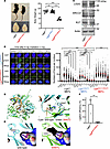 Figure 1
Lig4W447C/W447C mice displayed typical LIG4 syndrome features. (A) Appearance and brain of 6-week-old Lig4+/+ and Lig4W447C/W447C littermates. The graph depicts the weight of 6-week-old female mice (Lig4+/+, n = 6; Lig4+/W447C, n = 13; Lig4W447C/W447C, n = 6). P values were calculated using 1-way ANOVA followed by Tukey’s post hoc multiple-comparison test. (B) Immunofluorescence analysis of γH2AX foci in MEFs after 1 Gy irradiation. Left: Representative images of γH2AX foci in the nuclei of MEFs at the indicated time points. Right: Quantification of γH2AX foci in plateau-phase MEFs after 1 Gy irradiation. Sample sizes ranged from 119 to 391 cells per group. Within each genotype, 1-way ANOVA was conducted, followed by Tukey’s multiple-comparison test. Mann-Whitney U test was used to compare baseline measurements among different genotypes. Original magnification: ×100. (C) Western blots of indicated protein expressions on MEFs. Numbers below the representative bands indicate the ratio of signal intensity relative to Lig4+/+ after normalization to β-actin. (D) Left: Mouse LIGIV protein structures in wild-type and W447C mutant mice. Right: The overall LIGIV protein structure, with DNA-binding and enzymatic domains (residues 1–620), is depicted as a ribbon model. W447 and C447 binding to AMP is shown in stick representation (cyan and green, respectively). (E) Structural comparison between W447 (left, cyan) and C447 (right, green). AlphaFold 3–predicted side chains related to AMP binding are substantially different between them. Consequently, W447C leads to enlargement of the AMP-binding pocket (dashed red circle in right panel), suggesting destabilization of the AMP-binding site during DNA repair processes (black region). (F) Ligation efficiency in an in vitro plasmid-based DNA end-joining assay. Ligation efficiency was quantified by quantitative PCR detection of re-ligated circular plasmid DNA in MEF extracts. Data represent results of 3 independent experiments. In A, B, and F, data are presented as mean ± SEM. **P < 0.01, ***P < 0.001.
Expression of LIGIV protein in Lig4W447C/W447C MEFs was at comparable levels to that in wild-type MEFs, indicating that the mutation does not affect the amounts of LIGIV protein (Figure 1C). In addition, the expression levels of XRCC4 and XLF, which assist LIGIV in its DNA repair function, were also unchanged between Lig4W447C/W447C and wild-type MEFs (Figure 1C). We therefore performed in silico analysis on wild-type and the W447C mutant of LIGIV. W447 was localized en face to AMP in the enzymatic domain. The W447C mutant did not affect overall structures, but it resulted in a decrease of the AMP-binding interface area (wild-type 2.66 Å2, W447C mutant 1.98 Å2) and enlargement of the AMP-binding pocket mainly due to lack of an indole ring (Figure 1, D and E) (23). We also analyzed the R278H and Y288C mutants of LIGIV (Supplemental Figure 1, C and D), but neither mutant altered overall structures of LIGIV. Y288 did not face to AMP, while R278 was in contact with AMP and the R278H mutant decreased the interface area (wild-type 27.84 Å2, R278H mutant 18.46 Å2) but did not significantly affect the structure of the AMP-binding pocket.
We next investigated functional consequences of W447C mutation through the in vitro plasmid-based DNA end-joining assay. Lig4+/+ MEF extracts exhibited robust DNA end-joining activity (31.9% ± 11.3%), whereas Lig4W447C/W447C MEF extracts showed markedly reduced activity (1.04% ± 0.2%) (Figure 1F). The results indicate that the Lig4 W447C mutation severely impairs the LIGIV-dependent final ligation step of NHEJ.
Lig4W447C/W447C mice exhibit severe adaptive immunodeficiency. We then analyzed the phenotype of lymphocytes in Lig4+/+ and Lig4W447C/W447C mice. Lig4W447C/W447C mice had half the number of BM cells of Lig4+/+ mice (Figure 2, A and B). In the BM of Lig4W447C/W447C mice, B220+CD43+IgM− pro–B cells were decreased, and there was marked reduction in B220+CD43−IgM− pre–B cells, B220loCD43−IgM+ immature B cells, and B220hiCD43−IgM+ mature B cells (Figure 2, C and D). Meanwhile, the proportion of pro–B cells among B220+ cells was significantly increased, indicating incomplete block at the pro–B cell stage, which was consistent with impaired rearrangement of the IgH locus (Figure 2, C and D). In the spleens of Lig4W447C/W447C mice, follicular formation was severely impaired, and the numbers of splenocytes and B220+ cells were severely decreased (Figure 2, E–H). Furthermore, in Lig4W447C/W447C mice, serum IgM, IgG1, and IgA levels were severely decreased (Figure 2I). Notably, serum IgE levels remained low, similar to those of Lig4+/+ mice (Figure 2I). In addition, the serum levels of anti-dsDNA antibody in Lig4W447C/W447C mice were lower than those in Lig4+/+ mice (Supplemental Figure 2A). Thus, B cell generation was defective in Lig4W447C/W447C mice.
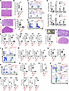 Figure 2
B and T cell differentiation was impaired in Lig4W447C/W447C mice. (A) H&E staining revealed hypocellular marrow in BM of Lig4W447C/W447C mice compared with that of Lig4+/+ mice. Scale bar: 60 μm. (B) Number of BM cells (Lig4+/+, n = 7; Lig4W447C/W447C, n = 7; 7–23 weeks). (C) FACS analysis of BM cells labeled with B220, IgM, and CD43 antibodies. (D) Number and percentage of B cells in BM (Lig4+/+, n = 5; Lig4W447C/W447C, n = 5; 8–30 weeks). (E) H&E staining of spleen. Scale bar: 300 μm. (F) Splenocyte numbers (Lig4+/+, n = 15; Lig4W447C/W447C, n = 15; 7–20 weeks). (G) FACS analysis of splenocytes labeled with CD3 and B220 antibodies. (H) Absolute number and percentage of B220+ splenocytes analyzed by FACS (Lig4+/+, n = 8; Lig4W447C/W447C, n = 8; 7–20 weeks). (I) Serum Ig levels (Lig4+/+, n = 6; Lig4W447C/W447C, n = 6; 9–12 weeks). (J) Thymic hypoplasia in a 6-week-old Lig4W447C/W447C mouse. The graph depicts the number of thymocytes (Lig4+/+, n = 7; Lig4W447C/W447C, n = 7; 8–23 weeks). (K) H&E staining of thymus. Scale bar: 300 μm. (L) FACS analysis of thymocytes labeled with CD3, CD4, and CD8 antibodies. (M) T cell subset numbers in thymocytes, including DN, DP, and SP (Lig4+/+, n = 4; Lig4W447C/W447C, n = 4; 7–16 weeks). (N) Number and percentage of CD3+ splenocytes (Lig4+/+, n = 7; Lig4W447C/W447C, n = 7; 8–11 weeks). (O) FACS analysis of splenocytes labeled with CD3, CD4, and CD8 antibodies. (P) Absolute numbers of CD4+ and CD8+ splenocytes (Lig4+/+, n = 7; Lig4W447C/W447C, n = 7; 8–32 weeks). (Q) CD4/CD8 ratio of CD3+ spleen T cells (Lig4+/+, n = 7; Lig4W447C/W447C, n = 7; 8–32 weeks). (R) FACS analysis of CD44 and CD62L in splenic CD4+ and CD8+ T cells. (S) Percentages of naive, central memory (CMT), and effector memory (EMT) T cell subsets (Lig4+/+, n = 5; Lig4W447C/W447C, n = 5; 9–10 weeks). Data are presented as mean ± SEM. **P < 0.01, ***P < 0.001.
In Lig4W447C/W447C mice, the thymus was very small (Figure 2J), and histological analysis showed unclear corticomedullary boundary (Figure 2K). The proportion of CD3+ cells in thymocytes of Lig4W447C/W447C mice was severely decreased compared with that of Lig4+/+ mice (1.7% vs. 19%) (Figure 2L). Furthermore, there were very few T-lineage cells, such as double-negative (DN) CD4−CD8−, double-positive (DP) CD4+CD8+, and single-positive (SP) CD4+ or CD8+ thymocytes (Figure 2M). The percentages of DN thymocytes in the Lig4W447C/W447C mice were increased, whereas those of DP and SP CD4+ thymocytes were decreased (Figure 2L and Supplemental Figure 2B). In the spleen, CD3+CD4+ and CD3+CD8+ T cells were severely decreased in Lig4W447C/W447C mice (Figure 2, G and N–P). Notably, the CD4/CD8 ratio was higher in Lig4W447C/W447C mice than in Lig4+/+ mice (Figure 2Q). The majority of CD4+ and CD8+ T cells remaining in the mutant mice were effector memory T cells (CD44hiCD62Llo), and naive T cells (CD44loCD62Lhi) were hardly detected (Figure 2, R and S). Concerning splenic Foxp3+ Tregs in CD4+ T cells, the percentages were lower in Lig4W447C/W447C than in Lig4+/+ mice (Supplemental Figure 2, C and D). Taken together, Lig4W447C/W447C mice showed severe defects in both B and T cell generation, i.e., severe adaptive immunodeficiency.
Lig4W447C/W447C mice develop severe intestinal inflammation. After weaning, Lig4W447C/W447C mice exhibited varying degrees of wasting, diarrhea, and rectal prolapse and showed higher mortality than Lig4+/+ and Lig4W447C/+ mice when housed in specific pathogen–free conditions. Kaplan-Meier analysis revealed that Lig4W447C/W447C mice had a median overall survival of 92 days (Figure 3A). In the macro-anatomical study, marked edema and shortening of the large intestine were found in almost all Lig4W447C/W447C mice around 10 weeks of age (Figure 3B). Histological examination of the large intestines revealed epithelial hyperplasia, goblet cell disappearance, and infiltration of inflammatory cells in the lamina propria and submucosa in Lig4W447C/W447C mice (Figure 3C). Mouse colitis histology index (MCHI) of the Lig4W447C/W447C mice was significantly higher than that of Lig4+/+ mice (Figure 3D). Moreover, crypt abscesses, which are formed by active inflammation and are a hallmark of ulcerative colitis, were often present in the Lig4W447C/W447C mice (Figure 3C, arrowhead). Immunohistochemical analysis of the colons further showed that infiltrated cells in Lig4W447C/W447C mice consisted mainly of T cells, especially CD4+ T cells, and F4/80+ macrophages (Figure 3E). These pathological findings were also observed in the small intestines of Lig4W447C/W447C mice (Supplemental Figure 3, A and B). In Lig4W447C/W447C mice, increased T cells were mainly TCRαβ+ cells rather than TCRγδ+ cells, although their ratio of CD4 to CD8 was similar to that of T cells in Lig4+/+ mice (Supplemental Figure 3, C and D). Meanwhile, B220+ cells were hardly detected (Figure 3E and Supplemental Figure 3A). Lig4W447C/W447C mice therefore showed severe inflammation in the small and large intestines with infiltration mainly of CD4+ T cells and macrophages into the mucosa and submucosa.
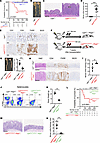 Figure 3
Hematopoietic cells caused severe intestinal inflammation in Lig4W447C/W447C mice. (A) Kaplan-Meier survival curves of Lig4+/+ (n = 13), Lig4+/W447C (n = 24), and Lig4W447C/W447C (n = 13) mice. Statistical analysis was performed using the log-rank test. (B) Macroscopic features of the Lig4W447C/W447C mouse colon revealed an edematous, thickened, and shortened colon. (C) Representative H&E staining of colons. White arrowhead indicates a crypt abscess. Scale bar: 200 μm. (D) Mouse colitis histology index (MCHI) (Lig4+/+, n = 15; Lig4W447C/W447C, n = 15; 7–13 weeks). (E) Immunohistochemistry of colons with CD3, CD4, F4/80, and B220 antibodies. Scale bars: 60 μm. (F) Scheme of BM transplantation experiment. BM cells from Lig4+/+ or Lig4W447C/W447C mice were transplanted into irradiated Rag2−/− mice. (G) A shortened and edematous colon from a Lig4+/+→Rag2−/− mouse and a Lig4W447C/W447C→Rag2−/− mouse. The graph depicts the length of colons. (Lig4+/+→Rag2−/−, n = 4; Lig4W447C/W447C→Rag2−/−, n = 5). (H) H&E staining and immunohistochemistry with CD4, F4/80, and B220 antibodies of colons from a Lig4+/+→Rag2−/− mouse and a Lig4W447C/W447C→Rag2−/− mouse. Scale bars: 100 μm. (I) MCHI of Lig4+/+→Rag2−/− and Lig4W447C/W447C→Rag2−/− mice (Lig4+/+→Rag2−/−, n = 5; Lig4W447C/W447C→Rag2−/−, n = 7). (J) FACS analysis of CD19 and TCRβ expression in splenocytes. (K) Weight of female mice (Lig4+/+ Rag2+/+, n = 3; Lig4W447C/W447C Rag2−/−, n = 4; 14–16 weeks). (L) Kaplan-Meier survival curves of Lig4+/+ Rag2+/+ (n = 21), Lig4W447C/W447C Rag2+/+ (n = 13), and Lig4W447C/W447C Rag2−/− (n = 12) mice. Statistical analysis was performed using the log-rank test. (M) Representative H&E staining of colons. Scale bar: 200 μm. (N) MCHI (Lig4W447C/W447C Rag2+/+, n = 17; Lig4W447C/W447C Rag2−/−, n = 5; 7–18 weeks). Data are presented as mean ± SEM. **P < 0.01, ***P < 0.001.
Intestinal inflammation in Lig4W447C/W447C mice is dependent on lymphocytes. Next, to determine whether hematopoietic cells were sufficient for the development of colitis in the Lig4W447C/W447C mice, we generated BM chimeric mice by transferring BM cells from Lig4+/+ and Lig4W447C/W447C mice into irradiated Rag2−/− mice, which are hereafter referred to as Lig4+/+→Rag2−/− and Lig4W447C/W447C→Rag2−/− mice, respectively (Figure 3F). Compared with Lig4+/+→Rag2−/− mice, Lig4W447C/W447C→Rag2−/− mice showed severe intestinal manifestations, including diarrhea, colonic shortness, and edema (Figure 3G). Furthermore, in the colons of Lig4W447C/W447C→Rag2−/− mice, mainly CD4+ T cells and macrophages infiltrated, as observed in the colons of Lig4W447C/W447C mice (Figure 3, H and I). These results suggest that hematopoietic cells are responsible for the development of intestinal inflammation in Lig4W447C/W447C mice. Meanwhile, Lig4W447C/W447C mice were sensitive to and died from irradiation. Therefore, we could not generate BM chimeric mice by using Lig4W447C/W447C mice as recipients. Thus, we could not assess the roles of non-hematopoietic cells in intestinal inflammation in Lig4W447C/W447C mice.
To further investigate the involvement of lymphocytes in intestinal inflammation in the Lig4W447C/W447C mice, Lig4W447C/W447C Rag2−/− mice were generated by crossing of Lig4W447C/W447C mice with Rag2−/− mice. Splenic T cells were still detected in Lig4W447C/W447C mice, but they were absent in Lig4W447C/W447C Rag2−/− mice (Figure 3J). The double-mutant mice had growth disturbances and microcephaly but showed significantly longer survival than Lig4W447C/W447C mice (Figure 3, K and L, and Supplemental Figure 4). Notably, Lig4W447C/W447C Rag2−/− mice developed no signs of intestinal inflammation (Figure 3, M and N). These results suggest that lymphocytes are required for the development of intestinal inflammation in Lig4W447C/W447C mice.
Innate lymphoid cells (ILCs) are innate immune cells that lack antigen-specific receptors and are classified into ILC1, ILC2, and ILC3 subsets, which produce cytokines similar to those produced by Th1, Th2, and Th17 cells, respectively (24). These subsets can contribute to various inflammatory conditions, including colitis. We therefore analyzed ILC subsets using the lineage-defining transcription factors T-bet, GATA3, and RORγt, which include ILC1, ILC2, and ILC3, respectively. In Lig4W447C/W447C mice, the percentages of T-bet+ and RORγt+ cells as well as the expression levels of these transcription factors were reduced (Supplemental Figure 5, A and B).
Expression of IFN-γ–inducible genes is enhanced in the colon of Lig4W447C/W447C mice. To characterize intestinal inflammation in Lig4W447C/W447C mice, RNA sequencing (RNA-seq) analysis was performed on colonic tissues from Lig4+/+ and Lig4W447C/W447C mice. In total, 25,706 genes were differentially expressed between Lig4+/+ and Lig4W447C/W447C mice (Supplemental Table 1). Expression of 2,278 and 3,728 genes was up- and downregulated, respectively, more than 1.5-fold. Unbiased gene set enrichment analysis (GSEA) of these genes revealed up- and downregulation of 24 and 2 gene sets, respectively (nominal P value < 0.05, false discovery rate q value < 0.25) (Figure 4A and Supplemental Table 2). The most upregulated gene set was “interferon-gamma response” (normalized enrichment score = 2.97; P < 0.001) (Figure 4, A–C). Expression of 15 representative IFN-γ–inducible genes such as Ciita or Cxcl9 was more than 5-fold higher in the colons of Lig4W447C/W447C mice than in those of Lig4+/+ mice (Figure 4B and Supplemental Table 1). Furthermore, expression of these genes was more prominently enhanced in the colons from Lig4W447C/W447C mice with severe manifestations (MCHI: #3, score 17; #4, score 20) versus relatively mild manifestations (MCHI: #1, score 9; #2, score 3) (Figure 4D). Thus, intestinal inflammation in Lig4W447C/W447C mice is characterized by enhanced production of IFN-γ.
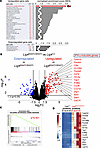 Figure 4
Bulk RNA-seq analysis of the colon of Lig4W447C/W447C mice. (A) Gene set enrichment analysis (GSEA) revealed the top 25 upregulated (top) and top 4 downregulated (bottom) gene sets in the colons of Lig4W447C/W447C mice compared with Lig4+/+ mice, based on normalized enrichment scores. Gene sets associated with “interferon-gamma response,” indicated by the red arrow, represent the focus of this study. NOM p-val, nominal P value; FDR q-val, false discovery rate q value. (B) Relative expression levels in the colons of eight 10-week-old mice (Lig4+/+, n = 4; Lig4W447C/W447C, n = 4) analyzed by RNA-seq analysis. Dashed lines indicate log2 fold-change thresholds of −1.5 and +1.5 (x axis) and P = 0.05 (y axis). (C) GSEA enrichment plot for “interferon-gamma response” comparing the colons of 4 Lig4W447C/W447C mice and 4 Lig4+/+ mice. “p” indicates nominal P value. (D) Heatmap shows the changes in gene expression in the IFN-γ–inducible genes within the “interferon-gamma response” gene sets. Numbers with # represent individual mice.
Loss of IFN-γ expression in Lig4W447C/W447C mice evokes Th2/Th17-skewed intestinal inflammation. IFN-γ is a key cytokine expressed in Th1 cells among lymphocytes. We therefore compared the expression of various Th cell subset signature genes. Th1 cell signature genes including Ifng were expressed at significantly higher levels in Lig4W447C/W447C colons than in Lig4+/+ colons (Figure 5A). Expression of Th2 cell and Treg signature genes was also increased, whereas expression of some Th17 cell signature genes was rather decreased, in the mutant colons (Figure 5A).
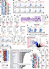 Figure 5
Analysis of Lig4W447C/W447C Ifng−/− mice. (A) Heatmap of Th cell subset signature genes in colons of 10-week-old mice (Lig4+/+, n = 4; Lig4W447C/W447C, n = 4) analyzed by bulk RNA-seq. (B) Intracellular cytokine staining of CD4+ T cells in splenocytes, MLNs, and intestinal LP. (C) Percentages of IFN-γ–producing CD4+ T cells (Lig4+/+: spleen and MLNs, n = 6; LP, n = 4; Lig4W447C/W447C: spleen and MLNs, n = 6; LP, n = 3). (D) Ratio of IFN-γ/IL-17A–producing CD4+ T cells in LP. (E) H&E staining of colons. The square in the middle panel is magnified in the right panel. Dashed circle, infiltrated eosinophils; arrow, multinucleated giant cell. Scale bars: 200 μm (left and middle), 50 μm (right). (F) MCHI (Lig4W447C/W447C Ifng+/+, n = 15; Lig4W447C/W447C Ifng−/−, n = 13; 7–13 weeks). (G) Quantification of myeloid cells (Lig4W447C/W447C Ifng+/+, n = 13; Lig4W447C/W447C Ifng−/−, n = 13; F4/80+ cells, n = 8 each). (H) FACS analysis of CD19 and TCRβ expression in splenocytes. (I) Intracellular cytokine staining of CD4+ T cells in intestinal LP. (J) Percentages of IFN-γ+ and IL-17A+ CD4+ T cells in intestinal LP and their ratio (Lig4W447C/W447C Ifng+/+, n = 5; Lig4W447C/W447C Ifng−/−, n = 3). (K) Relative expression levels in colons of 10-week-old mice (Lig4W447C/W447C Ifng+/+, n = 4; Lig4W447C/W447C Ifng−/−, n = 3). Dashed lines indicate log2 fold-change thresholds of −1.5 and +1.5 (x axis) and P = 0.05 (y axis). (L) Heatmap of IFN-γ–inducible genes in the “interferon-gamma response” gene set in colons. (M) GSEA revealed the top 24 downregulated and top 7 upregulated gene sets in colons of Lig4W447C/W447C Ifng−/− mice (n = 3) versus Lig4W447C/W447C Ifng+/+ mice (n = 4). Blue arrow, “interferon-gamma response” gene sets. NOM p-val, nominal P value; FDR q-val, false discovery rate q value. (N) GSEA enrichment plot for “interferon-gamma response” comparing colons of Lig4W447C/W447C Ifng+/+ mice (n = 4) and Lig4W447C/W447C Ifng−/− mice (n = 3). “p” indicates nominal P value. (O) Heatmap of Th cell subset signature genes in colons. (P) Reads per kilobase per million (RPKM) levels of indicated genes in colons. Data are presented as mean ± SEM. *P < 0.05, **P < 0.01, ***P < 0.001.
We also performed intracellular cytokine staining analysis in CD4+ T cells from Lig4+/+ and Lig4W447C/W447C mice. IFN-γ–expressing CD4+ T cells were higher in frequency in the spleen, mesenteric lymph nodes (MLNs), lamina propria (LP), and intraepithelial compartment of Lig4W447C/W447C mice than in those of Lig4+/+ mice (Figure 5, B and C, and Supplemental Figure 6, A and B). Notably, almost half of splenic CD4+ T cells expressed IFN-γ in Lig4W447C/W447C mice. Meanwhile, IL-4–expressing CD4+ T cells were comparable (Figure 5B). The frequency of IL-17A–expressing CD4+ T cells was increased among MLN lymphocytes, LP lymphocytes, and intraepithelial lymphocytes in Lig4W447C/W447C mice compared with wild-type mice, although it remained below 10% (Figure 5B and Supplemental Figure 6A). Among LP CD4+ T cells, IL-17A–expressing cells were dominant in Lig4+/+ mice, whereas IFN-γ–expressing cells were dominant in the mutant mice (Figure 5D). Dominance of IFN-γ–expressing cells was more prominent in the spleen and MLN CD4+ T cells in Lig4W447C/W447C mice. These results suggest that Th1 cells producing IFN-γ are mainly activated in Lig4W447C/W447C mice.
To examine whether altered thymic egress might be associated with the Th1-skewed responses in Lig4W447C/W447C mice, we examined thymic egress by analyzing expression of thymic egress markers in SP CD4+ thymocytes. SP CD4+ thymocytes mature from CD69+MHC class I– semimature (SM) through CD69+MHC class I+ mature 1 (M1) to CD69–MHC class I+ mature 2 (M2) cells, which also express Qa-2 and S1PR1 (25). Compared with those from Lig4+/+ mice, most SP CD4+ cells were CD69–MHC class I+S1PR1–Qa-2+ in Lig4W447C/W447C mice (Supplemental Figure 7). Notably, the expression levels of MHC class I and Qa-2 in CD69– SP CD4+ cells were higher in Lig4W447C/W447C mice than in Lig4+/+ mice. Considering that Qa-2 and MHC class I are upregulated during thymocyte maturation and are highly expressed in mature peripheral T cells (26), the results suggest that SP CD4+ cells from Lig4W447C/W447C mice may represent recirculating peripheral T cells rather than developing thymocytes.
To determine whether and how IFN-γ is involved in the development of intestinal inflammation in Lig4W447C/W447C mice, we generated Lig4W447C/W447C Ifng−/− mice. The double-mutant mice showed developmental defect and microcephaly, similar to Lig4W447C/W447C mice. The double-mutant mice had inflammatory cell infiltration in the intestine, with a similar inflammatory score to that of the Lig4W447C/W447C mice (Figure 5, E and F). Although crypt abscesses were not observed, neutrophils were more markedly increased, while macrophages (F4/80+ cells) were increased to a comparable extent, relative to Lig4W447C/W447C mice (Figure 5, E and G). Furthermore, in the double-mutant mice, eosinophils tended to increase (Figure 5E, dashed circle, and Figure 5G), and multinucleated giant cells, which could be potentially derived from activated macrophages, were occasionally found (Figure 5E, arrow, and Figure 5G). Meanwhile, mast cells were hardly detected in both Lig4W447C/W447C and Lig4W447C/W447C Ifng−/− mice. Flow cytometric analysis revealed residual T cells in the splenocytes of Lig4W447C/W447C Ifng−/− mice (Figure 5H). Intracellular cytokine staining analysis revealed that the residual splenic T cells showed no IFN-γ expression but increased IL-4 and IL-17A expression (Supplemental Figure 8). IL-17A–expressing T cells were also increased in the LP and intraepithelial compartment of Lig4W447C/W447C Ifng−/− mice (Figure 5, I and J, and Supplemental Figure 9, A and B). The transcriptome analysis also revealed that expression of IFN-γ–inducible genes was profoundly decreased in the colons of Lig4W447C/W447C Ifng−/− mice compared with Lig4W447C/W447C mice (Figure 5, K and L, and Supplemental Table 1). GSEA comparing Lig4W447C/W447C Ifng−/− and Lig4W447C/W447C mice showed downregulation and upregulation of 18 and 5 gene sets, respectively (nominal P value < 0.05, false discovery rate q value < 0.25), and confirmed that the most downregulated gene set was “interferon-gamma response” (Figure 5, M and N, and Supplemental Table 2). Furthermore, we compared the expression of various Th cell subset signature genes. Expression of Th1 cell signature genes in the Lig4W447C/W447C Ifng−/− colons was returned to comparable levels to those in the Lig4+/+ Ifng+/+ colons (Supplemental Table 1). Meanwhile, expression of Th17 (Il17a and Cxcl1) and Th2 (Ccl17 and Ccl24) cell signature genes was upregulated in Lig4W447C/W447C Ifng−/− colons compared with Lig4+/+ Ifng+/+ and Lig4W447C/W447C Ifng+/+ colons (Figure 5, O and P, and Supplemental Table 1). Expression of Treg signature genes was comparable among these mice (Figure 5O and Supplemental Table 1). Taken together, IFN-γ deficiency ameliorated Th1-skewed intestinal inflammation but instead deteriorated Th2/Th17-skewed intestinal inflammation in Lig4W447C/W447C mice.
Lig4W447C/W447C mice exhibit a biased TCR repertoire in the spleen. We then performed a comprehensive bulk-based analysis of a diverse repertoire of T cell receptors in the spleen and the MLN to investigate whether T cell repertoire shows some skewing in Lig4W447C/W447C mice. The T cell repertoire was quite heterogeneous in Lig4+/+ mice, while certain T cell receptors were abundantly expressed in Lig4W447C/W447C mice (Supplemental Figure 10, A–D, and Supplemental Tables 3 and 4). A close examination of the 3′ (proximal) Vα segments revealed a striking difference in their usages among Lig4+/+ and Lig4W447C/W447C mice (Supplemental Figure 10, A and B). TCR Vα usage in the spleen and the MLN of Lig4W447C/W447C mice was strongly skewed to the 3′ (proximal) end. Meanwhile, as for Vβ, a slight bias in the utilization frequency of segments was observed in Lig4W447C/W447C mice, regardless of their position (Supplemental Figure 10, C and D).
Next, we performed single-cell RNA-seq (scRNA-seq) analysis to investigate which pairs of T cell receptors are expressed in the spleens of Lig4+/+ and Lig4W447C/W447C mice. Consistent with flow cytometry analysis, T cells were severely decreased, and the residual T cells mainly consisted of Th1 cells in the spleens of the mutant mice (Figure 6, A and B, Supplemental Figure 11, and Supplemental Table 5). Lig4+/+ T cells from 2,015 barcodes included 2,002 clonotypes, which contained the clones limited to two at most, and all but the top 6 clonotypes were represented by one cell. Meanwhile, 667 Lig4W447C/W447C T cells consisted of 235 clonotypes, and the top 10 clonotypes each occupied more than 10 T cells, 1.5% of the total T cells (experiment 1 in Figure 6C). Those 10 clones contained 4 CD4+ cells, 5 CD8+ cells, and 1 CD4−CD8− T cell. Seven of 10 clones did not express Sell, indicating that most of them were activated. Two CD4+ T cell clonotypes, which were activated and expressed Trbv3/Trbj1-1 or Trbv16/Trbj2-3, showed high expression of Ifng (experiment 1 in Figure 6, D and E). Meanwhile, neither of these 10 T cell clonotypes expressed Il4 or Il17a (experiment 1 in Figure 6D). Furthermore, we analyzed the T cell repertoire diversity in 2 additional Lig4W447C/W447C spleens (experiments 2 and 3) by scRNA-seq analysis. In experiment 2 and 3 mutant mice, 1,265 and 1,610 T cells consisted of 366 and 416 clonotypes, respectively (Figure 6C). These top 10 clonotypes each occupied 1.26% and 1.30% of the total T cells (experiments 2 and 3 in Figure 6C). In mutant mouse #2, two CD4+ T cell clonotypes expressing Ifng represented Trbv3/Trbj2-3/Trav14-3/Traj56 or Trbv15/Trbj2-5/Trav13-1/Traj58 transcripts (experiment 2 in Figure 6, D and E). Meanwhile, in mutant mouse #3, all the top 9 clonotypes were effector memory CD4+ T cells. However, none of them expressed Ifng, Il4, or Il17a (experiment 3 in Figure 6, D and E). Therefore, several T cell clonotypes, which may include clonotypes with pathogenic potential, were skewed and expanded in the mutant mice.
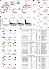 Figure 6
Single-cell RNA-seq of splenocyte samples. (A) The t-distributed stochastic neighbor embedding (t-SNE) plots of splenocytes from Lig4+/+ and Lig4W447C/W447C mice based on 17,377 cells (Lig4+/+, 5,289 cells; Lig4W447C/W447C, 12,088 cells). Splenocytes were classified into 24 clusters. t-SNE plots show the data of total (left), Lig4+/+ (middle), and Lig4W447C/W447C (right). Cells are grouped into 6 major cell populations: T cells, B-lineage cells, myeloid cells, erythroid cells, mast cells, and unclassified cells. A detailed cluster annotation is shown in Supplemental Figure 11. (B) Feature plots of Cd3e, Cd4, Tbx21, and Ifng. The color bar, from yellow to brown, reveals gradual expression intensity differences from low to high. (C) The clonotype frequencies of splenocytes from Lig4+/+ mice and Lig4W447C/W447C mice (n = 3; experiments [exp] 1, 2, and 3). (D) Violin plots of the expression of the listed gene in the respective clonotype of each Lig4W447C/W447C mouse (n = 3; experiments 1, 2, and 3). (E) Clonotype summary of each Lig4W447C/W447C mouse (n = 3; experiments 1, 2, and 3).
Moreover, we quantitated usage of TCR V and J gene segments (Vα, Jα, Vβ, and Jβ) in CDR3 sequences by scRNA-seq analysis in splenic T cells of one Lig4+/+ and three Lig4W447C/W447C mice (Figure 7, A–F). The Vα repertoire in T cells from 3 mutant mice was dominated by the most 3′ (proximal) segments from Trav16 to Trav21-dv12 segments (Figure 7, A and E). Conversely, the Jα repertoire in the mutant T cells was dominated by the most 5′ (proximal) segments from Traj58 to Traj49 (Figure 7, B and E). Moreover, Vβ and Jβ segments in the mutant T cells were evenly distributed, although usage of some segments, including Trbv1, Trbv3, and Trbj1-1, was increased (Figure 7, C, D, and F).
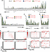 Figure 7
TCR repertoire analysis by single-cell RNA-seq of splenocyte samples. (A–D) The bar graphs depict the utilization frequencies of TCR gene segments in Lig4+/+ mice (n = 1) and Lig4W447C/W447C mice (n = 3; experiments [exp] 1, 2, and 3). (A) Trav segments, arranged in chromosomal order from the 5′ end (left) to the 3′ end (right) of the Trav locus. (B) Traj segments, arranged in chromosomal order from the 5′ end (left) to the 3′ end (right) of the Traj locus. (C) Trbv segments, arranged in chromosomal order from the 5′ end (left) to the 3′ end (right) of the Trbv locus. (D) Trbj segments, arranged in chromosomal order from the 5′ end (left) to the 3′ end (right) of the Trbj locus. (E and F) Heatmaps showing the usage of Trav-Traj (E) and Trbv-Trbj (F) combinations. Splenocytes were prepared from individual mice (Lig4+/+, n = 1; Lig4W447C/W447C, n = 3; experiments 1, 2, and 3). (E) The x and y axes represent Trav and Traj segments, respectively, arranged from 5′ to 3′ end of the locus. The color gradient ranges from red to white, indicating high to low frequency of each Trav-Traj pair, respectively. The scale is shown on the right. (F) The x and y axes show Trbv and Trbj segments, respectively, arranged from 5′ to 3′ end of the locus. The same color gradient as shown in E is used.
We next analyzed TCR Vα and Vβ segment usage in CD4+ T cells (Supplemental Figure 12, A–C) and examined expression of selected Vα and Vβ chains by flow cytometry (Supplemental Figure 12D). In mutant mice, Vα2+ T cells, which express the TCR Vα chain encoded by the proximal segments, the Trav14 family, were significantly increased. Vβ11 (Trbv16)+ T cells also tended to increase, whereas Vβ8 (Trbv13 family)+ T cells were comparable to those in wild-type mice. These flow cytometric data were consistent with the scRNA-seq results (Supplemental Figure 12).


