
Research ArticleEndocrinologyInflammationPulmonology
Open Access |  10.1172/jci.insight.196812
Pathophysiological and therapeutic implications of C-type natriuretic peptide/cyclic GMP signaling in pulmonary fibrosis
Rene Weyer,1 Katharina Völker,1 Tamara Potapenko,1 Lisa Krebes,1 Marco Abeßer,1 Anna-Lena Friedrich,1 Eva Lessmann,1 Ali Khadim,2 Clemens Ruppert,2 Elie El Agha,2 Dalia Sheta,3 Andreas Beilhack,3 Daniel V. Santi,4 Eric L. Schneider,4 Michaela Kuhn,1 and Swati Dabral1
1Institute of Physiology, Julius-Maximilians University of Würzburg, Würzburg, Germany.
2Excellence Cluster Cardio-Pulmonary Institute (CPI), Universities of Giessen and Marburg Lung Center (UGMLC), Justus-Liebig-University Giessen, German Center for Lung Research (DZL), Giessen, Germany.
3Department of Internal Medicine II, Interdisciplinary Center for Clinical Research, University Hospital of Würzburg, Würzburg, Germany.
4ProLynx, Inc., San Francisco, California, USA.
Address correspondence to: Swati Dabral or Michaela Kuhn, Institute of Physiology, University of Würzburg, Röntgenring 9, D-97070 Würzburg, Germany, Phone: 0049.931.3182721; Email: swati.dabral@uni-wuerzburg.de (SD); michaela.kuhn@uni-wuerzburg.de (MK).
Authorship note: MK and SD contributed equally to this work.
Find articles by Weyer, R. in: PubMed | Google Scholar
1Institute of Physiology, Julius-Maximilians University of Würzburg, Würzburg, Germany.
2Excellence Cluster Cardio-Pulmonary Institute (CPI), Universities of Giessen and Marburg Lung Center (UGMLC), Justus-Liebig-University Giessen, German Center for Lung Research (DZL), Giessen, Germany.
3Department of Internal Medicine II, Interdisciplinary Center for Clinical Research, University Hospital of Würzburg, Würzburg, Germany.
4ProLynx, Inc., San Francisco, California, USA.
Address correspondence to: Swati Dabral or Michaela Kuhn, Institute of Physiology, University of Würzburg, Röntgenring 9, D-97070 Würzburg, Germany, Phone: 0049.931.3182721; Email: swati.dabral@uni-wuerzburg.de (SD); michaela.kuhn@uni-wuerzburg.de (MK).
Authorship note: MK and SD contributed equally to this work.
Find articles by Völker, K. in: PubMed | Google Scholar
1Institute of Physiology, Julius-Maximilians University of Würzburg, Würzburg, Germany.
2Excellence Cluster Cardio-Pulmonary Institute (CPI), Universities of Giessen and Marburg Lung Center (UGMLC), Justus-Liebig-University Giessen, German Center for Lung Research (DZL), Giessen, Germany.
3Department of Internal Medicine II, Interdisciplinary Center for Clinical Research, University Hospital of Würzburg, Würzburg, Germany.
4ProLynx, Inc., San Francisco, California, USA.
Address correspondence to: Swati Dabral or Michaela Kuhn, Institute of Physiology, University of Würzburg, Röntgenring 9, D-97070 Würzburg, Germany, Phone: 0049.931.3182721; Email: swati.dabral@uni-wuerzburg.de (SD); michaela.kuhn@uni-wuerzburg.de (MK).
Authorship note: MK and SD contributed equally to this work.
Find articles by Potapenko, T. in: PubMed | Google Scholar
1Institute of Physiology, Julius-Maximilians University of Würzburg, Würzburg, Germany.
2Excellence Cluster Cardio-Pulmonary Institute (CPI), Universities of Giessen and Marburg Lung Center (UGMLC), Justus-Liebig-University Giessen, German Center for Lung Research (DZL), Giessen, Germany.
3Department of Internal Medicine II, Interdisciplinary Center for Clinical Research, University Hospital of Würzburg, Würzburg, Germany.
4ProLynx, Inc., San Francisco, California, USA.
Address correspondence to: Swati Dabral or Michaela Kuhn, Institute of Physiology, University of Würzburg, Röntgenring 9, D-97070 Würzburg, Germany, Phone: 0049.931.3182721; Email: swati.dabral@uni-wuerzburg.de (SD); michaela.kuhn@uni-wuerzburg.de (MK).
Authorship note: MK and SD contributed equally to this work.
Find articles by Krebes, L. in: PubMed | Google Scholar
1Institute of Physiology, Julius-Maximilians University of Würzburg, Würzburg, Germany.
2Excellence Cluster Cardio-Pulmonary Institute (CPI), Universities of Giessen and Marburg Lung Center (UGMLC), Justus-Liebig-University Giessen, German Center for Lung Research (DZL), Giessen, Germany.
3Department of Internal Medicine II, Interdisciplinary Center for Clinical Research, University Hospital of Würzburg, Würzburg, Germany.
4ProLynx, Inc., San Francisco, California, USA.
Address correspondence to: Swati Dabral or Michaela Kuhn, Institute of Physiology, University of Würzburg, Röntgenring 9, D-97070 Würzburg, Germany, Phone: 0049.931.3182721; Email: swati.dabral@uni-wuerzburg.de (SD); michaela.kuhn@uni-wuerzburg.de (MK).
Authorship note: MK and SD contributed equally to this work.
Find articles by Abeßer, M. in: PubMed | Google Scholar
1Institute of Physiology, Julius-Maximilians University of Würzburg, Würzburg, Germany.
2Excellence Cluster Cardio-Pulmonary Institute (CPI), Universities of Giessen and Marburg Lung Center (UGMLC), Justus-Liebig-University Giessen, German Center for Lung Research (DZL), Giessen, Germany.
3Department of Internal Medicine II, Interdisciplinary Center for Clinical Research, University Hospital of Würzburg, Würzburg, Germany.
4ProLynx, Inc., San Francisco, California, USA.
Address correspondence to: Swati Dabral or Michaela Kuhn, Institute of Physiology, University of Würzburg, Röntgenring 9, D-97070 Würzburg, Germany, Phone: 0049.931.3182721; Email: swati.dabral@uni-wuerzburg.de (SD); michaela.kuhn@uni-wuerzburg.de (MK).
Authorship note: MK and SD contributed equally to this work.
Find articles by Friedrich, A. in: PubMed | Google Scholar
1Institute of Physiology, Julius-Maximilians University of Würzburg, Würzburg, Germany.
2Excellence Cluster Cardio-Pulmonary Institute (CPI), Universities of Giessen and Marburg Lung Center (UGMLC), Justus-Liebig-University Giessen, German Center for Lung Research (DZL), Giessen, Germany.
3Department of Internal Medicine II, Interdisciplinary Center for Clinical Research, University Hospital of Würzburg, Würzburg, Germany.
4ProLynx, Inc., San Francisco, California, USA.
Address correspondence to: Swati Dabral or Michaela Kuhn, Institute of Physiology, University of Würzburg, Röntgenring 9, D-97070 Würzburg, Germany, Phone: 0049.931.3182721; Email: swati.dabral@uni-wuerzburg.de (SD); michaela.kuhn@uni-wuerzburg.de (MK).
Authorship note: MK and SD contributed equally to this work.
Find articles by Lessmann, E. in: PubMed | Google Scholar
1Institute of Physiology, Julius-Maximilians University of Würzburg, Würzburg, Germany.
2Excellence Cluster Cardio-Pulmonary Institute (CPI), Universities of Giessen and Marburg Lung Center (UGMLC), Justus-Liebig-University Giessen, German Center for Lung Research (DZL), Giessen, Germany.
3Department of Internal Medicine II, Interdisciplinary Center for Clinical Research, University Hospital of Würzburg, Würzburg, Germany.
4ProLynx, Inc., San Francisco, California, USA.
Address correspondence to: Swati Dabral or Michaela Kuhn, Institute of Physiology, University of Würzburg, Röntgenring 9, D-97070 Würzburg, Germany, Phone: 0049.931.3182721; Email: swati.dabral@uni-wuerzburg.de (SD); michaela.kuhn@uni-wuerzburg.de (MK).
Authorship note: MK and SD contributed equally to this work.
Find articles by Khadim, A. in: PubMed | Google Scholar
1Institute of Physiology, Julius-Maximilians University of Würzburg, Würzburg, Germany.
2Excellence Cluster Cardio-Pulmonary Institute (CPI), Universities of Giessen and Marburg Lung Center (UGMLC), Justus-Liebig-University Giessen, German Center for Lung Research (DZL), Giessen, Germany.
3Department of Internal Medicine II, Interdisciplinary Center for Clinical Research, University Hospital of Würzburg, Würzburg, Germany.
4ProLynx, Inc., San Francisco, California, USA.
Address correspondence to: Swati Dabral or Michaela Kuhn, Institute of Physiology, University of Würzburg, Röntgenring 9, D-97070 Würzburg, Germany, Phone: 0049.931.3182721; Email: swati.dabral@uni-wuerzburg.de (SD); michaela.kuhn@uni-wuerzburg.de (MK).
Authorship note: MK and SD contributed equally to this work.
Find articles by Ruppert, C. in: PubMed | Google Scholar
1Institute of Physiology, Julius-Maximilians University of Würzburg, Würzburg, Germany.
2Excellence Cluster Cardio-Pulmonary Institute (CPI), Universities of Giessen and Marburg Lung Center (UGMLC), Justus-Liebig-University Giessen, German Center for Lung Research (DZL), Giessen, Germany.
3Department of Internal Medicine II, Interdisciplinary Center for Clinical Research, University Hospital of Würzburg, Würzburg, Germany.
4ProLynx, Inc., San Francisco, California, USA.
Address correspondence to: Swati Dabral or Michaela Kuhn, Institute of Physiology, University of Würzburg, Röntgenring 9, D-97070 Würzburg, Germany, Phone: 0049.931.3182721; Email: swati.dabral@uni-wuerzburg.de (SD); michaela.kuhn@uni-wuerzburg.de (MK).
Authorship note: MK and SD contributed equally to this work.
Find articles by
El Agha, E.
in:
PubMed
|
Google Scholar
|

1Institute of Physiology, Julius-Maximilians University of Würzburg, Würzburg, Germany.
2Excellence Cluster Cardio-Pulmonary Institute (CPI), Universities of Giessen and Marburg Lung Center (UGMLC), Justus-Liebig-University Giessen, German Center for Lung Research (DZL), Giessen, Germany.
3Department of Internal Medicine II, Interdisciplinary Center for Clinical Research, University Hospital of Würzburg, Würzburg, Germany.
4ProLynx, Inc., San Francisco, California, USA.
Address correspondence to: Swati Dabral or Michaela Kuhn, Institute of Physiology, University of Würzburg, Röntgenring 9, D-97070 Würzburg, Germany, Phone: 0049.931.3182721; Email: swati.dabral@uni-wuerzburg.de (SD); michaela.kuhn@uni-wuerzburg.de (MK).
Authorship note: MK and SD contributed equally to this work.
Find articles by Sheta, D. in: PubMed | Google Scholar
1Institute of Physiology, Julius-Maximilians University of Würzburg, Würzburg, Germany.
2Excellence Cluster Cardio-Pulmonary Institute (CPI), Universities of Giessen and Marburg Lung Center (UGMLC), Justus-Liebig-University Giessen, German Center for Lung Research (DZL), Giessen, Germany.
3Department of Internal Medicine II, Interdisciplinary Center for Clinical Research, University Hospital of Würzburg, Würzburg, Germany.
4ProLynx, Inc., San Francisco, California, USA.
Address correspondence to: Swati Dabral or Michaela Kuhn, Institute of Physiology, University of Würzburg, Röntgenring 9, D-97070 Würzburg, Germany, Phone: 0049.931.3182721; Email: swati.dabral@uni-wuerzburg.de (SD); michaela.kuhn@uni-wuerzburg.de (MK).
Authorship note: MK and SD contributed equally to this work.
Find articles by
Beilhack, A.
in:
PubMed
|
Google Scholar
|
1Institute of Physiology, Julius-Maximilians University of Würzburg, Würzburg, Germany.
2Excellence Cluster Cardio-Pulmonary Institute (CPI), Universities of Giessen and Marburg Lung Center (UGMLC), Justus-Liebig-University Giessen, German Center for Lung Research (DZL), Giessen, Germany.
3Department of Internal Medicine II, Interdisciplinary Center for Clinical Research, University Hospital of Würzburg, Würzburg, Germany.
4ProLynx, Inc., San Francisco, California, USA.
Address correspondence to: Swati Dabral or Michaela Kuhn, Institute of Physiology, University of Würzburg, Röntgenring 9, D-97070 Würzburg, Germany, Phone: 0049.931.3182721; Email: swati.dabral@uni-wuerzburg.de (SD); michaela.kuhn@uni-wuerzburg.de (MK).
Authorship note: MK and SD contributed equally to this work.
Find articles by Santi, D. in: PubMed | Google Scholar
1Institute of Physiology, Julius-Maximilians University of Würzburg, Würzburg, Germany.
2Excellence Cluster Cardio-Pulmonary Institute (CPI), Universities of Giessen and Marburg Lung Center (UGMLC), Justus-Liebig-University Giessen, German Center for Lung Research (DZL), Giessen, Germany.
3Department of Internal Medicine II, Interdisciplinary Center for Clinical Research, University Hospital of Würzburg, Würzburg, Germany.
4ProLynx, Inc., San Francisco, California, USA.
Address correspondence to: Swati Dabral or Michaela Kuhn, Institute of Physiology, University of Würzburg, Röntgenring 9, D-97070 Würzburg, Germany, Phone: 0049.931.3182721; Email: swati.dabral@uni-wuerzburg.de (SD); michaela.kuhn@uni-wuerzburg.de (MK).
Authorship note: MK and SD contributed equally to this work.
Find articles by Schneider, E. in: PubMed | Google Scholar
1Institute of Physiology, Julius-Maximilians University of Würzburg, Würzburg, Germany.
2Excellence Cluster Cardio-Pulmonary Institute (CPI), Universities of Giessen and Marburg Lung Center (UGMLC), Justus-Liebig-University Giessen, German Center for Lung Research (DZL), Giessen, Germany.
3Department of Internal Medicine II, Interdisciplinary Center for Clinical Research, University Hospital of Würzburg, Würzburg, Germany.
4ProLynx, Inc., San Francisco, California, USA.
Address correspondence to: Swati Dabral or Michaela Kuhn, Institute of Physiology, University of Würzburg, Röntgenring 9, D-97070 Würzburg, Germany, Phone: 0049.931.3182721; Email: swati.dabral@uni-wuerzburg.de (SD); michaela.kuhn@uni-wuerzburg.de (MK).
Authorship note: MK and SD contributed equally to this work.
Find articles by Kuhn, M. in: PubMed | Google Scholar
1Institute of Physiology, Julius-Maximilians University of Würzburg, Würzburg, Germany.
2Excellence Cluster Cardio-Pulmonary Institute (CPI), Universities of Giessen and Marburg Lung Center (UGMLC), Justus-Liebig-University Giessen, German Center for Lung Research (DZL), Giessen, Germany.
3Department of Internal Medicine II, Interdisciplinary Center for Clinical Research, University Hospital of Würzburg, Würzburg, Germany.
4ProLynx, Inc., San Francisco, California, USA.
Address correspondence to: Swati Dabral or Michaela Kuhn, Institute of Physiology, University of Würzburg, Röntgenring 9, D-97070 Würzburg, Germany, Phone: 0049.931.3182721; Email: swati.dabral@uni-wuerzburg.de (SD); michaela.kuhn@uni-wuerzburg.de (MK).
Authorship note: MK and SD contributed equally to this work.
Find articles by Dabral, S. in: PubMed | Google Scholar
Published January 7, 2026 - More info
JCI Insight. 2026;11(4):e196812. https://doi.org/10.1172/jci.insight.196812.
© 2026 Weyer et al. This work is licensed under the Creative Commons Attribution 4.0 International License. To view a copy of this license, visit http://creativecommons.org/licenses/by/4.0/.
Received: June 16, 2025; Accepted: December 19, 2025


-
Results
Among various types of lung cells, GC-B expression is greatest in fibroblasts. The CNP receptor, GC-B, is expressed in several types of cells involved in the development of PF, including vascular and immune cells (5). Supporting our focus on fibroblasts, scRNA-seq data of the Human Lung Cell Atlas (HLCA; https://cellxgene.cziscience.com/collections/6f6d381a-7701-4781-935c-db10d30de293) (11) reveal that GC-B (gene name: NPR2) is mainly expressed in stromal cells and at much lower levels in endothelial, epithelial and immune cells (Supplemental Figure 1, A–C; supplemental material available online with this article; https://doi.org/10.1172/jci.insight.196812DS1). Subclustering the stromal compartment into distinct fibroblast subtypes demonstrated highest NPR2 expression in subpleural fibroblasts, followed by peribronchial and adventitial fibroblasts (Supplemental Figure 1, D and E). To validate this expression pattern at the protein level, we separated different cell populations from murine lungs using antibodies against PDGFR-α, NG2, and CD144 and magnetic-assisted cell sorting. Immunoblot analysis confirmed the separation and enrichment of PDGFR-α+ fibroblasts, PDGFR-β+ smooth muscle cells/pericytes, and CD31+ endothelial cells (Supplemental Figure 1F). In line with the human scRNA-seq data, GC-B protein expression was high in lung fibroblasts and low in endothelial cells.
CNP stimulates antifibrotic GC-B/cGMP signaling in cultured lung fibroblasts from patients with IPF. To assess the functional and possible clinical relevance of the GC-B expression data, we studied CNP/GC-B/cGMP signaling in cultured human lung fibroblasts. Healthy “control” lung fibroblasts were derived from unused explanted donor lungs or from histologically unaltered lung regions surrounding resected tumors. “IPF fibroblasts” were derived from end-stage patients undergoing lung transplantation. In comparison with control fibroblasts, IPF fibroblasts had unaltered baseline proliferation rates, as shown by bromodeoxyuridine (BrdU) incorporation (Supplemental Figure 2A). However, in agreement with published studies (12), the proliferative effect of platelet-derived growth factor BB (PDGF-BB; 50 ng/mL, 24 hours) was enhanced (Supplemental Figure 2A). To study migration, fibroblasts were seeded in 24-well plates and grown until confluency. A scratch was made followed by PDGF-BB (50 ng/mL) or PBS (vehicle) treatment and wound closure was monitored for 24 hours. As shown, IPF fibroblasts showed accelerated baseline and PDGF-BB–stimulated migration (Supplemental Figure 2B). Hence, cultured IPF fibroblasts “intrinsically” exhibit increased migration and proliferation (present studies) as well as differentiation into a contractile phenotype (12), with enhanced responses to growth factors such as TGF-β and PDGF-BB. Notably, the expression levels of the GC-B receptor and of its downstream target cGMP-dependent Protein Kinase I (cGKI) were similar in control and IPF fibroblasts (immunoblots in Figure 1A). Accordingly, in both groups, CNP raised intracellular cGMP levels in a similar concentration-dependent manner (Figure 1B).
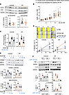 Figure 1
CNP, via GC-B/cGMP signaling, attenuates the profibrotic and proinflammatory activation of cultured human lung fibroblasts, and such effects are preserved in IPF fibroblasts. (A) GC-B and cGKI expression in control and IPF fibroblasts (immunoblots). (B) Effects of CNP (0.1–100 nM, 15 minutes) on intracellular cGMP contents (detected by radioimmunoassay) of control and IPF lung fibroblasts in presence of the PDE inhibitor 3-Isobutyl-1-methylxanthin (0.5 mM IBMX). (C–E) Effects of CNP pretreatment (30 minutes) on PDGF-BB–induced (50 ng/mL, 24 hours) proliferation (BrdU incorporation; C), migration (scratch assay; D), and expression levels of collagen 1 and MMP-9 (immunoblots; E). (F) Effects of CNP pretreatment (30 minutes) on TGF-β–induced (2 ng/mL, 24 hours) and TNF-α–induced (10 ng/mL, 24 hours) expression of TNF-α and IL-6, respectively (immunoblots). The sample number for each experiment (n) varied between 3 and 16 and is indicated by the number of data points in each histogram. The scratch assays were performed with 12–16 wells from 3 biological replicates from each group. Significance was determined by unpaired Student’s t test (A), 1-way ANOVA (B, C, E, and F), and 2-way ANOVA (D). *P < 0.05 versus PBS (–), #P < 0.05 versus PDGF-BB, TGF-β, or TNF-α.
To evaluate whether CNP moderates the profibrotic effects of growth factors and cytokines, fibroblasts were pretreated with CNP (100 nM, 30 minutes) prior to stimulation for 24 hours. CNP significantly reduced the proliferative and promigratory actions of PDGF-BB (50 ng/mL), and these inhibitory CNP effects were preserved and even slightly enhanced in IPF fibroblasts (Figure 1, C and D). The PDGF-BB–stimulated expression of the ECM proteins collagen 1 and matrix metalloproteinase-9 (MMP-9) was also attenuated by 10 and 100 nM CNP in both control and IPF cells (immunoblots in Figure 1E). Moreover, in both groups, CNP (100 nM, 30 minutes pretreatment) attenuated the TGF-β–induced expression (2 ng/mL, 24 hours) of the myofibroblast marker, collagen triple helix repeat containing protein 1 (Cthrc1) (Supplemental Figure 3). Lastly, we examined the effect of CNP on the expression of the proinflammatory “fibrokines” TNF-α and IL-6. As shown in Figure 1F, TGF-β (2 ng/mL, 24 hours) and TNF-α (10 ng/mL, 24 hours) augmented the expression of TNF-α and IL-6, respectively, in control and even more in IPF fibroblasts. CNP (100 nM, 30 minutes pretreatment) reduced such proinflammatory activation in both groups. We conclude that the CNP/GC-B/cGMP pathway exerts antifibrotic and antiinflammatory effects in cultured human lung fibroblasts. Such effects are preserved in fibroblasts from patients with IPF in vitro.
Development and characterization of a fibroblast-specific GC-B–KO mouse line. To unravel whether endogenous paracrine CNP/GC-B signaling counterregulates the pathological activation of lung fibroblasts, we utilized a recently reported transgenic mouse line with tamoxifen-induced fibroblast-restricted GC-B deletion (Fibro GC-B KO: Npr2fl/fl;Col1a2-CreERT2 mice) (8). To study the efficiency of the genetic GC-B deletion in lung fibroblasts, such cells were isolated from tamoxifen-treated Fibro GC-B KO and Cre-negative littermate mice (controls: Npr2fl/fl). Cultured lung fibroblasts were passaged twice and then plated for experiments. Western blotting demonstrated an approximately 50% reduction of GC-B expression in GC-B–KO lung fibroblasts (Figure 2A). CNP (100 nM) increased cGMP contents of control fibroblasts by approximately 6-fold and by only approximately 2-fold in KO fibroblasts (Figure 2B). To investigate how inhibited CNP/GC-B/cGMP signaling affects the interactions with PDGF-BB, we performed functional assays. PDGF-BB (50 ng/mL, 24 hours) enhanced the proliferation, migration, and collagen 1 as well as MMP-9 expression levels of murine lung fibroblasts without genotype-dependent differences (Figure 2, C–E). As shown, CNP significantly attenuated these effects of PDGF-BB in control but not in GC-B–KO fibroblasts.
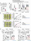 Figure 2
Comparison of GC-B expression and signaling in lung fibroblasts isolated from tamoxifen-treated Npr2fl/fl (controls) and Npr2fl/fl;Col1a2-CreERT2 littermate mice (Fibro GC-B KO). Cultured fibroblasts were studied at passages 2 and 3. (A) Immunoblot: Expression of GC-B was normalized to β-tubulin and calculated as x-fold from the average of controls. (B) Effects of CNP on intracellular cGMP contents were determined by radioimmunoassay and calculated as x-fold versus PBS. (C–E) Effect of CNP pretreatment (30 minutes) on PDGF-BB–induced (50 ng/mL, 24 hours) proliferation (C), migration (D), and collagen 1 as well as MMP-9 expression (E) of such control and GC-B–deficient murine lung fibroblasts. The sample number for each experiment (n) varied between 3 and 14 and is indicated by the number of data points in each histogram. The scratch assays were performed with 8–14 wells from 3 biological replicates from each group. Significance was determined by unpaired 2-tailed Student’s t test (A), 2-way ANOVA (B–D), and 1-way ANOVA (E). *P < 0.05 versus PBS ([–], in all panels), #P < 0.05 versus control mice (A and B); and ƗP < 0.05 versus PDGF-BB (C–E).
As recently published (8), in contrast to mice with global GC-B deletion (5), such Fibro GC-B–KO mice have normal Mendelian inheritance, life span, and skeletal growth. Under baseline and sham conditions, arterial blood pressure, cardiac and lung function, and cardiac and lung interstitial collagen fractions were not different between control and KO littermates from both sexes.
Mice with fibroblast-restricted GC-B inhibition show enhanced early lung inflammation in response to bleomycin. To explore the effects of a fibroblast-specific inhibition of CNP signaling on pathological lung inflammation and subsequent fibrosis, control and Fibro GC-B–KO mice received bleomycin (0.75 IU/kg bodyweight [BW]) by intratracheal administration under anesthesia. Bleomycin injures the airway epithelium and, thereby, provokes a rapid inflammatory response, followed by subsequent reactive interstitial fibrosis (7, 13). Thereby, this model resembles inflammation-driven fibrosis in patients.
In the first series of experiments, mice were euthanized 3 and 7 days after bleomycin administration (scheme in Figure 3A), to characterize the inflammatory phase through analyses of bronchoalveolar lavage fluid (BALF) (13), as well as the molecular signatures of resident fibroblasts isolated from the lung parenchyma. BALF samples were used to quantify extravasated proteins (i.e., albumin) with a regular BCA assay (Figure 3, B and C) and by immunoblotting (Figure 3C). As shown in Figure 3, B and C, the bleomycin-treated mice exhibited greater protein, especially albumin, levels in BALF than saline-treated mice. In control mice, such “plasma leakage” was increased by 3-fold on day 3 and by 4- to 5-fold on day 7 after bleomycin instillation (as compared with saline). These inflammatory responses were increased in Fibro GC-B–KO mice (Figure 3, B and C). The findings were corroborated by immunoblot analyses of Mac-2, a marker for macrophages (Figure 3C). Mac-2 was barely detectable in BALF from vehicle-treated mice, but the levels rose by nearly 20-fold in BALF from bleomycin-treated control mice (day 7), suggesting a strong activation and/or transepithelial migration of tissue macrophages. Notably, in BALF of bleomycin-treated Fibro GC-B–KO mice, the levels of Mac-2 in BALF rose nearly 40-fold (Figure 3C).
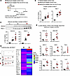 Figure 3
Fibroblast-restricted GC-B deletion exacerbates bleomycin-induced lung inflammation in mice. (A) Schematic illustration of these studies performed in control and Fibro GC-B KO littermate mice 3 and 7 days after bleomycin administration. (B) Plasma leakage was evaluated 3 and 7 days after bleomycin treatment by determination of the protein content in bronchoalveolar lavage fluid (BALF) samples (by BCA assay). (C) Albumin and Mac-2 levels were evaluated in equal volumes of BALF obtained 7 days after bleomycin treatment (immunoblot). (D and E) Cytokine contents of BALF samples obtained 7 days after bleomycin treatment were evaluated with a commercially available mouse cytokine array. D shows representative cytokine array membranes probed with BALF from saline- or bleomycin-treated control mice as well as from bleomycin-treated Fibro GC-B–KO littermate mice. The heatmap depicts cytokines that were detected at markedly different levels in BALF from the 2 genotypes (mean-normalized values are shown, with red color indicating high, and blue indicating low values). (E) Quantitative evaluation of pixel densities of representative cytokines differing between bleomycin-treated control and KO mice. In B and C, each data point represents an individual study mouse; E shows replicates from 3 mice per group. Significance was tested by 1-way ANOVA (B and C) and Student’s unpaired t test (E). *P < 0.05 versus vehicle; #P < 0.05 versus control mice.
To further characterize the inflammatory response 7 days after bleomycin treatment, we used a cytokine array detecting 40 different cytokines (ARY006, R&D Systems). As illustrated in Figure 3D, BALF from bleomycin-treated control mice contained numerous proinflammatory cytokines and chemokines, which were all undetectable in saline-instilled mice. This response to bleomycin was much greater in the Fibro GC-B–KO mice (Figure 3D). Supplemental Figure 4 provides an overview of all changed cytokines. For a more comprehensive appreciation of the genotype-dependent changes, Figure 3E depicts quantitative evaluations of the cytokines, which are known to be secreted by activated myofibroblasts: CXCL13, GM-CSF, IL-1α, IL-6, monocyte chemoattractant protein-1 (MCP-1)/CCL2, stromal cell-derived factor 1 (SDF-1/CXCL12), IL-1 receptor antagonist (IL-1Ra) and TNF-α (4, 13–22). Notably, the levels of all these fibrokines were significantly greater in the BALF of bleomycin-treated Fibro GC-B–KO mice than in BALF from their control littermates. Collectively, these results confirm that bleomycin-induced pulmonary inflammation was exacerbated in Fibro GC-B–KO mice.
Mice with fibroblast-restricted GC-B inhibition show unaltered lung fibrosis in response to bleomycin. To characterize the postinflammatory fibrotic phase, mice were studied 21 days after bleomycin administration (scheme in Figure 4A) (22). Masson’s trichrome staining of lung sections together with immunoblot studies revealed significant interstitial collagen deposition in lungs from bleomycin-treated mice. Unexpectedly, we did not observe differences between control and Fibro GC-B–KO mice (Figure 4, B and C). Together with collagen 1, pulmonary Cthrc1 expression was also upregulated in bleomycin-treated mice, without differences between genotypes. In fact, levels trended lower in the KO mice (Figure 4C). Pulmonary α-SMA expression was not different between treatment groups or genotypes (Figure 4C). We assume that the expression of α-SMA in vascular and airway smooth muscle cells masked the bleomycin-induced increase in fibroblast-specific α-SMA.
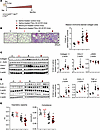 Figure 4
Fibroblast-restricted GC-B deletion does not significantly alter bleomycin-induced lung fibrosis and dysfunction in mice. (A) Schematic illustration of these studies in control and Fibro GC-B–KO littermate mice 21 days after bleomycin administration. (B) Collagen deposition was determined by Masson’s trichrome staining of lung paraffin sections, followed by quantification with ORBIT. The mean value from quantification of 2 sections per mouse was taken (10–15 fields per lung were evaluated). Collagen areas (in percent from corresponding total section areas) are presented as x-fold from the average value of saline-instilled control mice. (C and D) Immunoblots: pulmonary collagen 1, Cthrc1, α-SMA (C), IL-6, CXCL-1, and TNF-α expression levels (D) were normalized to β-actin and calculated as X-fold from the average value of saline-treated control mice. (E) Lung inspiratory capacity and compliance were evaluated in anesthetized mice with a Flexivent system. For B and E: n = 6 (Controls, saline), 4–5 (KO, saline), 10 (Controls, bleomycin), and 11 mice (KO, bleomycin). For C and D: n = 3 (Controls, saline), 3 (KO, saline), 6 (Controls, bleomycin) and 6 mice (KO, bleomycin). Significance was tested by 2-way ANOVA. *P < 0.05 versus saline.
To characterize the inflammatory response, we studied the pulmonary expression levels of IL-6, CXCL-1, and TNF-α by immunoblot. As shown in Figure 4D, all 3 cytokines were similarly increased in lungs from bleomycin-treated control and KO mice.
The effect on lung function was evaluated with a Flexivent system (in anesthetized mice, immediately before necropsy). In line with the histology, no differences in lung capacity and compliance were observed between saline-treated control and Fibro GC-B–KO mice (Figure 4E), demonstrating that the GC-B knockdown did not affect baseline lung morphology and function. Bleomycin-treated mice showed a significant decline of inspiratory capacity and compliance. These functional changes were slightly more pronounced in Fibro GC-B–KO mice than in controls, but this difference did not reach statistical significance (Figure 4E).
Bleomycin-induced lung inflammation impairs the CNP/GC-B signaling pathway in resident lung myofibroblasts. The main results of the previous sections can be summarized as follows: (a) synthetic CNP, via GC-B/cGMP signaling, inhibits the profibrotic activation of cultured human and murine lung fibroblasts; (b) transgenic mice with inhibited CNP/GC-B signaling in fibroblasts react to bleomycin with enhanced lung inflammation; and (c) despite this, such Fibro GC-B–KO mice do not have exacerbated reactive lung fibrosis and dysfunction. In view of this discrepancy, we hypothesized that bleomycin-induced lung inflammation inhibits the CNP/GC-B signaling pathway and/or the expression and activity of cGMP-modulated or -modulating proteins in resident lung (myo)fibroblasts. CNP/GC-B, via cGMP, can activate cGKI and the dual cGMP/cAMP-degrading phosphodiesterase (PDE) 2A. In addition, cGMP inhibits the cAMP-degrading PDE 3A (5). Furthermore, apart from GC-B, CNP binds with high affinity to the Natriuretic Peptide Receptor C (NPR-C), a “clearance” receptor mediating the cellular internalization and degradation of natriuretic peptides (5). Figure 5A provides a scheme of these pathways. To evaluate the expression of these signaling proteins in resident lung (myo)fibroblasts, the lungs from control mice euthanized 3 or 7 days after bleomycin or vehicle treatment were enzymatically digested, fibroblasts were enriched with magnetic beads coated with antibodies against the fibroblast protein PDGFR-α, and they were immediately lysed with RIPA buffer for immunoblot studies (scheme in Figure 5B).
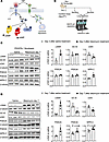 Figure 5
Bleomycin-induced lung inflammation impairs the CNP/GC-B signaling pathway in resident lung myofibroblasts. (A–D) Schematic illustrations of the CNP/GC-B/cGMP signaling cascade (A) and of the animal protocol (B) used for these studies in control (Npr2fl/fl) mice 3 days (C) and 7 days (D) after bleomycin or vehicle administration. Lungs were digested with dispase and the isolated pulmonary PDGFR-α+ fibroblasts were enriched for immunoblot studies. Due to low cell yield, eventually, lung fibroblasts from 2 mice had to be combined for target protein determinations. The expression levels of α-SMA, GC-B, cGKI, PDE2A, PDE3A, and NPR-C were normalized to β-tubulin and calculated as the average value from vehicle-treated (saline-treated) mice (n = 3–4 samples from 6–8 mice per group). Significance was tested by Student’s unpaired t test. *P < 0.05 versus vehicle.
As shown in Figure 5C, in PDGFR-α+ fibroblasts isolated 3 days after bleomycin treatment, the expression of α-SMA was increased by approximately 3-fold (as compared with vehicle), indicating the starting differentiation to myofibroblasts. At this early point, the expression levels of GC-B, PDE2A, and PDE3A were not different between fibroblasts from vehicle- and bleomycin-treated mice. However, NPR-C expression was significantly increased, and cGKI expression levels were mildly but significantly reduced.
In activated α-SMA+ lung myofibroblasts obtained from mice 1 week after bleomycin instillation, such alterations of cGKI and NPR-C levels were maintained and accompanied by altered expression of the whole CNP signaling machinery (Figure 5D). PDE3A and especially PDE2A levels were upregulated. Most importantly, GC-B expression was significantly reduced by about 50% (compared with saline). In fact, the reduction of fibroblast GC-B expression observed in the bleomycin-treated control mice was like the genetic reduction achieved in the Fibro GC-B–KO mice (please compare Figure 5D and Figure 2A).
Collectively, these data reveal that, together with the inflammatory process, fibroblast-to-myofibroblast activation progresses from day 3 to day 7 after bleomycin instillation. While on day 3, the CNP signaling pathway in resident (myo)fibroblasts was mostly preserved, on day 7, the expression of molecules mediating CNP/cGMP effects (GC-B and cGKI) was diminished. Moreover, pathways that degrade CNP (NPR-C) and cGMP as well as cAMP (PDEs 2A and 3A) were induced. This indicates “loss-of-CNP-function” in activated lung myofibroblasts of bleomycin-treated control mice and may explain why bleomycin-induced lung fibrosis did not differ between control and Fibro GC-B KO littermates.
Cytokines involved in lung inflammation and fibrosis impair the CNP/GC-B signaling pathway in cultured human lung fibroblasts. To characterize the upstream factors altering the CNP/GC-B signaling pathway, we tested the effect of relevant cytokines (1, 13, 15, 18) on cultured normal human lung fibroblasts. Cells were treated with TGF-β, TNF-α, MCP-1, IL-1β, or IL-6 (all at 10 ng/mL; except IL-6, used at 50 IU/mL) for 48 hours and then lysed for immunoblot studies. Supplemental Figure 5 illustrates that TGF-β and TNF-α reduced the expression levels of GC-B and cGKI; all cytokines greatly increased the expression of PDE2A, and TGF-β additionally elevated the levels of PDE3A; TNF-α and IL-1β robustly induced the expression of NPR-C. Considering that, in pulmonary inflammation, the resident fibroblasts are exposed to a combination of these cytokines (13–22), this may account for the complex remodeling of the CNP signaling pathways in the bleomycin model during the inflammatory phase (Figure 5).
Supporting the hypothesis that cytokine-driven impairment of the antifibrotic effects of paracrine-acting endogenous CNP contributes to the pathogenesis and progression of lung fibrosis, qPCR studies of human lung tissue samples revealed that the mRNA expression levels of GC-B (mildly) and cGKI are diminished in IPF lungs (Figure 6A). Moreover, the pulmonary expression levels of PDE2A are significantly upregulated (Figure 6A). To determine whether these “global” tissue changes involve alterations within fibroblast populations, we used published scRNA sequencing data sets from the HLCA (11) to compare gene expression levels in the stromal cell clusters between “healthy” control lungs, ILD and IPF. The Uniform Manifold Approximation and Projection (UMAP) plots are shown in Figure 6B. As shown in the UMAP and the violin plots (Figure 6C), CTHRC1 was significantly upregulated in stromal lung cells from ILD and IPF. Concomitantly the expression levels of NPR2 (GC-B) and PKGI (cGKI) were significantly reduced. In contrast to the observations in whole lung samples (Figure 6A), stromal PDE2A expression levels were similar in control and IPF lungs, and they were even reduced in ILD (Figure 6C). This discrepancy warrants further investigation. In future studies, we will try to analyze PDE2A protein expression in specific lung fibroblast subpopulations to unravel potential cell type–specific alterations. However, taken together, these findings indicate that fibroblast CNP/GC-B signaling is altered in inflammation-driven experimental and clinical PF.
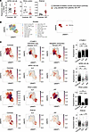 Figure 6
Altered mRNA expression of components of the CNP/GC-B signaling pathway in whole lung samples and lung stromal cells from patients with PF. (A) The mRNA expression levels of NPR2 (GC-B), PKGI (cGKI), and PDE2A in samples of healthy human lung tissue and lung samples from patients with IPF were studied by qPCR. The expression was normalized to porphobilinogen deaminase (PBGD). The sample number for each experiment (n) varied between 10 and 16 and is indicated by the number of data points in each graph. Significance was tested by Student’s unpaired t test. *P < 0.05 versus controls. (B) UMAP plots derived from integrating scRNA-seq data reported in the HLCA (11) reveal the stromal cell clusters (right panel). (C) UMAP visualization (left panels) and violin plots (right panels) compare the mRNA expression levels of CTHRC1 (as myofibroblast marker), NPR2 (GC-B), PKGI (cGKI), and PDE2A in stromal cell clusters from control lungs and lungs from patients with interstitial lung diseases (ILD) or IPF. Significance was tested with the Wilcoxon signed rank test; *P < 0.05.
A single s.c. injection of MS-conjugated long-acting CNP prevents bleomycin-induced lung inflammation and fibrosis. Exogenous high-dose CNP infusions exerted antiinflammatory and antifibrotic actions in LPS-induced acute lung injury and bleomycin-induced PF (6, 7) despite the here-observed receptor and postreceptor changes. Thus, impaired CNP signaling might be overcome by high CNP concentrations. The short 3-minute half-life of CNP and consequently derived need for constant or repeated CNP infusions hamper the translation of such preclinical observations into clinical trials. The stabilized CNP analog vosoritide has recently been approved for treatment of achondroplasia in children (23). However, this requires daily s.c. injections. Recently, a long-acting stabilized CNP analog MS~[Gln6,14]CNP-38 was developed, with release rates allowing once-weekly and once-monthly s.c. injections (10), a regimen that would be acceptable in patients with chronic PF. This compound was generated using a tunable, releasable linker system to attach a stabilized CNP analog, [Gln6,14]CNP-38, to a long-lived MS carrier. Injection of MS~[Gln6,14]CNP-38 creates a s.c. deposit that slowly releases the peptide (10). As proof of principle, first we compared the effects of [Gln6,14]CNP-38 and regular, unmodified short-acting CNP (CNP-22) in cultured human lung fibroblasts. As shown in Supplemental Figure 6, A and B, both peptides similarly enhanced intracellular cGMP levels and reduced PDGF-BB–stimulated fibroblast proliferation. Therefore, we tested the MS-conjugated long-acting CNP analog in experimental bleomycin-induced PF.
Study mice received a single s.c. injection of 120 mg/kg body weight (BW) MS~[Gln6,14]CNP-38 (in a volume of 9 mL/g BW) or carrier alone (9 mL/g BW MS, as vehicle) 10 minutes prior to the intratracheal instillation of bleomycin (0.75 IU/kg BW) (Figure 7). Three days later, the CNP plasma levels on average were 31-fold higher in the MS~[Gln6,14]CNP-38–treated mice (averaging 90 ng/mL, which corresponds to 41.3 nM) as compared with vehicle-treated mice (2.9 ng/mL corresponding to 1.3 nM; n = 4 per condition) The studies of lung morphology and function were performed 21 days after bleomycin treatment, and at this time, the circulating CNP levels were still approximately 4-fold higher in MS~[Gln6,14]CNP-38 as compared with vehicle-treated mice, which is consistent with the published pharmacokinetics (10).
![A single subcutaneous injection of long-acting MS conjugated MS~[Gln6,14]CN](//df6sxcketz7bb.cloudfront.net/manuscripts/196000/196812/small/jci.insight.196812.f7.gif "A single subcutaneous injection of long-acting MS conjugated MS~[Gln6,14]CNP-38 prevents bleomycin-induced lung inflammation.") Figure 7
Figure 7A single subcutaneous injection of long-acting MS conjugated MS~[Gln6,14]CNP-38 prevents bleomycin-induced lung inflammation. (A) Schematic illustration of these studies performed in control mice 21 days after bleomycin or vehicle (saline) administration. Bleomycin-instilled mice were pretreated with a single s.c. injection of either carrier (empty MS, as vehicle) or MS~[Gln6,14]CNP-38. (B and C) Plasma leakage was evaluated by determination of the protein and albumin contents of bronchoalveolar lavage fluid (BALF) samples by BCA assay (B) and immunoblotting (C). (D and E) Cytokine contents of BALF samples were evaluated with a commercial array. D shows representative cytokine array membranes probed with BALF from each study group. The heatmap depicts cytokines which were detected at markedly different levels in BALF from carrier (MS)- or MS~[Gln6,14]CNP-38–pretreated bleomycin-treated mice (mean-normalized values are shown, with red color indicating high, and blue indicating low values). (E) Quantitative evaluation of pixel densities of representative cytokines differing between these 2 groups. In B and C, each data point represents an individual study mouse; E shows replicates from 4–5 mice per group. Significance was tested by 1-way ANOVA (B and C), and Student’s unpaired t test (D). *P < 0.05 versus vehicle-instilled mice; #P < 0.05 versus carrier-pretreated/bleomycin-treated mice.
Pulmonary inflammation was evaluated by analyses of BALF. Figure 7, B and C, illustrates that bleomycin-evoked plasma protein — i.e., albumin extravasation at this late time point (21 days) — was like that observed 7 days after bleomycin administration (see previous Figure 3, B and C), indicating persistent inflammation. As also shown, this response was significantly prevented by a single s.c. injection of MS~[Gln6,14]CNP-38. Further characterization of the BALF with a cytokine array confirmed that numerous proinflammatory cytokines and chemokines were increased in BALF from vehicle (“empty” MS) pretreated bleomycin-instilled mice (Figure 7D), although the pattern at this late time point was different to that observed at earlier time points (Figure 3D). As shown, this response was blunted in the MS~[Gln6,14]CNP-38–treated mice. Figure 7E depicts the cytokines that were mostly affected by this treatment. Notably, the BALF levels of proinflammatory cytokines such as IL-17 and CXCL9 (16, 24) were reduced by the CNP analog, whereas the levels of antiinflammatory, antifibrotic factors such as G-CSF, IL-3, and IL-4 (14, 25, 26) were increased. Additionally, [Gln6,14]CNP-38 almost abolished the bleomycin-induced increases of tissue inhibitor of metalloproteinase 1 (TIMP-1), which may improve MMP-mediated interstitial collagen degradation (Figure 7E).
Indeed, Masson′s trichrome staining of lung sections together with immunoblot analyses of lung samples revealed that bleomycin-induced interstitial collagen accumulation was reduced by MS~[Gln6,14]CNP-38 (Figure 8, A and B). Accordingly, in vehicle-pretreated mice, the administration of bleomycin increased the pulmonary expression levels of the myofibroblast markers periostin and Cthrc1, phosphoglycerate dehydrogenase (PHDGH, involved in the early steps of L-serine synthesis for collagen), and MMP-9. Pretreatment of study mice with [Gln6,14]CNP-38 abrogated all these changes (Figure 8B). Notably, MS~[Gln6,14]CNP-38 also attenuated the bleomycin-induced expression of the proinflammatory cytokines IL-6, TNF-α, and (less) CXCL1 (Figure 8C). Similar to the previous studies (Figure 4C), whole-lung α-SMA levels were not different between bleomycin- and vehicle-treated mice (Figure 8B).
![A single s.c. injection of long-acting MS-conjugated MS~[Gln6,14]CNP-38 pre](//df6sxcketz7bb.cloudfront.net/manuscripts/196000/196812/small/jci.insight.196812.f8.gif "A single s.c. injection of long-acting MS-conjugated MS~[Gln6,14]CNP-38 prevents bleomycin-induced lung fibrosis.") Figure 8
Figure 8A single s.c. injection of long-acting MS-conjugated MS~[Gln6,14]CNP-38 prevents bleomycin-induced lung fibrosis. The animal protocol was illustrated in Figure 7. (A) Collagen deposition was determined by Masson’s trichrome staining of lung paraffin sections, followed by quantification with ORBIT. The mean value from 2 sections per mouse was taken (10–15 fields per section were evaluated). Collagen areas (in percent from corresponding total section areas) are presented as x-fold from the average value of control mice. (B and C) The pulmonary expression levels of fibrosis and myofibroblast markers Collagen 1, Periostin, Cthrc-1, α-SMA, MMP-9, and PHDGH (B) as well as of proinflammatory cytokines IL-6, CXCL-1, and TNF-α (C) were studied by immunoblotting and normalized to β-actin; values were calculated as x-fold from the average value of control mice. (D) Lung inspiratory capacity and compliance were evaluated in anesthetized mice with a Flexivent system. n = 6 (Carrier/saline), 6 (Carrier/bleomycin) and 5 mice (MS~[Gln6,14]CNP-38/bleomycin). Significance was tested by 1-way ANOVA. *P < 0.05 versus carrier/saline-treated mice; #P < 0.05 versus carrier/bleomycin-treated mice.
The effect on lung function was evaluated with the Flexivent system (Figure 8D). In vehicle-pretreated mice, bleomycin induced a significant decline of inspiratory capacity and compliance. These functional changes were fully prevented by MS~[Gln6,14]CNP-38. Collectively, our results demonstrate that a single dose of this long-acting CNP analog led to long-lasting elevations of plasma CNP levels, which blunted bleomycin-induced lung inflammation, fibrosis, and dysfunction.
CNP reverts the profibrotic activation of cultured PF fibroblasts in vitro and in situ. In the previously illustrated experiments in vitro and in vivo, we tested the “preventive” antifibrotic effects of CNP. To further explore the therapeutic potential, lastly, we performed treatment studies in 2 models of cultured human PF fibroblasts. In vitro, cultured IPF lung fibroblasts were prestimulated with TGF-β for 24 hours to induce profibrotic changes, followed by treatment with vehicle (PBS) or CNP (100 nM) for additional 24 hours (Supplemental Figure 7A provides a scheme). TGF-β stimulation significantly upregulated the expression of Collagen 1 and Cthrc 1. Notably, subsequent CNP treatment significantly reduced these changes (Supplemental Figure 7A). In a second experimental series, we studied cultured precision-cut lung slices (PCLS) from a patient with end-stage PF caused by HP (2). As illustrated in Supplemental Figure 7B, the slices were cultured in the presence of CNP (100 nM, daily addition) or vehicle (PBS) for 7 days. At the end of the experiment, the slices were lysed in RIPA buffer and the extracted proteins were analyzed by immunoblotting. Notably, in CNP-treated PCLS, the expression of Collagen 1 and Cthrc 1 was reduced (in comparison with PBS-treated PCLS; see Supplemental Figure 7B). Together, these results indicate that CNP can prevent and even revert the profibrotic phenotype of human lung fibroblasts in vitro and in situ (in PCLS), supporting further studies to investigate its therapeutic potential.
![A single subcutaneous injection of long-acting MS conjugated MS~[Gln6,14]CN](http://df6sxcketz7bb.cloudfront.net/manuscripts/196000/196812/small/jci.insight.196812.f7.gif "A single subcutaneous injection of long-acting MS conjugated MS~[Gln6,14]CNP-38 prevents bleomycin-induced lung inflammation.")
![A single s.c. injection of long-acting MS-conjugated MS~[Gln6,14]CNP-38 pre](http://df6sxcketz7bb.cloudfront.net/manuscripts/196000/196812/small/jci.insight.196812.f8.gif "A single s.c. injection of long-acting MS-conjugated MS~[Gln6,14]CNP-38 prevents bleomycin-induced lung fibrosis.")


