
Research ArticleBone biologyInflammation
Open Access |  10.1172/jci.insight.196750
Innate immune sensor NOD2 promotes cartilage degradation and osteoarthritis progression by stabilizing TRAF6 in chondrocytes
Yuting Wang,1,2,3 Song Li,1 Yonghui Dong,4 Jiaming Zhang,5 Jian Liu,1 Zhenggang Wang,1 Shuang Liang,1 Nathan R. Martinez,2,3 Hongxu Pu,1 Peng Cheng,1 Anmin Chen,1 Qing Yang,1 Charles K.F. Chan,2,3 Wen Jiang,6 Jun Xiao,1 Fengjing Guo,1 and Liming Zhao1,2,3
1Department of Orthopaedics, Tongji Hospital, Tongji Medical College, Huazhong University of Science and Technology, Wuhan, Hubei, China.
2Institute for Stem Cell Biology and Regenerative Medicine and
3Department of Surgery, Division of Plastic and Reconstructive Surgery, Stanford University School of Medicine, Stanford, California, USA.
4Department of Orthopedics, Henan Provincial People’s Hospital, People’s Hospital of Zhengzhou University, Zhengzhou, Henan, China.
5Clinical Innovation and Research Center, Shenzhen Hospital, Southern Medical University, Shenzhen, Guangdong, China.
6Department of Radiation Oncology, The University of Texas MD Anderson Cancer Center, Houston, Texas, USA.
Authorship note: YW and SL contributed equally to this work and are co–first authors. CKFC is deceased.
Find articles by Wang, Y. in: PubMed | Google Scholar
1Department of Orthopaedics, Tongji Hospital, Tongji Medical College, Huazhong University of Science and Technology, Wuhan, Hubei, China.
2Institute for Stem Cell Biology and Regenerative Medicine and
3Department of Surgery, Division of Plastic and Reconstructive Surgery, Stanford University School of Medicine, Stanford, California, USA.
4Department of Orthopedics, Henan Provincial People’s Hospital, People’s Hospital of Zhengzhou University, Zhengzhou, Henan, China.
5Clinical Innovation and Research Center, Shenzhen Hospital, Southern Medical University, Shenzhen, Guangdong, China.
6Department of Radiation Oncology, The University of Texas MD Anderson Cancer Center, Houston, Texas, USA.
Authorship note: YW and SL contributed equally to this work and are co–first authors. CKFC is deceased.
Find articles by Li, S. in: PubMed | Google Scholar
1Department of Orthopaedics, Tongji Hospital, Tongji Medical College, Huazhong University of Science and Technology, Wuhan, Hubei, China.
2Institute for Stem Cell Biology and Regenerative Medicine and
3Department of Surgery, Division of Plastic and Reconstructive Surgery, Stanford University School of Medicine, Stanford, California, USA.
4Department of Orthopedics, Henan Provincial People’s Hospital, People’s Hospital of Zhengzhou University, Zhengzhou, Henan, China.
5Clinical Innovation and Research Center, Shenzhen Hospital, Southern Medical University, Shenzhen, Guangdong, China.
6Department of Radiation Oncology, The University of Texas MD Anderson Cancer Center, Houston, Texas, USA.
Authorship note: YW and SL contributed equally to this work and are co–first authors. CKFC is deceased.
Find articles by Dong, Y. in: PubMed | Google Scholar
1Department of Orthopaedics, Tongji Hospital, Tongji Medical College, Huazhong University of Science and Technology, Wuhan, Hubei, China.
2Institute for Stem Cell Biology and Regenerative Medicine and
3Department of Surgery, Division of Plastic and Reconstructive Surgery, Stanford University School of Medicine, Stanford, California, USA.
4Department of Orthopedics, Henan Provincial People’s Hospital, People’s Hospital of Zhengzhou University, Zhengzhou, Henan, China.
5Clinical Innovation and Research Center, Shenzhen Hospital, Southern Medical University, Shenzhen, Guangdong, China.
6Department of Radiation Oncology, The University of Texas MD Anderson Cancer Center, Houston, Texas, USA.
Authorship note: YW and SL contributed equally to this work and are co–first authors. CKFC is deceased.
Find articles by
Zhang, J.
in:
PubMed
|
Google Scholar
|

1Department of Orthopaedics, Tongji Hospital, Tongji Medical College, Huazhong University of Science and Technology, Wuhan, Hubei, China.
2Institute for Stem Cell Biology and Regenerative Medicine and
3Department of Surgery, Division of Plastic and Reconstructive Surgery, Stanford University School of Medicine, Stanford, California, USA.
4Department of Orthopedics, Henan Provincial People’s Hospital, People’s Hospital of Zhengzhou University, Zhengzhou, Henan, China.
5Clinical Innovation and Research Center, Shenzhen Hospital, Southern Medical University, Shenzhen, Guangdong, China.
6Department of Radiation Oncology, The University of Texas MD Anderson Cancer Center, Houston, Texas, USA.
Authorship note: YW and SL contributed equally to this work and are co–first authors. CKFC is deceased.
Find articles by Liu, J. in: PubMed | Google Scholar
1Department of Orthopaedics, Tongji Hospital, Tongji Medical College, Huazhong University of Science and Technology, Wuhan, Hubei, China.
2Institute for Stem Cell Biology and Regenerative Medicine and
3Department of Surgery, Division of Plastic and Reconstructive Surgery, Stanford University School of Medicine, Stanford, California, USA.
4Department of Orthopedics, Henan Provincial People’s Hospital, People’s Hospital of Zhengzhou University, Zhengzhou, Henan, China.
5Clinical Innovation and Research Center, Shenzhen Hospital, Southern Medical University, Shenzhen, Guangdong, China.
6Department of Radiation Oncology, The University of Texas MD Anderson Cancer Center, Houston, Texas, USA.
Authorship note: YW and SL contributed equally to this work and are co–first authors. CKFC is deceased.
Find articles by Wang, Z. in: PubMed | Google Scholar
1Department of Orthopaedics, Tongji Hospital, Tongji Medical College, Huazhong University of Science and Technology, Wuhan, Hubei, China.
2Institute for Stem Cell Biology and Regenerative Medicine and
3Department of Surgery, Division of Plastic and Reconstructive Surgery, Stanford University School of Medicine, Stanford, California, USA.
4Department of Orthopedics, Henan Provincial People’s Hospital, People’s Hospital of Zhengzhou University, Zhengzhou, Henan, China.
5Clinical Innovation and Research Center, Shenzhen Hospital, Southern Medical University, Shenzhen, Guangdong, China.
6Department of Radiation Oncology, The University of Texas MD Anderson Cancer Center, Houston, Texas, USA.
Authorship note: YW and SL contributed equally to this work and are co–first authors. CKFC is deceased.
Find articles by
Liang, S.
in:
PubMed
|
Google Scholar
|
1Department of Orthopaedics, Tongji Hospital, Tongji Medical College, Huazhong University of Science and Technology, Wuhan, Hubei, China.
2Institute for Stem Cell Biology and Regenerative Medicine and
3Department of Surgery, Division of Plastic and Reconstructive Surgery, Stanford University School of Medicine, Stanford, California, USA.
4Department of Orthopedics, Henan Provincial People’s Hospital, People’s Hospital of Zhengzhou University, Zhengzhou, Henan, China.
5Clinical Innovation and Research Center, Shenzhen Hospital, Southern Medical University, Shenzhen, Guangdong, China.
6Department of Radiation Oncology, The University of Texas MD Anderson Cancer Center, Houston, Texas, USA.
Authorship note: YW and SL contributed equally to this work and are co–first authors. CKFC is deceased.
Find articles by Martinez, N. in: PubMed | Google Scholar
1Department of Orthopaedics, Tongji Hospital, Tongji Medical College, Huazhong University of Science and Technology, Wuhan, Hubei, China.
2Institute for Stem Cell Biology and Regenerative Medicine and
3Department of Surgery, Division of Plastic and Reconstructive Surgery, Stanford University School of Medicine, Stanford, California, USA.
4Department of Orthopedics, Henan Provincial People’s Hospital, People’s Hospital of Zhengzhou University, Zhengzhou, Henan, China.
5Clinical Innovation and Research Center, Shenzhen Hospital, Southern Medical University, Shenzhen, Guangdong, China.
6Department of Radiation Oncology, The University of Texas MD Anderson Cancer Center, Houston, Texas, USA.
Authorship note: YW and SL contributed equally to this work and are co–first authors. CKFC is deceased.
Find articles by Pu, H. in: PubMed | Google Scholar
1Department of Orthopaedics, Tongji Hospital, Tongji Medical College, Huazhong University of Science and Technology, Wuhan, Hubei, China.
2Institute for Stem Cell Biology and Regenerative Medicine and
3Department of Surgery, Division of Plastic and Reconstructive Surgery, Stanford University School of Medicine, Stanford, California, USA.
4Department of Orthopedics, Henan Provincial People’s Hospital, People’s Hospital of Zhengzhou University, Zhengzhou, Henan, China.
5Clinical Innovation and Research Center, Shenzhen Hospital, Southern Medical University, Shenzhen, Guangdong, China.
6Department of Radiation Oncology, The University of Texas MD Anderson Cancer Center, Houston, Texas, USA.
Authorship note: YW and SL contributed equally to this work and are co–first authors. CKFC is deceased.
Find articles by Cheng, P. in: PubMed | Google Scholar
1Department of Orthopaedics, Tongji Hospital, Tongji Medical College, Huazhong University of Science and Technology, Wuhan, Hubei, China.
2Institute for Stem Cell Biology and Regenerative Medicine and
3Department of Surgery, Division of Plastic and Reconstructive Surgery, Stanford University School of Medicine, Stanford, California, USA.
4Department of Orthopedics, Henan Provincial People’s Hospital, People’s Hospital of Zhengzhou University, Zhengzhou, Henan, China.
5Clinical Innovation and Research Center, Shenzhen Hospital, Southern Medical University, Shenzhen, Guangdong, China.
6Department of Radiation Oncology, The University of Texas MD Anderson Cancer Center, Houston, Texas, USA.
Authorship note: YW and SL contributed equally to this work and are co–first authors. CKFC is deceased.
Find articles by Chen, A. in: PubMed | Google Scholar
1Department of Orthopaedics, Tongji Hospital, Tongji Medical College, Huazhong University of Science and Technology, Wuhan, Hubei, China.
2Institute for Stem Cell Biology and Regenerative Medicine and
3Department of Surgery, Division of Plastic and Reconstructive Surgery, Stanford University School of Medicine, Stanford, California, USA.
4Department of Orthopedics, Henan Provincial People’s Hospital, People’s Hospital of Zhengzhou University, Zhengzhou, Henan, China.
5Clinical Innovation and Research Center, Shenzhen Hospital, Southern Medical University, Shenzhen, Guangdong, China.
6Department of Radiation Oncology, The University of Texas MD Anderson Cancer Center, Houston, Texas, USA.
Authorship note: YW and SL contributed equally to this work and are co–first authors. CKFC is deceased.
Find articles by Yang, Q. in: PubMed | Google Scholar
1Department of Orthopaedics, Tongji Hospital, Tongji Medical College, Huazhong University of Science and Technology, Wuhan, Hubei, China.
2Institute for Stem Cell Biology and Regenerative Medicine and
3Department of Surgery, Division of Plastic and Reconstructive Surgery, Stanford University School of Medicine, Stanford, California, USA.
4Department of Orthopedics, Henan Provincial People’s Hospital, People’s Hospital of Zhengzhou University, Zhengzhou, Henan, China.
5Clinical Innovation and Research Center, Shenzhen Hospital, Southern Medical University, Shenzhen, Guangdong, China.
6Department of Radiation Oncology, The University of Texas MD Anderson Cancer Center, Houston, Texas, USA.
Authorship note: YW and SL contributed equally to this work and are co–first authors. CKFC is deceased.
Find articles by Chan, C. in: PubMed | Google Scholar
1Department of Orthopaedics, Tongji Hospital, Tongji Medical College, Huazhong University of Science and Technology, Wuhan, Hubei, China.
2Institute for Stem Cell Biology and Regenerative Medicine and
3Department of Surgery, Division of Plastic and Reconstructive Surgery, Stanford University School of Medicine, Stanford, California, USA.
4Department of Orthopedics, Henan Provincial People’s Hospital, People’s Hospital of Zhengzhou University, Zhengzhou, Henan, China.
5Clinical Innovation and Research Center, Shenzhen Hospital, Southern Medical University, Shenzhen, Guangdong, China.
6Department of Radiation Oncology, The University of Texas MD Anderson Cancer Center, Houston, Texas, USA.
Authorship note: YW and SL contributed equally to this work and are co–first authors. CKFC is deceased.
Find articles by
Jiang, W.
in:
PubMed
|
Google Scholar
|
1Department of Orthopaedics, Tongji Hospital, Tongji Medical College, Huazhong University of Science and Technology, Wuhan, Hubei, China.
2Institute for Stem Cell Biology and Regenerative Medicine and
3Department of Surgery, Division of Plastic and Reconstructive Surgery, Stanford University School of Medicine, Stanford, California, USA.
4Department of Orthopedics, Henan Provincial People’s Hospital, People’s Hospital of Zhengzhou University, Zhengzhou, Henan, China.
5Clinical Innovation and Research Center, Shenzhen Hospital, Southern Medical University, Shenzhen, Guangdong, China.
6Department of Radiation Oncology, The University of Texas MD Anderson Cancer Center, Houston, Texas, USA.
Authorship note: YW and SL contributed equally to this work and are co–first authors. CKFC is deceased.
Find articles by Xiao, J. in: PubMed | Google Scholar
1Department of Orthopaedics, Tongji Hospital, Tongji Medical College, Huazhong University of Science and Technology, Wuhan, Hubei, China.
2Institute for Stem Cell Biology and Regenerative Medicine and
3Department of Surgery, Division of Plastic and Reconstructive Surgery, Stanford University School of Medicine, Stanford, California, USA.
4Department of Orthopedics, Henan Provincial People’s Hospital, People’s Hospital of Zhengzhou University, Zhengzhou, Henan, China.
5Clinical Innovation and Research Center, Shenzhen Hospital, Southern Medical University, Shenzhen, Guangdong, China.
6Department of Radiation Oncology, The University of Texas MD Anderson Cancer Center, Houston, Texas, USA.
Authorship note: YW and SL contributed equally to this work and are co–first authors. CKFC is deceased.
Find articles by Guo, F. in: PubMed | Google Scholar
1Department of Orthopaedics, Tongji Hospital, Tongji Medical College, Huazhong University of Science and Technology, Wuhan, Hubei, China.
2Institute for Stem Cell Biology and Regenerative Medicine and
3Department of Surgery, Division of Plastic and Reconstructive Surgery, Stanford University School of Medicine, Stanford, California, USA.
4Department of Orthopedics, Henan Provincial People’s Hospital, People’s Hospital of Zhengzhou University, Zhengzhou, Henan, China.
5Clinical Innovation and Research Center, Shenzhen Hospital, Southern Medical University, Shenzhen, Guangdong, China.
6Department of Radiation Oncology, The University of Texas MD Anderson Cancer Center, Houston, Texas, USA.
Authorship note: YW and SL contributed equally to this work and are co–first authors. CKFC is deceased.
Find articles by Zhao, L. in: PubMed | Google Scholar
Published August 24, 2026 - More info
JCI Insight. 2026;11(16):e196750. https://doi.org/10.1172/jci.insight.196750.
© 2026 Wang et al. This work is licensed under the Creative Commons Attribution 4.0 International License. To view a copy of this license, visit http://creativecommons.org/licenses/by/4.0/.
Received: June 13, 2025; Accepted: June 25, 2026

-
Results
NOD2 is activated in both human and mouse osteoarthritic cartilage. To gain a more comprehensive understanding of its role in OA pathophysiology, we first examined NOD2 level in human and mouse osteoarthritic cartilage. Human osteoarthritic cartilage was obtained from patients undergoing total knee arthroplasty (Kellgren-Lawrence grade 3–4) (19), while healthy control cartilage was collected from donors undergoing amputation due to trauma or tumor (Kellgren-Lawrence grade 0). As summarized in Supplemental Table 1 (supplemental material available online with this article; https://doi.org/10.1172/jci.insight.196750DS1), demographic factors including age, sex, and BMI were comparable between the 2 groups. IHC staining of medial tibial plateau sections revealed significantly increased NOD2 protein level in osteoarthritic cartilage compared with healthy controls (65.33% ± 12.10% versus 11.67% ± 7.02%, P = 0.0027) (Figure 1, A and B). Similarly, in a mouse OA model induced by surgical destabilization of the medial meniscus (DMM) (20), NOD2 protein level was significantly elevated in articular cartilage (55.33% ± 14.01% versus 2.67% ± 2.08%, P = 0.0030) (Figure 1, C and D). Given that inflammatory cytokines such as IL-1β and TNF-α are key mediators of chondrocyte catabolism and cartilage degradation (22), we next investigated whether they regulate NOD2 expression. In primary human chondrocytes, IL-1β stimulation induced a dose-dependent increase in NOD2 expression at both RNA and protein levels, accompanied by upregulation of the catabolic enzyme matrix metalloproteinase-3 (MMP3). Peak mRNA and protein expression occurred at 12–24 hours and 48–72 hours after treatment, respectively (Figure 1, E–G). Consistently, IL-1β treatment also induced Nod2 expression in primary mouse chondrocytes (Figure 1, H–J). In addition, TNF-α robustly upregulated both Nod2 and catabolic enzyme expression (Figure 1, K–N). To confirm NOD2 activation, we treated mouse chondrocytes with MDP, a bacterial peptidoglycan-derived ligand known to activate NOD2 (22), and we observed increased Nod2 expression (Figure 1O). These findings demonstrate that NOD2 expression is elevated in osteoarthritic cartilage and is inducible by proinflammatory stimuli, suggesting a potential role for NOD2 in chondrocyte catabolism and cartilage homeostasis during OA progression.
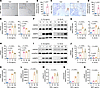 Figure 1
NOD2 is activated in both human and mouse osteoarthritic cartilage. (A) Representative IHC images of NOD2 in healthy and osteoarthritic human cartilage. Scale bar: 100 μm. (B) Quantification of NOD2+ chondrocytes. n = 3 donors/group. Two-tailed, unpaired Student’s t test. (C) Representative IHC images of NOD2 in healthy and osteoarthritic mouse cartilage. Scale bar: 50 μm. (D) Quantification of NOD2+ chondrocytes. n = 3 mice/group. Two-tailed, unpaired Student’s t test. (E) qPCR analysis of NOD2 mRNA in primary human chondrocytes treated with IL-1β at indicated concentrations (for 24 h) and durations (using 5 ng/mL of IL-1β). n = 3 independent experiments. One-way ANOVA with Tukey’s multiple-comparison test. (F and G) Western blot analysis and densitometric quantification of NOD2 protein in primary human chondrocytes treated with IL-1β at indicated concentrations (for 48 h) and durations (using 5 ng/mL of IL-1β). n = 3 independent experiments. One-way ANOVA with Tukey’s multiple-comparison test. (H) qPCR analysis of Nod2 mRNA in primary mouse chondrocytes treated with IL-1β at indicated concentrations (for 24 h) and durations (using 5 ng/mL of IL-1β). n = 3 independent experiments. One-way ANOVA with Tukey’s multiple-comparison test. (I and J) Western blot analysis and densitometric quantification of NOD2 protein in primary mouse chondrocytes treated with IL-1β at indicated concentrations (for 72 h) and durations (using 5 ng/mL of IL-1β). n = 3 independent experiments. One-way ANOVA with Tukey’s multiple-comparison test. (K–N) qPCR analysis of Nod2, Mmp3, Mmp13, and ADAMTS5 mRNA levels in primary mouse chondrocytes treated with TNF-α at indicated concentrations (for 24 h). n = 3 independent experiments. One-way ANOVA with Tukey’s multiple-comparison test. (O) qPCR analysis of Nod2 mRNA levels in primary mouse chondrocytes treated with muramyl dipeptide (MDP) at indicated concentrations (for 24 h). n = 3 independent experiments. One-way ANOVA with Tukey’s multiple-comparison test. Data are presented as mean ± SD.
Knockdown of Nod2 inhibits inflammatory cytokine-induced chondrocyte catabolism and apoptosis in vitro. We next investigated whether NOD2 contributes to chondrocyte catabolism and apoptosis under inflammatory conditions. Small interfering RNA (siRNA) was used to knock down Nod2 in primary mouse chondrocytes. As expected, IL-1β treatment induced catabolic activity, as indicated by increased mRNA and protein levels of matrix-degrading enzymes, including Mmp3, Mmp13, and ADAMTS5. However, Nod2 knockdown significantly attenuated this inflammatory catabolic response (Figure 2, A–C). To further validate the protective effect of Nod2 knockdown on cartilage matrix metabolism, we utilized a high-density micromass culture model of primary chondrocytes. Alcian blue staining demonstrated that Nod2 knockdown preserved ECM content in IL-1β–treated cultures compared with control siRNA-treated groups (Figure 2, D and E). Given that IL-1β–induced chondrocyte apoptosis also contributes to ECM loss, we next examined whether Nod2 knockdown could mitigate apoptosis. Indeed, Nod2 knockdown significantly reduced IL-1β–induced chondrocyte apoptosis (10.66% ± 1.01% versus 19.99% ± 1.50%, P < 0.0001) (Figure 2F). Based on these results, we next sought to explore the in vivo role of NOD2 in OA progression using mouse models.
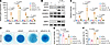 Figure 2
Nod2 knockdown attenuates inflammation-induced chondrocyte catabolism and apoptosis in vitro. (A) qPCR analysis of Nod2, Mmp3, Mmp13, and ADAMTS5 mRNA levels in primary mouse chondrocytes following Nod2 knockdown and IL-1β treatment (5 ng/mL for 24 h). n = 3 independent experiments. One-way ANOVA with Tukey’s multiple-comparison test. (B and C) Western blot analysis and densitometric quantification of NOD2, MMP3, MMP13, and ADAMTS5 protein levels following Nod2 knockdown and IL-1β treatment (5 ng/mL for 72 h). n = 3 independent experiments. One-way ANOVA with Tukey’s multiple-comparison test. (D) Alcian blue staining of micromass cultures of primary mouse chondrocytes following Nod2 knockdown and IL-1β treatment (5 ng/mL for 2 weeks). (E) Quantification of proteoglycan content by absorbance of dissolved Alcian blue at 595 nm. n = 3 independent experiments. One-way ANOVA with Tukey’s multiple-comparison test. (F) Flow cytometric analysis of apoptosis in primary mouse chondrocytes following Nod2 knockdown and IL-1β treatment (5 ng/mL for 24 h). n = 3 independent experiments. One-way ANOVA with Tukey’s multiple-comparison test. Data are presented as mean ± SD.
Adenovirus-mediated Nod2 knockdown attenuates cartilage degradation in mouse OA joint. To investigate the role of NOD2 in vivo, we generated adenoviruses expressing shRNA targeting Nod2 for intraarticular injection into mouse knee joints. One week following adenoviral delivery, DMM surgery was performed to induce OA (20). As subchondral bone sclerosis and osteophyte formation are hallmarks of OA progression (23), we used μCT and 3D reconstruction to evaluate structural changes in the joint. Compared with sham operated controls, DMM surgery led to densification of the tibial subchondral bone plate and the development of discernible osteophytes at the joint margins (Figure 3, A and B). The increased bone volume per total volume (BV/TV) further confirmed the occurrence of subchondral bone sclerosis in the DMM group. Notably, Nod2 knockdown reversed these pathological changes, resulting in both lower BV/TV ratios and qualitatively reduced osteophyte formation (BV/TV: 0.38 ± 0.10 versus 0.52 ± 0.08, P = 0.0023). These findings suggest that Nod2 deficiency exerts a protective effect on subchondral bone remodeling during OA progression. Histological analysis using Safranin O/Fast Green staining revealed substantial cartilage degradation at 8 weeks after DMM surgery, whereas Nod2 knockdown significantly preserved cartilage integrity (Figure 3C). Quantification using the OARSI scoring system demonstrated a significant reduction in cartilage damage in the shNod2 group compared with the shCon group after surgery (6.63 ± 3.66 versus 12.50 ± 3.21, P = 0.0007) (24) (Figure 3D). IHC staining confirmed efficient Nod2 knockdown in joint tissues (Figure 3, E and F). Furthermore, IHC revealed that catabolic enzymes elevated by DMM surgery, such as MMP3, MMP13, and ADAMTS5, were markedly reduced in the Nod2 knockdown group (Figure 3, G–J). Although NOD2 levels were increased in the synovium following DMM surgery, and intraarticular injection of adenoviruses expressing Nod2-shRNA successfully suppressed its expression, synovial inflammation was not markedly altered. Specifically, the synovitis score, as assessed by H&E staining (25), as well as the DMM-induced infiltration of CD45+ leukocytes and the expression of the inflammatory mediator IL-1β, showed no significant differences between the control and knockdown groups (Supplemental Figure 1). Together, these findings indicate that intraarticular Nod2 knockdown by adenoviruses attenuates cartilage degradation and subchondral bone remodeling in OA, without significantly altering synovial inflammation.
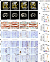 Figure 3
Adenovirus-mediated Nod2 knockdown attenuates cartilage degradation in mouse OA joints. (A) Reconstructed μCT images of mouse knee joints following Nod2 knockdown and DMM surgery. Yellow overlays indicate the sectional plane in the 3D reconstruction. Scale bar: 1 mm. (B) Quantification of bone volume/total volume (BV/TV) in tibial subchondral bone. n = 8 mice/group. One-way ANOVA with Tukey’s multiple-comparison test. (C) Safranin O/Fast Green staining of knee joint sections. Scale bar: 100 μm. (D) OARSI scores for cartilage degradation based on Safranin O/Fast Green staining. n = 8 mice/group. One-way ANOVA with Tukey’s multiple-comparison test. (E) IHC staining of knee sections for NOD2. Scale bar: 50 μm. (F) Quantification of positively stained chondrocytes for NOD2. n = 8 mice/group. One-way ANOVA with Tukey’s multiple-comparison test. (G) IHC staining of knee sections for MMP3, MMP13, and ADAMTS5. Scale bar: 50 μm. (H–J) Quantification of positively stained chondrocytes for MMP3, MMP13, and ADAMTS5. n = 8 mice/group. One-way ANOVA with Tukey’s multiple-comparison test. Data are presented as mean ± SD.
cKO of Nod2 in chondrocytes prevents OA development. Given the elevated expression of NOD2 in both osteoarthritic synovium and cartilage, and the high immunogenicity of adenoviruses, which may incite nonspecific synovial inflammation, we further investigated its functional role using a chondrocyte-specific cKO model. Nod2fl/fl mice were crossed with Col2a1-CreER mice to generate chondrocyte-specific Nod2-cKO. Both Nod2-cKO and WT littermates (Nod2-WT) were treated with tamoxifen at 3 weeks of age to achieve optimal Nod2 KO in articular cartilage (26), followed by DMM surgeries at 12 weeks of age to induce OA. At 8 weeks after surgery, Nod2-WT mice displayed characteristic OA pathology, including tibial subchondral bone densification (Figure 4, A and B) and significant cartilage degradation (Figure 4, C and D), accompanied by increased expression of catabolic enzymes such as MMP3, MMP13, and ADAMTS5 (Figure 4, E–J). In contrast, Nod2-cKO mice were protected from these pathological changes, maintaining subchondral bone structure (BV/TV: 0.36 ± 0.05 versus 0.48 ± 0.10, P = 0.0160) and cartilage integrity (OARSI score: 8.67 ± 3.67 versus 15.17 ± 3.71, P = 0.0032). IHC staining confirmed efficient NOD2 deletion in chondrocytes and revealed a corresponding reduction in catabolic enzyme expression in Nod2-cKO mice. We further examined the synovial pathology in Nod2-cKO mice. This was prompted by the observation that adenovirus-mediated Nod2 knockdown did not significantly alter DMM-induced synovitis, a result that may have been confounded by the inherent immunogenicity typically associated with viral vectors. Surprisingly, the specific ablation of Nod2 in chondrocytes resulted in a significant reduction in synovial inflammation compared with control Nod2-WT mice. Specifically, Nod2-cKO mice exhibited attenuated synovial hyperplasia and decreased infiltration of inflammatory cells (synovitis score: 1.50 ± 0.55 versus 2.50 ± 0.55, P = 0.0012) (Figure 5, A–H). These findings suggest that targeted ablation of Nod2 in chondrocytes was associated with a reduction in synovial inflammation. This protective effect potentially results from decreased release of cartilage-derived DAMPs into the joint space, thereby attenuating the catabolic crosstalk between cartilage and synovium that drives OA progression.
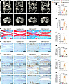 Figure 4
Conditional KO of Nod2 in chondrocytes prevents OA development. (A) Reconstructed μCT images of mouse knee joints following Nod2 KO and DMM surgery. Scale bar: 1 mm. (B) Quantification of bone volume/total volume (BV/TV) in tibial subchondral bone. n = 6 mice/group. One-way ANOVA with Tukey’s multiple-comparison test. (C) Safranin O/Fast Green staining of knee joint sections. Scale bar: 100 μm. (D) OARSI scores for cartilage degradation based on Safranin O/Fast Green staining. n = 6 mice/group. One-way ANOVA with Tukey’s multiple-comparison test. (E) IHC staining of knee sections for NOD2. Scale bar: 100 μm. (F) Quantification of positively stained chondrocytes for NOD2. n = 6 mice/group. One-way ANOVA with Tukey’s multiple-comparison test. (G) IHC staining of knee sections for MMP3, MMP13, and ADAMTS5. Scale bar: 100 μm. (H–J) Quantification of positively stained chondrocytes for MMP3, MMP13, and ADAMTS5. n = 6 mice/group. One-way ANOVA with Tukey’s multiple-comparison test. Data are presented as mean ± SD.
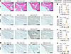 Figure 5
Conditional KO of Nod2 in chondrocytes attenuates synovial inflammation. (A) H&E staining of mouse knee joints following Nod2 knockout and DMM surgery. Scale bar: 200 μm. (B) Quantification of synovitis grading scores based on H&E staining. n = 6 mice/group. One-way ANOVA with Tukey’s multiple-comparison test. (C) IHC staining of knee sections for NOD2. Scale bar: 100 μm. (D) Quantification of positively stained synoviocytes for NOD2. n = 6 mice/group. One-way ANOVA with Tukey’s multiple-comparison test. (E) IHC staining of knee sections for IL-1β. Scale bar: 100 μm. (F) Quantification of positively stained synoviocytes for IL-1β. n = 6 mice/group. One-way ANOVA with Tukey’s multiple-comparison test. (G) IHC staining of knee sections for CD45. Scale bar: 100 μm. (H) Quantification of positively stained synoviocytes for CD45. n = 6 mice/group. One-way ANOVA with Tukey’s multiple-comparison test. Data are presented as mean ± SD.
Activation of NOD2 by MDP is not sufficient to promote cartilage degradation. As we have observed earlier, MDP alone can activate NOD2 in chondrocytes (Figure 1O); we investigated whether MDP-mediated NOD2 activation is sufficient to drive cartilage degradation. As expected, MDP treatment significantly increased NOD2 protein levels in primary mouse chondrocytes. However, MDP stimulation did not efficiently induce the expression of catabolic enzymes, including MMP3, MMP13, or ADAMTS5 (Figure 6, A and B). In high-density chondrocyte micromass cultures, MDP treatment failed to promote ECM degradation, as shown by Alcian blue staining (Figure 6, C and D). Furthermore, intra-articular injection of MDP following DMM surgery in the mouse OA model did not exacerbate cartilage damage. Although MDP increased NOD2 expression in sham-operated joints, its effect was minimal compared with the robust NOD2 activation induced by DMM surgery, as indicated by IHC staining (Figure 6, E and F). Safranin O/Fast Green staining and OARSI scoring revealed no significant differences in cartilage integrity between MDP- and vehicle-treated groups (OARSI score: 12.83 ± 3.37 versus 12.17 ± 3.92, P = 0.9768) (Figure 6, G and H). To further investigate whether NOD2 requires a proinflammatory context to exert its pathological effects, we performed costimulation experiments using low-dose IL-1β (0.1 ng/mL) and MDP in primary mouse chondrocytes. While MDP alone had minimal effects, the combination of IL-1β and MDP led to a synergistic increase in the expression of catabolic enzymes, including MMP3 and ADAMTS5, compared with IL-1β treatment alone (Figure 6, I and J). Consistent with these molecular changes, Alcian blue staining demonstrated a more pronounced loss of proteoglycan content in the costimulated group (Figure 6, K and L). These results indicate that MDP-mediated NOD2 activation alone is insufficient to trigger cartilage degradation and NOD2 functions as an inflammatory amplifier, thereby driving cartilage matrix degradation through a synergistic mechanism.
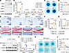 Figure 6
Activation of NOD2 by MDP alone is insufficient to drive cartilage degradation. (A and B) Western blot analysis and densitometric quantification of NOD2, MMP3, MMP13, and ADAMTS5 protein levels in primary mouse chondrocytes treated with MDP (0–10 μg/mL) for 72 h. n = 3 independent experiments. One-way ANOVA with Tukey’s multiple-comparison test. (C) Alcian blue staining of micromass cultures of primary mouse chondrocytes treated with MDP (0–10 μg/mL) for 2 weeks. (D) Quantification of proteoglycan content by absorbance of dissolved Alcian blue at 595 nm. n = 3 independent experiments. One-way ANOVA with Tukey’s multiple-comparison test. (E) IHC staining of knee sections for NOD2. Scale bar: 50 μm. (F) Quantification of positively stained chondrocytes for NOD2. n = 6 mice/group. One-way ANOVA with Tukey’s multiple-comparison test. (G) Safranin O/Fast Green staining of knee joint sections. Scale bar: 100 μm. (H) OARSI scores for cartilage degradation based on Safranin O/Fast Green staining. n = 6 mice/group. One-way ANOVA with Tukey’s multiple-comparison test. (I and J) Western blot analysis and densitometric quantification of MMP3 and ADAMTS5 protein levels following indicated concentrations of MDP and IL-1β treatment (0.1 ng/mL for 72 h). n = 3 independent experiments. One-way ANOVA with Tukey’s multiple-comparison test. (K) Alcian blue staining of micromass cultures of primary mouse chondrocytes treated with indicated concentrations of MDP and IL-1β treatment (0.1 ng/mL) for 2 weeks. (L) Quantification of proteoglycan content by absorbance of dissolved Alcian blue at 595 nm. n = 3 independent experiments. One-way ANOVA with Tukey’s multiple-comparison test. Data are presented as mean ± SD.
NOD2 amplifies inflammatory signals by regulating TRAF6 stability and ubiquitination. As an intracellular pattern recognition receptor, NOD2 is typically localized in the cytoplasm under basal conditions. However, its recruitment to membrane compartments is essential for initiating downstream inflammatory responses (27). In primary mouse chondrocytes treated with IL-1β, we observed rapid translocation of NOD2 to the plasma membrane, where it colocalized with the type I IL-1 receptor (IL-1R1), suggesting a potential role for NOD2 in initiating the IL-1β signaling cascade (Figure 7A). IL-1β signaling in chondrocytes involves the activation of NF-κB and MAPK pathways through shared kinase cascades involving tumor necrosis factor receptor–associated factor 6 (TRAF6), TGF-β–activated kinase 1 binding protein 1 (TAB1), and TGF-β–activated kinase 1 (TAK1) (28). To elucidate the role of NOD2 in this pathway, we analyzed key components downstream of IL-1β stimulation. Knockdown of Nod2 in primary chondrocytes led to reduced TRAF6 protein levels and decreased phosphorylation of TAB1 and TAK1. Consistently, activation of both NF-κB and MAPK pathways was suppressed, as shown by reduced phosphorylation of IKKs, P65, JNK, and P38 at 10 minutes after IL-1β treatment (Figure 7B). Since NOD2 protein levels did not markedly increase until 72 hours after IL-1β treatment (Figure 1, I and J), we hypothesized that the basal level of NOD2 is sufficient to mediate the early cellular response to IL-1β stimulation.
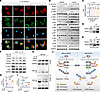 Figure 7
NOD2 amplifies inflammatory signals by regulating TRAF6 stability and ubiquitination. (A) Immunofluorescence images showing the subcellular localization of NOD2 and IL-1R1 in primary mouse chondrocytes stimulated with IL-1β (5 ng/mL) over the indicated time course. Scale bar: 10 μm. (B) Western blot analysis of NOD2 and IL-1β signaling pathway activation (TRAF6, p-TAB2, p-TAK1, p-IKK, p-p65, p-AKT, p-JNK, and p-p38) in primary mouse chondrocytes following Nod2 knockdown and IL-1β treatment (5 ng/mL) for the indicated durations. (C) qPCR analysis of Traf6 mRNA levels in primary mouse chondrocytes. n = 3 independent experiments. One-way ANOVA with Tukey’s multiple-comparison test. (D and E) Coimmunoprecipitation (Co-IP) and Western blot analysis of endogenous and exogenous interactions between TRAF6 and NOD2. (F) Western blot analysis of NOD2 and TRAF6 protein levels in chondrocytes treated with cycloheximide (CHX) and IL-1β (5 ng/mL), with or without Nod2 knockdown and MG132. (G) IP and Western blot analysis of TRAF6 ubiquitination status. (H and I) Densitometric quantification of K63-linked and K48-linked ubiquitination of TRAF6. n = 3 independent experiments. One-way ANOVA with Tukey’s multiple-comparison test. (J) Schematic illustration showing that NOD2 stabilizes TRAF6 and amplifies IL-1β signaling by modulating K63- and K48-linked ubiquitination. Data are presented as mean ± SD.
TRAF6 is a central adaptor protein involved in transducing signals from multiple inflammatory cytokines, including IL-1β and TNF-α, and functions upstream of several kinase cascades (29). We next examined whether NOD2 regulates IL-1β signaling by modulating TRAF6 activity. qPCR analysis confirmed that neither IL-1β stimulation nor Nod2 knockdown altered Traf6 mRNA expression (Figure 7C), suggesting posttranscriptional regulation. At the protein level, coimmunoprecipitation of both endogenous and exogenous proteins revealed that NOD2 interacts with TRAF6 upon IL-1β treatment (Figure 7, D and E). The activity and stability of TRAF6 are regulated by distinct ubiquitin linkages; K63-linked ubiquitination promotes signal transduction, while K48-linked ubiquitination targets TRAF6 for proteasomal degradation (30). To determine whether NOD2 influences TRAF6 stability, we treated cells with cycloheximide (CHX) to block new protein synthesis and MG132 to inhibit proteasomal degradation. Nod2 knockdown reduced IL-1β–induced TRAF6 protein levels, whereas this effect was negated in the presence of MG132, suggesting that NOD2 protects TRAF6 from proteasome-dependent degradation (Figure 7F). Notably, MG132-mediated restoration of TRAF6 protein levels failed to rescue downstream TAK1 activation, highlighting the critical role of proper posttranslational modification in enabling TRAF6 signaling. We next assessed the ubiquitination status of TRAF6. Following IL-1β stimulation in the presence of MG132, we immunoprecipitated TRAF6 and examined K63- and K48-linked ubiquitination. IL-1β increased K63-linked and decreased K48-linked ubiquitination of TRAF6, whereas Nod2 knockdown inhibited K63-linked ubiquitination (relative level: 1.83 ± 0.21 versus 2.35 ± 0.11, P = 0.0094) and enhanced K48-linked ubiquitination (relative level: 0.37 ± 0.03 versus 0.24 ± 0.02, P = 0.0220) of TRAF6 (Figure 7, G–I). Together, these findings demonstrate that NOD2 binds to TRAF6 upon IL-1β stimulation and promotes its activation by enhancing K63-linked ubiquitination while suppressing K48-linked degradation signals. This dual regulatory mechanism stabilizes TRAF6 and amplifies IL-1β induced inflammatory signaling in chondrocytes (Figure 7J).
NOD2 is a potential therapeutic target for OA. To rigorously assess the therapeutic potential of targeting NOD2 in established OA, we evaluated the effects of post-DMM intervention using 3 complementary approaches: adenovirus-mediated knockdown, inducible chondrocyte-specific knockout, and pharmacological inhibition with small-molecule antagonists. The mouse OA model was induced via DMM surgery, and all therapeutic interventions were initiated 1 week after DMM, a time point reflecting early-stage OA pathology. At 8 weeks after DMM, adenovirus-mediated Nod2 knockdown significantly attenuated further cartilage structural deterioration and suppressed the expression of key catabolic enzymes, including MMP3, MMP13, and ADAMTS5 (Supplemental Figure 2). Similarly, the specific KO of Nod2 in chondrocytes post-DMM yielded robust chondroprotective effects, characterized by preserved cartilage morphology (OARSI score: 7.17 ± 3.37 versus 15.50 ± 3.21, P < 0.0001) and reduced catabolic enzymes (Figure 8, A–J). Two independent inhibitors were tested: NOD-IN-1, a nonselective inhibitor targeting both NOD1 and NOD2, and GSK717, a selective NOD2 inhibitor (31, 32). The mouse OA model was induced via DMM surgery, and inhibitors were intraarticularly injected twice a week for 7 weeks after surgery. Histological analysis of knee sections using Safranin O/Fast Green staining revealed that both NOD-IN-1 and GSK717 significantly attenuated cartilage degradation compared with vehicle-treated controls. This was evidenced by increased cartilage thickness, reduced cartilage damage, and lower OARSI histological scores (7.00 ± 3.23 versus 14.50 ± 3.45, P = 0.0019 for NOD-IN-1 versus vehicle; 6.67 ± 3.67 versus 14.50 ± 3.45, P = 0.0012 for GSK717 versus vehicle) (Figure 8, K and L). These findings support NOD2 as a promising therapeutic target for the prevention and treatment of OA.
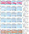 Figure 8
Post-DMM interventions targeting NOD2 attenuate OA development. (A) Safranin O/Fast Green staining of knee joint sections following DMM surgery and Nod2 KO. Scale bar: 100 μm. (B) OARSI scores for cartilage degradation based on Safranin O/Fast Green staining. n = 6 mice/group. One-way ANOVA with Tukey’s multiple-comparison test. (C) IHC staining of knee sections for NOD2. Scale bar: 100 μm. (D) Quantification of positively stained chondrocytes for NOD2. n = 6 mice/group. One-way ANOVA with Tukey’s multiple-comparison test. (E–J) IHC staining of knee sections and quantification of positively stained chondrocytes for MMP3, MMP13, and ADAMTS5. Scale bar: 100 μm. n = 6 mice/group. One-way ANOVA with Tukey’s multiple-comparison test. (K) Safranin O/Fast Green staining of knee joint sections following DMM surgery and NOD2 antagonists. Scale bar: 100 μm. (L) OARSI scores for cartilage degradation based on Safranin O/Fast Green staining. n = 6 mice/group. One-way ANOVA with Tukey’s multiple-comparison test. Data are presented as mean ± SD.


