
Research ArticleCell biologyOncology
Open Access |  10.1172/jci.insight.205126
RAD52 is a therapeutic vulnerability for acquired PARP inhibitor resistance in ovarian cancer
Yukihide Ota,1,2,3 Maia L. Elizagaray,1,2 Vijayalaxmi Gupta,1,2 Bisiayo E. Fashemi,1,2 Moreniola Akande,1,2 Preedia Babu,1,2 Prasanth Kumar Thuthika,1,2 Elena Lomonosova,1,2 Joshua Brill,1,2 Lulu Sun,4 Brooke Sanders,1 Lindsay M. Kuroki,1 Carolyn K. McCourt,1 Andrea R. Hagemann,1 Ian S. Hagemann,1,5 Premal H. Thaker,1 David G. Mutch,1 Matthew A. Powell,1 Krzysztof Hyrc,6 Priyanka Verma,5 John Krais,5 Benjamin G. Bitler,7 Mary M. Mullen,1,2 and Dineo Khabele1,2
1Division of Gynecologic Oncology, Department of Obstetrics and Gynecology, Alvin J. Siteman Cancer Center, Washington University School of Medicine, St. Louis, Missouri, USA.
2Center for Reproductive Health Sciences, Department of Obstetrics and Gynecology, Washington University, St. Louis, Missouri, USA.
3Department of Obstetrics and Gynecology, Yokohama City University Graduate School of Medicine, Yokohama, Kanagawa, Japan.
4Division of Anatomic and Molecular Pathology, Department of Pathology and Immunology, Washington University School of Medicine, St. Louis, Missouri, USA.
5Division of Oncology, Department of Medicine, Alvin J. Siteman Cancer Center, Washington University School of Medicine, St. Louis, Missouri, USA.
6Department of Neurology, Washington University School of Medicine, St. Louis, Missouri, USA.
7Division of Reproductive Sciences, University of Colorado, Aurora, Colorado, USA.
Address for correspondence. Dineo Khabele, The Mitchell and Elaine Yanow Professor and Chair of the Department of Obstetrics and Gynecology, Department of Obstetrics and Gynecology, Washington University in St. Louis School of Medicine, St. Louis, Missouri 63110, USA. Email: khabeled@wustl.edu.
Find articles by Ota, Y. in: PubMed | Google Scholar
1Division of Gynecologic Oncology, Department of Obstetrics and Gynecology, Alvin J. Siteman Cancer Center, Washington University School of Medicine, St. Louis, Missouri, USA.
2Center for Reproductive Health Sciences, Department of Obstetrics and Gynecology, Washington University, St. Louis, Missouri, USA.
3Department of Obstetrics and Gynecology, Yokohama City University Graduate School of Medicine, Yokohama, Kanagawa, Japan.
4Division of Anatomic and Molecular Pathology, Department of Pathology and Immunology, Washington University School of Medicine, St. Louis, Missouri, USA.
5Division of Oncology, Department of Medicine, Alvin J. Siteman Cancer Center, Washington University School of Medicine, St. Louis, Missouri, USA.
6Department of Neurology, Washington University School of Medicine, St. Louis, Missouri, USA.
7Division of Reproductive Sciences, University of Colorado, Aurora, Colorado, USA.
Address for correspondence. Dineo Khabele, The Mitchell and Elaine Yanow Professor and Chair of the Department of Obstetrics and Gynecology, Department of Obstetrics and Gynecology, Washington University in St. Louis School of Medicine, St. Louis, Missouri 63110, USA. Email: khabeled@wustl.edu.
Find articles by Elizagaray, M. in: PubMed | Google Scholar
1Division of Gynecologic Oncology, Department of Obstetrics and Gynecology, Alvin J. Siteman Cancer Center, Washington University School of Medicine, St. Louis, Missouri, USA.
2Center for Reproductive Health Sciences, Department of Obstetrics and Gynecology, Washington University, St. Louis, Missouri, USA.
3Department of Obstetrics and Gynecology, Yokohama City University Graduate School of Medicine, Yokohama, Kanagawa, Japan.
4Division of Anatomic and Molecular Pathology, Department of Pathology and Immunology, Washington University School of Medicine, St. Louis, Missouri, USA.
5Division of Oncology, Department of Medicine, Alvin J. Siteman Cancer Center, Washington University School of Medicine, St. Louis, Missouri, USA.
6Department of Neurology, Washington University School of Medicine, St. Louis, Missouri, USA.
7Division of Reproductive Sciences, University of Colorado, Aurora, Colorado, USA.
Address for correspondence. Dineo Khabele, The Mitchell and Elaine Yanow Professor and Chair of the Department of Obstetrics and Gynecology, Department of Obstetrics and Gynecology, Washington University in St. Louis School of Medicine, St. Louis, Missouri 63110, USA. Email: khabeled@wustl.edu.
Find articles by Gupta, V. in: PubMed | Google Scholar
1Division of Gynecologic Oncology, Department of Obstetrics and Gynecology, Alvin J. Siteman Cancer Center, Washington University School of Medicine, St. Louis, Missouri, USA.
2Center for Reproductive Health Sciences, Department of Obstetrics and Gynecology, Washington University, St. Louis, Missouri, USA.
3Department of Obstetrics and Gynecology, Yokohama City University Graduate School of Medicine, Yokohama, Kanagawa, Japan.
4Division of Anatomic and Molecular Pathology, Department of Pathology and Immunology, Washington University School of Medicine, St. Louis, Missouri, USA.
5Division of Oncology, Department of Medicine, Alvin J. Siteman Cancer Center, Washington University School of Medicine, St. Louis, Missouri, USA.
6Department of Neurology, Washington University School of Medicine, St. Louis, Missouri, USA.
7Division of Reproductive Sciences, University of Colorado, Aurora, Colorado, USA.
Address for correspondence. Dineo Khabele, The Mitchell and Elaine Yanow Professor and Chair of the Department of Obstetrics and Gynecology, Department of Obstetrics and Gynecology, Washington University in St. Louis School of Medicine, St. Louis, Missouri 63110, USA. Email: khabeled@wustl.edu.
Find articles by Fashemi, B. in: PubMed | Google Scholar
1Division of Gynecologic Oncology, Department of Obstetrics and Gynecology, Alvin J. Siteman Cancer Center, Washington University School of Medicine, St. Louis, Missouri, USA.
2Center for Reproductive Health Sciences, Department of Obstetrics and Gynecology, Washington University, St. Louis, Missouri, USA.
3Department of Obstetrics and Gynecology, Yokohama City University Graduate School of Medicine, Yokohama, Kanagawa, Japan.
4Division of Anatomic and Molecular Pathology, Department of Pathology and Immunology, Washington University School of Medicine, St. Louis, Missouri, USA.
5Division of Oncology, Department of Medicine, Alvin J. Siteman Cancer Center, Washington University School of Medicine, St. Louis, Missouri, USA.
6Department of Neurology, Washington University School of Medicine, St. Louis, Missouri, USA.
7Division of Reproductive Sciences, University of Colorado, Aurora, Colorado, USA.
Address for correspondence. Dineo Khabele, The Mitchell and Elaine Yanow Professor and Chair of the Department of Obstetrics and Gynecology, Department of Obstetrics and Gynecology, Washington University in St. Louis School of Medicine, St. Louis, Missouri 63110, USA. Email: khabeled@wustl.edu.
Find articles by Akande, M. in: PubMed | Google Scholar
1Division of Gynecologic Oncology, Department of Obstetrics and Gynecology, Alvin J. Siteman Cancer Center, Washington University School of Medicine, St. Louis, Missouri, USA.
2Center for Reproductive Health Sciences, Department of Obstetrics and Gynecology, Washington University, St. Louis, Missouri, USA.
3Department of Obstetrics and Gynecology, Yokohama City University Graduate School of Medicine, Yokohama, Kanagawa, Japan.
4Division of Anatomic and Molecular Pathology, Department of Pathology and Immunology, Washington University School of Medicine, St. Louis, Missouri, USA.
5Division of Oncology, Department of Medicine, Alvin J. Siteman Cancer Center, Washington University School of Medicine, St. Louis, Missouri, USA.
6Department of Neurology, Washington University School of Medicine, St. Louis, Missouri, USA.
7Division of Reproductive Sciences, University of Colorado, Aurora, Colorado, USA.
Address for correspondence. Dineo Khabele, The Mitchell and Elaine Yanow Professor and Chair of the Department of Obstetrics and Gynecology, Department of Obstetrics and Gynecology, Washington University in St. Louis School of Medicine, St. Louis, Missouri 63110, USA. Email: khabeled@wustl.edu.
Find articles by Babu, P. in: PubMed | Google Scholar
1Division of Gynecologic Oncology, Department of Obstetrics and Gynecology, Alvin J. Siteman Cancer Center, Washington University School of Medicine, St. Louis, Missouri, USA.
2Center for Reproductive Health Sciences, Department of Obstetrics and Gynecology, Washington University, St. Louis, Missouri, USA.
3Department of Obstetrics and Gynecology, Yokohama City University Graduate School of Medicine, Yokohama, Kanagawa, Japan.
4Division of Anatomic and Molecular Pathology, Department of Pathology and Immunology, Washington University School of Medicine, St. Louis, Missouri, USA.
5Division of Oncology, Department of Medicine, Alvin J. Siteman Cancer Center, Washington University School of Medicine, St. Louis, Missouri, USA.
6Department of Neurology, Washington University School of Medicine, St. Louis, Missouri, USA.
7Division of Reproductive Sciences, University of Colorado, Aurora, Colorado, USA.
Address for correspondence. Dineo Khabele, The Mitchell and Elaine Yanow Professor and Chair of the Department of Obstetrics and Gynecology, Department of Obstetrics and Gynecology, Washington University in St. Louis School of Medicine, St. Louis, Missouri 63110, USA. Email: khabeled@wustl.edu.
Find articles by Thuthika, P. in: PubMed | Google Scholar
1Division of Gynecologic Oncology, Department of Obstetrics and Gynecology, Alvin J. Siteman Cancer Center, Washington University School of Medicine, St. Louis, Missouri, USA.
2Center for Reproductive Health Sciences, Department of Obstetrics and Gynecology, Washington University, St. Louis, Missouri, USA.
3Department of Obstetrics and Gynecology, Yokohama City University Graduate School of Medicine, Yokohama, Kanagawa, Japan.
4Division of Anatomic and Molecular Pathology, Department of Pathology and Immunology, Washington University School of Medicine, St. Louis, Missouri, USA.
5Division of Oncology, Department of Medicine, Alvin J. Siteman Cancer Center, Washington University School of Medicine, St. Louis, Missouri, USA.
6Department of Neurology, Washington University School of Medicine, St. Louis, Missouri, USA.
7Division of Reproductive Sciences, University of Colorado, Aurora, Colorado, USA.
Address for correspondence. Dineo Khabele, The Mitchell and Elaine Yanow Professor and Chair of the Department of Obstetrics and Gynecology, Department of Obstetrics and Gynecology, Washington University in St. Louis School of Medicine, St. Louis, Missouri 63110, USA. Email: khabeled@wustl.edu.
Find articles by Lomonosova, E. in: PubMed | Google Scholar
1Division of Gynecologic Oncology, Department of Obstetrics and Gynecology, Alvin J. Siteman Cancer Center, Washington University School of Medicine, St. Louis, Missouri, USA.
2Center for Reproductive Health Sciences, Department of Obstetrics and Gynecology, Washington University, St. Louis, Missouri, USA.
3Department of Obstetrics and Gynecology, Yokohama City University Graduate School of Medicine, Yokohama, Kanagawa, Japan.
4Division of Anatomic and Molecular Pathology, Department of Pathology and Immunology, Washington University School of Medicine, St. Louis, Missouri, USA.
5Division of Oncology, Department of Medicine, Alvin J. Siteman Cancer Center, Washington University School of Medicine, St. Louis, Missouri, USA.
6Department of Neurology, Washington University School of Medicine, St. Louis, Missouri, USA.
7Division of Reproductive Sciences, University of Colorado, Aurora, Colorado, USA.
Address for correspondence. Dineo Khabele, The Mitchell and Elaine Yanow Professor and Chair of the Department of Obstetrics and Gynecology, Department of Obstetrics and Gynecology, Washington University in St. Louis School of Medicine, St. Louis, Missouri 63110, USA. Email: khabeled@wustl.edu.
Find articles by Brill, J. in: PubMed | Google Scholar
1Division of Gynecologic Oncology, Department of Obstetrics and Gynecology, Alvin J. Siteman Cancer Center, Washington University School of Medicine, St. Louis, Missouri, USA.
2Center for Reproductive Health Sciences, Department of Obstetrics and Gynecology, Washington University, St. Louis, Missouri, USA.
3Department of Obstetrics and Gynecology, Yokohama City University Graduate School of Medicine, Yokohama, Kanagawa, Japan.
4Division of Anatomic and Molecular Pathology, Department of Pathology and Immunology, Washington University School of Medicine, St. Louis, Missouri, USA.
5Division of Oncology, Department of Medicine, Alvin J. Siteman Cancer Center, Washington University School of Medicine, St. Louis, Missouri, USA.
6Department of Neurology, Washington University School of Medicine, St. Louis, Missouri, USA.
7Division of Reproductive Sciences, University of Colorado, Aurora, Colorado, USA.
Address for correspondence. Dineo Khabele, The Mitchell and Elaine Yanow Professor and Chair of the Department of Obstetrics and Gynecology, Department of Obstetrics and Gynecology, Washington University in St. Louis School of Medicine, St. Louis, Missouri 63110, USA. Email: khabeled@wustl.edu.
Find articles by
Sun, L.
in:
PubMed
|
Google Scholar
|

1Division of Gynecologic Oncology, Department of Obstetrics and Gynecology, Alvin J. Siteman Cancer Center, Washington University School of Medicine, St. Louis, Missouri, USA.
2Center for Reproductive Health Sciences, Department of Obstetrics and Gynecology, Washington University, St. Louis, Missouri, USA.
3Department of Obstetrics and Gynecology, Yokohama City University Graduate School of Medicine, Yokohama, Kanagawa, Japan.
4Division of Anatomic and Molecular Pathology, Department of Pathology and Immunology, Washington University School of Medicine, St. Louis, Missouri, USA.
5Division of Oncology, Department of Medicine, Alvin J. Siteman Cancer Center, Washington University School of Medicine, St. Louis, Missouri, USA.
6Department of Neurology, Washington University School of Medicine, St. Louis, Missouri, USA.
7Division of Reproductive Sciences, University of Colorado, Aurora, Colorado, USA.
Address for correspondence. Dineo Khabele, The Mitchell and Elaine Yanow Professor and Chair of the Department of Obstetrics and Gynecology, Department of Obstetrics and Gynecology, Washington University in St. Louis School of Medicine, St. Louis, Missouri 63110, USA. Email: khabeled@wustl.edu.
Find articles by Sanders, B. in: PubMed | Google Scholar
1Division of Gynecologic Oncology, Department of Obstetrics and Gynecology, Alvin J. Siteman Cancer Center, Washington University School of Medicine, St. Louis, Missouri, USA.
2Center for Reproductive Health Sciences, Department of Obstetrics and Gynecology, Washington University, St. Louis, Missouri, USA.
3Department of Obstetrics and Gynecology, Yokohama City University Graduate School of Medicine, Yokohama, Kanagawa, Japan.
4Division of Anatomic and Molecular Pathology, Department of Pathology and Immunology, Washington University School of Medicine, St. Louis, Missouri, USA.
5Division of Oncology, Department of Medicine, Alvin J. Siteman Cancer Center, Washington University School of Medicine, St. Louis, Missouri, USA.
6Department of Neurology, Washington University School of Medicine, St. Louis, Missouri, USA.
7Division of Reproductive Sciences, University of Colorado, Aurora, Colorado, USA.
Address for correspondence. Dineo Khabele, The Mitchell and Elaine Yanow Professor and Chair of the Department of Obstetrics and Gynecology, Department of Obstetrics and Gynecology, Washington University in St. Louis School of Medicine, St. Louis, Missouri 63110, USA. Email: khabeled@wustl.edu.
Find articles by Kuroki, L. in: PubMed | Google Scholar
1Division of Gynecologic Oncology, Department of Obstetrics and Gynecology, Alvin J. Siteman Cancer Center, Washington University School of Medicine, St. Louis, Missouri, USA.
2Center for Reproductive Health Sciences, Department of Obstetrics and Gynecology, Washington University, St. Louis, Missouri, USA.
3Department of Obstetrics and Gynecology, Yokohama City University Graduate School of Medicine, Yokohama, Kanagawa, Japan.
4Division of Anatomic and Molecular Pathology, Department of Pathology and Immunology, Washington University School of Medicine, St. Louis, Missouri, USA.
5Division of Oncology, Department of Medicine, Alvin J. Siteman Cancer Center, Washington University School of Medicine, St. Louis, Missouri, USA.
6Department of Neurology, Washington University School of Medicine, St. Louis, Missouri, USA.
7Division of Reproductive Sciences, University of Colorado, Aurora, Colorado, USA.
Address for correspondence. Dineo Khabele, The Mitchell and Elaine Yanow Professor and Chair of the Department of Obstetrics and Gynecology, Department of Obstetrics and Gynecology, Washington University in St. Louis School of Medicine, St. Louis, Missouri 63110, USA. Email: khabeled@wustl.edu.
Find articles by McCourt, C. in: PubMed | Google Scholar
1Division of Gynecologic Oncology, Department of Obstetrics and Gynecology, Alvin J. Siteman Cancer Center, Washington University School of Medicine, St. Louis, Missouri, USA.
2Center for Reproductive Health Sciences, Department of Obstetrics and Gynecology, Washington University, St. Louis, Missouri, USA.
3Department of Obstetrics and Gynecology, Yokohama City University Graduate School of Medicine, Yokohama, Kanagawa, Japan.
4Division of Anatomic and Molecular Pathology, Department of Pathology and Immunology, Washington University School of Medicine, St. Louis, Missouri, USA.
5Division of Oncology, Department of Medicine, Alvin J. Siteman Cancer Center, Washington University School of Medicine, St. Louis, Missouri, USA.
6Department of Neurology, Washington University School of Medicine, St. Louis, Missouri, USA.
7Division of Reproductive Sciences, University of Colorado, Aurora, Colorado, USA.
Address for correspondence. Dineo Khabele, The Mitchell and Elaine Yanow Professor and Chair of the Department of Obstetrics and Gynecology, Department of Obstetrics and Gynecology, Washington University in St. Louis School of Medicine, St. Louis, Missouri 63110, USA. Email: khabeled@wustl.edu.
Find articles by Hagemann, A. in: PubMed | Google Scholar
1Division of Gynecologic Oncology, Department of Obstetrics and Gynecology, Alvin J. Siteman Cancer Center, Washington University School of Medicine, St. Louis, Missouri, USA.
2Center for Reproductive Health Sciences, Department of Obstetrics and Gynecology, Washington University, St. Louis, Missouri, USA.
3Department of Obstetrics and Gynecology, Yokohama City University Graduate School of Medicine, Yokohama, Kanagawa, Japan.
4Division of Anatomic and Molecular Pathology, Department of Pathology and Immunology, Washington University School of Medicine, St. Louis, Missouri, USA.
5Division of Oncology, Department of Medicine, Alvin J. Siteman Cancer Center, Washington University School of Medicine, St. Louis, Missouri, USA.
6Department of Neurology, Washington University School of Medicine, St. Louis, Missouri, USA.
7Division of Reproductive Sciences, University of Colorado, Aurora, Colorado, USA.
Address for correspondence. Dineo Khabele, The Mitchell and Elaine Yanow Professor and Chair of the Department of Obstetrics and Gynecology, Department of Obstetrics and Gynecology, Washington University in St. Louis School of Medicine, St. Louis, Missouri 63110, USA. Email: khabeled@wustl.edu.
Find articles by
Hagemann, I.
in:
PubMed
|
Google Scholar
|
1Division of Gynecologic Oncology, Department of Obstetrics and Gynecology, Alvin J. Siteman Cancer Center, Washington University School of Medicine, St. Louis, Missouri, USA.
2Center for Reproductive Health Sciences, Department of Obstetrics and Gynecology, Washington University, St. Louis, Missouri, USA.
3Department of Obstetrics and Gynecology, Yokohama City University Graduate School of Medicine, Yokohama, Kanagawa, Japan.
4Division of Anatomic and Molecular Pathology, Department of Pathology and Immunology, Washington University School of Medicine, St. Louis, Missouri, USA.
5Division of Oncology, Department of Medicine, Alvin J. Siteman Cancer Center, Washington University School of Medicine, St. Louis, Missouri, USA.
6Department of Neurology, Washington University School of Medicine, St. Louis, Missouri, USA.
7Division of Reproductive Sciences, University of Colorado, Aurora, Colorado, USA.
Address for correspondence. Dineo Khabele, The Mitchell and Elaine Yanow Professor and Chair of the Department of Obstetrics and Gynecology, Department of Obstetrics and Gynecology, Washington University in St. Louis School of Medicine, St. Louis, Missouri 63110, USA. Email: khabeled@wustl.edu.
Find articles by Thaker, P. in: PubMed | Google Scholar
1Division of Gynecologic Oncology, Department of Obstetrics and Gynecology, Alvin J. Siteman Cancer Center, Washington University School of Medicine, St. Louis, Missouri, USA.
2Center for Reproductive Health Sciences, Department of Obstetrics and Gynecology, Washington University, St. Louis, Missouri, USA.
3Department of Obstetrics and Gynecology, Yokohama City University Graduate School of Medicine, Yokohama, Kanagawa, Japan.
4Division of Anatomic and Molecular Pathology, Department of Pathology and Immunology, Washington University School of Medicine, St. Louis, Missouri, USA.
5Division of Oncology, Department of Medicine, Alvin J. Siteman Cancer Center, Washington University School of Medicine, St. Louis, Missouri, USA.
6Department of Neurology, Washington University School of Medicine, St. Louis, Missouri, USA.
7Division of Reproductive Sciences, University of Colorado, Aurora, Colorado, USA.
Address for correspondence. Dineo Khabele, The Mitchell and Elaine Yanow Professor and Chair of the Department of Obstetrics and Gynecology, Department of Obstetrics and Gynecology, Washington University in St. Louis School of Medicine, St. Louis, Missouri 63110, USA. Email: khabeled@wustl.edu.
Find articles by Mutch, D. in: PubMed | Google Scholar
1Division of Gynecologic Oncology, Department of Obstetrics and Gynecology, Alvin J. Siteman Cancer Center, Washington University School of Medicine, St. Louis, Missouri, USA.
2Center for Reproductive Health Sciences, Department of Obstetrics and Gynecology, Washington University, St. Louis, Missouri, USA.
3Department of Obstetrics and Gynecology, Yokohama City University Graduate School of Medicine, Yokohama, Kanagawa, Japan.
4Division of Anatomic and Molecular Pathology, Department of Pathology and Immunology, Washington University School of Medicine, St. Louis, Missouri, USA.
5Division of Oncology, Department of Medicine, Alvin J. Siteman Cancer Center, Washington University School of Medicine, St. Louis, Missouri, USA.
6Department of Neurology, Washington University School of Medicine, St. Louis, Missouri, USA.
7Division of Reproductive Sciences, University of Colorado, Aurora, Colorado, USA.
Address for correspondence. Dineo Khabele, The Mitchell and Elaine Yanow Professor and Chair of the Department of Obstetrics and Gynecology, Department of Obstetrics and Gynecology, Washington University in St. Louis School of Medicine, St. Louis, Missouri 63110, USA. Email: khabeled@wustl.edu.
Find articles by Powell, M. in: PubMed | Google Scholar
1Division of Gynecologic Oncology, Department of Obstetrics and Gynecology, Alvin J. Siteman Cancer Center, Washington University School of Medicine, St. Louis, Missouri, USA.
2Center for Reproductive Health Sciences, Department of Obstetrics and Gynecology, Washington University, St. Louis, Missouri, USA.
3Department of Obstetrics and Gynecology, Yokohama City University Graduate School of Medicine, Yokohama, Kanagawa, Japan.
4Division of Anatomic and Molecular Pathology, Department of Pathology and Immunology, Washington University School of Medicine, St. Louis, Missouri, USA.
5Division of Oncology, Department of Medicine, Alvin J. Siteman Cancer Center, Washington University School of Medicine, St. Louis, Missouri, USA.
6Department of Neurology, Washington University School of Medicine, St. Louis, Missouri, USA.
7Division of Reproductive Sciences, University of Colorado, Aurora, Colorado, USA.
Address for correspondence. Dineo Khabele, The Mitchell and Elaine Yanow Professor and Chair of the Department of Obstetrics and Gynecology, Department of Obstetrics and Gynecology, Washington University in St. Louis School of Medicine, St. Louis, Missouri 63110, USA. Email: khabeled@wustl.edu.
Find articles by Hyrc, K. in: PubMed | Google Scholar
1Division of Gynecologic Oncology, Department of Obstetrics and Gynecology, Alvin J. Siteman Cancer Center, Washington University School of Medicine, St. Louis, Missouri, USA.
2Center for Reproductive Health Sciences, Department of Obstetrics and Gynecology, Washington University, St. Louis, Missouri, USA.
3Department of Obstetrics and Gynecology, Yokohama City University Graduate School of Medicine, Yokohama, Kanagawa, Japan.
4Division of Anatomic and Molecular Pathology, Department of Pathology and Immunology, Washington University School of Medicine, St. Louis, Missouri, USA.
5Division of Oncology, Department of Medicine, Alvin J. Siteman Cancer Center, Washington University School of Medicine, St. Louis, Missouri, USA.
6Department of Neurology, Washington University School of Medicine, St. Louis, Missouri, USA.
7Division of Reproductive Sciences, University of Colorado, Aurora, Colorado, USA.
Address for correspondence. Dineo Khabele, The Mitchell and Elaine Yanow Professor and Chair of the Department of Obstetrics and Gynecology, Department of Obstetrics and Gynecology, Washington University in St. Louis School of Medicine, St. Louis, Missouri 63110, USA. Email: khabeled@wustl.edu.
Find articles by Verma, P. in: PubMed | Google Scholar
1Division of Gynecologic Oncology, Department of Obstetrics and Gynecology, Alvin J. Siteman Cancer Center, Washington University School of Medicine, St. Louis, Missouri, USA.
2Center for Reproductive Health Sciences, Department of Obstetrics and Gynecology, Washington University, St. Louis, Missouri, USA.
3Department of Obstetrics and Gynecology, Yokohama City University Graduate School of Medicine, Yokohama, Kanagawa, Japan.
4Division of Anatomic and Molecular Pathology, Department of Pathology and Immunology, Washington University School of Medicine, St. Louis, Missouri, USA.
5Division of Oncology, Department of Medicine, Alvin J. Siteman Cancer Center, Washington University School of Medicine, St. Louis, Missouri, USA.
6Department of Neurology, Washington University School of Medicine, St. Louis, Missouri, USA.
7Division of Reproductive Sciences, University of Colorado, Aurora, Colorado, USA.
Address for correspondence. Dineo Khabele, The Mitchell and Elaine Yanow Professor and Chair of the Department of Obstetrics and Gynecology, Department of Obstetrics and Gynecology, Washington University in St. Louis School of Medicine, St. Louis, Missouri 63110, USA. Email: khabeled@wustl.edu.
Find articles by Krais, J. in: PubMed | Google Scholar
1Division of Gynecologic Oncology, Department of Obstetrics and Gynecology, Alvin J. Siteman Cancer Center, Washington University School of Medicine, St. Louis, Missouri, USA.
2Center for Reproductive Health Sciences, Department of Obstetrics and Gynecology, Washington University, St. Louis, Missouri, USA.
3Department of Obstetrics and Gynecology, Yokohama City University Graduate School of Medicine, Yokohama, Kanagawa, Japan.
4Division of Anatomic and Molecular Pathology, Department of Pathology and Immunology, Washington University School of Medicine, St. Louis, Missouri, USA.
5Division of Oncology, Department of Medicine, Alvin J. Siteman Cancer Center, Washington University School of Medicine, St. Louis, Missouri, USA.
6Department of Neurology, Washington University School of Medicine, St. Louis, Missouri, USA.
7Division of Reproductive Sciences, University of Colorado, Aurora, Colorado, USA.
Address for correspondence. Dineo Khabele, The Mitchell and Elaine Yanow Professor and Chair of the Department of Obstetrics and Gynecology, Department of Obstetrics and Gynecology, Washington University in St. Louis School of Medicine, St. Louis, Missouri 63110, USA. Email: khabeled@wustl.edu.
Find articles by Bitler, B. in: PubMed | Google Scholar
1Division of Gynecologic Oncology, Department of Obstetrics and Gynecology, Alvin J. Siteman Cancer Center, Washington University School of Medicine, St. Louis, Missouri, USA.
2Center for Reproductive Health Sciences, Department of Obstetrics and Gynecology, Washington University, St. Louis, Missouri, USA.
3Department of Obstetrics and Gynecology, Yokohama City University Graduate School of Medicine, Yokohama, Kanagawa, Japan.
4Division of Anatomic and Molecular Pathology, Department of Pathology and Immunology, Washington University School of Medicine, St. Louis, Missouri, USA.
5Division of Oncology, Department of Medicine, Alvin J. Siteman Cancer Center, Washington University School of Medicine, St. Louis, Missouri, USA.
6Department of Neurology, Washington University School of Medicine, St. Louis, Missouri, USA.
7Division of Reproductive Sciences, University of Colorado, Aurora, Colorado, USA.
Address for correspondence. Dineo Khabele, The Mitchell and Elaine Yanow Professor and Chair of the Department of Obstetrics and Gynecology, Department of Obstetrics and Gynecology, Washington University in St. Louis School of Medicine, St. Louis, Missouri 63110, USA. Email: khabeled@wustl.edu.
Find articles by Mullen, M. in: PubMed | Google Scholar
1Division of Gynecologic Oncology, Department of Obstetrics and Gynecology, Alvin J. Siteman Cancer Center, Washington University School of Medicine, St. Louis, Missouri, USA.
2Center for Reproductive Health Sciences, Department of Obstetrics and Gynecology, Washington University, St. Louis, Missouri, USA.
3Department of Obstetrics and Gynecology, Yokohama City University Graduate School of Medicine, Yokohama, Kanagawa, Japan.
4Division of Anatomic and Molecular Pathology, Department of Pathology and Immunology, Washington University School of Medicine, St. Louis, Missouri, USA.
5Division of Oncology, Department of Medicine, Alvin J. Siteman Cancer Center, Washington University School of Medicine, St. Louis, Missouri, USA.
6Department of Neurology, Washington University School of Medicine, St. Louis, Missouri, USA.
7Division of Reproductive Sciences, University of Colorado, Aurora, Colorado, USA.
Address for correspondence. Dineo Khabele, The Mitchell and Elaine Yanow Professor and Chair of the Department of Obstetrics and Gynecology, Department of Obstetrics and Gynecology, Washington University in St. Louis School of Medicine, St. Louis, Missouri 63110, USA. Email: khabeled@wustl.edu.
Find articles by Khabele, D. in: PubMed | Google Scholar
Published November 9, 2026 - More info
JCI Insight. 2026;11(21):e205126. https://doi.org/10.1172/jci.insight.205126.
© 2026 Ota et al. This work is licensed under the Creative Commons Attribution 4.0 International License. To view a copy of this license, visit http://creativecommons.org/licenses/by/4.0/.
Received: February 23, 2026; Accepted: September 24, 2026

-
Results
A Brca2-mutated PARPi-resistant mouse ovarian cancer cell line exhibits increased HR and SSA activity and elevated RAD52 expression. We analyzed 2 olaparib-resistant (OR) mouse ovarian cancer cell lines, HGS2-OR and ID8-OR, which were derived from the parental HGS2 (Trp53−/−, Brca2−/−, Pten−/−) and ID8-PB (Trp53−/−, Brca2−/−) cell lines (37, 38). Both OR cell lines showed a significantly higher olaparib IC50 value than their parental counterparts (Figure 1A). Next-generation sequencing confirmed that HGS2-OR retained the >4-kb deletion in Brca2 exon 11, and ID8-OR retained the exon 3 deletion and frameshift (Supplemental Figure 1; supplemental material available online with this article; https://doi.org/10.1172/jci.insight.205126DS1), demonstrating that the acquired PARPi resistance was not due to Brca2 reversion.
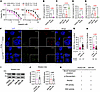 Figure 1
PARP inhibitor–resistant HGS2-OR cells exhibit increased HR/SSA repair activity and RAD52 upregulation. (A) Viability assays of parental (HGS2, ID8-PB) and olaparib-resistant (HGS2-OR, ID8-OR) ovarian cancer cells after 5 days of olaparib treatment. (B and C) HR activity in (B) HGS2-OR and HGS2 cells and (C) ID8-OR, ID8-PB, and ID8-P cells, normalized to the corresponding OR controls. (D and E) SSA activity in (D) HGS2-OR and HGS2 cells and (E) ID8-OR, ID8-PB, and ID8-P cells, normalized to the corresponding OR controls. (F) Representative immunofluorescence images showing RAD51 and γH2AX foci in HGS2 and HGS2-OR cells 4 hours after 10 Gy irradiation. Scale bar size: 10µm. Magnification insets: ×1.66. (G) Quantification of nuclear RAD51 foci. (H) Quantification of nuclear γH2AX foci. (I) Western blot analysis of RAD52 protein. (J) Quantification of RAD52 expression relative to β-actin and normalized to parental cells. (K) Summary of characteristics of cell lines. Data represent mean ± SD from ≥3 independent experiments; individual data points reflect biological replicates. Statistical analyses were performed using 2-sided Student’s t test (A, B, D, and J), 1-way ANOVA followed by Šídák’s correction (C and E), or Mann-Whitney test (G and H). Black and red numbers above graphs are non-significant and significant, respectively, P values. HR, homologous recombination; SSA, single-strand annealing.
To assess the relative contributions of upregulated DNA repair pathways to the development of acquired PARPi resistance in both models, we used reporter assays to measure HR and SSA activity. HGS2-OR cells exhibited significantly higher HR activity than HGS2 cells (Figure 1B and Supplemental Figure 2). ID8-OR cells had similar HR activity as ID8-PB cells and lower HR activity than ID8-P cells, an HR-proficient control (Figure 1C and Supplemental Figure 2). SSA activity was also significantly elevated in HGS2-OR cells but not in ID8-OR cells (Figure 1, D and E). After 10 Gy irradiation, HGS2-OR cells formed more RAD51 foci and fewer γH2AX foci than HGS2 cells, suggesting partial restoration of RAD51-mediated repair activity (Figure 1, F–H, and Supplemental Figure 3). Given that RAD52 can provide compensatory repair capacity through HR and SSA, we investigated RAD52 expression in both models. HGS2-OR cells, but not ID8-OR cells, had elevated RAD52 protein and mRNA (Figure 1, I and J, and Supplemental Figure 3). We assessed other methods of acquired resistance and found that both HGS2-OR and ID8-OR cells displayed elevated expression of P-glycoprotein, a common efflux-based resistance mechanism (Supplemental Figure 3). Conversely, expression of PAF1, which functionally cooperates with RAD52 to promote DNA repair capacity and contributes to therapeutic resistance (36), was non-significantly higher in HGS2-OR cells than in HGS2 cells (Supplemental Figure 3). Thus, although both models of acquired PARPi resistance had upregulation of P-glycoprotein, only HGS2-OR cells exhibited increased HR and SSA activity and elevated RAD52 expression. Characteristics of the cell models are summarized in Figure 1K.
Genetic depletion of Rad52 re-sensitizes Brca2-deficient mouse ovarian cancer cells to olaparib. To further evaluate RAD52 as a therapeutic target, we used short hairpin RNA (shRNA) to generate a stable Rad52 knockdown clone (KD#1) in HGS2-OR cells, and we used CRISPR/Cas9 to generate 2 Rad52-knockout clones (KO#1 and KO#2) in ID8-OR cells (Supplemental Figure 4). Depletion efficacy was confirmed by Western blotting, qRT-PCR, and amplicon sequencing (Figure 2, A–C, and Supplemental Figure 4). Rad52-depleted cells had significantly lower IC50 values and formed fewer colonies during olaparib treatment than scrambled (Scr) controls (Figure 2, D–G). HR and SSA activities were significantly decreased in all Rad52-depleted clones (Figure 2, H–K). After 72 hours of olaparib exposure, Rad52-KO ID8 cells accumulated significantly more γH2AX foci than Scr cells (Figure 2L and Supplemental Figure 5). Western blot analysis further confirmed olaparib-induced γH2AX elevation specifically in Rad52-deficient cells (Supplemental Figure 5). These findings demonstrate that Rad52 loss impairs HR and SSA capacity, leads to accumulation of DNA damage, and restores PARPi sensitivity in both Brca2-deficient, PARPi-resistant mouse HGSC cell lines.
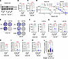 Figure 2
Genetic depletion of Rad52 re-sensitizes BRCA2-deficient ovarian cancer cells to olaparib. (A) Western blot validation of Rad52 knockdown in HGS2-OR and Rad52 knockout in ID8-OR cells. (B and C) qRT-PCR of Rad52 mRNA in HGS2-OR (B) and ID8-OR (C) cells. (D) Cell viability after 5 days of treatment with 0–20 μM olaparib. (E–G) Clonogenic assay and quantification. (E) Representative images after 8 days of 5 μM olaparib treatment. (J and G) Quantification of clonogenic survival in HGS2-OR (F) and ID8-OR (G) cells. (H and I) HR activity in HGS2-OR (H) and ID8-OR (I) cells. (J and K) SSA activity in HGS2-OR (J) and ID8-OR (K) cells. (L) Quantification of γH2AX foci after 72 hours of 5 μM olaparib. Data represent mean ± SD from ≥3 independent experiments, with individual data points shown. Two-sided Student’s t tests were used for 2-group comparisons (B, D, F, H, and J). One-way ANOVA with Šídák’s multiple-comparison test was used for 3-group comparisons (C, G, I, and K). Kruskal-Wallis followed by Dunn’s test was used for nonparametric data (L). Black and red numbers above graphs are non-significant and significant, respectively, P values. HR, homologous recombination; Ola, olaparib; SSA, single-strand annealing; Veh, vehicle.
Pharmacological RAD52 inhibition synergizes with olaparib to overcome PARPi resistance in mouse HGSC cells. To test whether pharmacological inhibition of RAD52 can overcome PARPi resistance, we treated cells with the selective small-molecule RAD52 inhibitor D-I03, which blocks RAD52-dependent SSA and D-loop formation (39). D-I03 significantly lowered olaparib IC50 values in both HGS2-OR and ID8-OR cells (Figure 3A and Supplemental Figure 6). Synergy between D-I03 and olaparib was demonstrated in both BLISS and HSA analysis (Figure 3B). Similar results were observed with mitoxantrone, an RPA-RAD52 interaction inhibitor (Supplemental Figure 6) (40). In clonogenic assays, D-I03 potentiated olaparib-induced suppression of colony formation in both resistant cell lines, whereas parental HGS2 and ID8-PB cells were intrinsically sensitive to olaparib alone (Figure 3, C–F). These data indicate that pharmacologic inhibition of RAD52 enhances the cytotoxic effects of olaparib in PARPi-resistant, Brca2-deficient mouse ovarian cancer models in vitro.
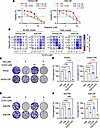 Figure 3
Pharmacological RAD52 inhibition synergizes with olaparib to overcome PARPi resistance. (A) Viability assay of cells treated with olaparib alone or in combination with D-I03 (2.5 μM for HGS2-OR, 1 μM for ID8-OR) for 5 days. (B) BLISS and HSA model of olaparib and D-I03 in HGS2-OR and ID8-OR cells. (C) Clonogenicity assay of HGS2, and HGS2-OR cells treated with the indicated concentrations of olaparib and D-I03. (D) Quantification of clonogenic assay results. (E) Clonogenicity assay of ID8-PB, and ID8-OR cells treated with the indicated concentrations of olaparib and D-I03. (F) Quantification of clonogenic assay results. Data represent mean ± SD from ≥3 independent experiments, with individual data points shown. One-way ANOVA with Šídák’s multiple-comparison test was used (D and F). Black and red numbers above graphs are non-significant and significant, respectively, P values. Ola, olaparib; Veh, vehicle.
RAD52 inhibition suppresses tumor progression and increases susceptibility to olaparib in mice. To determine whether targeting RAD52 confers benefit in vivo, we intraperitoneally injected ID8-Scr or ID8-KO#1 cells into mice, then treated the mice with daily olaparib or vehicle for 4 weeks (Figure 4A). Mice that received ID8-KO#1 cells developed lower tumor weight, nodule number, and ascites volume in both vehicle and olaparib groups (Figure 4, B–E, and Supplemental Figure 7). ID8-KO#1 tumors also displayed increased γH2AX positivity at baseline and further elevation with olaparib (Figure 4F and Supplemental Figure 7). Survival was significantly longer in mice bearing ID8-KO#1 tumors than in mice with ID8-Scr tumors (Figure 4G).
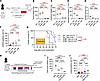 Figure 4
Genetic and pharmacological RAD52 inhibition suppresses tumor growth in a PARPi-resistant Brca2-deficient ID8-OR murine model. (A) Experimental scheme for the intraperitoneal xenograft model using ID8-OR–Scr and ID8-OR–Rad52-KO#1 cells. (B–E) Tumor burden metrics: (B) total tumor weight excluding the omentum, (C) weight of the omentum (including the pancreas and surrounding adipose tissue), (D) number of intraperitoneal tumor nodules (≥1 mm), and (E) ascites volume (n = 8 mice/group). (F) Quantification of γH2AX-positive nuclei (intensity > 2+ or 3+) in intraperitoneal tumor sections by immunohistochemistry (IHC). (G) Kaplan-Meier survival analysis comparing Scr-Veh, Scr-Ola, KO#1-Veh, and KO#1-Ola groups. A global log-rank test was followed by pairwise comparisons among 3 groups (Bonferroni-adjusted significance threshold P < 0.0167). Median survival (days): Scr-Veh 49, Scr-Ola 46.5, KO#1-Veh 67, KO#1-Ola not reached. (H) Experimental scheme for pharmacologic RAD52 inhibition. (I) Tumor weights. (J) Quantification of γH2AX-positive nuclei (intensity > 2+ or 3+) in intraperitoneal tumor sections by IHC. Data show mean ± SD of at least 3 independent experiments, with individual dots for each replicate. P values were determined by 1-way ANOVA followed by Šídák’s multiple-comparison test (B–F, and I). Black and red numbers above graphs are non-significant and significant, respectively, P values. i.p, intraperitoneal injection; Ola, olaparib; Veh, vehicle.
To test the effects of pharmacologic RAD52 inhibition in vivo, mice bearing ID8-OR tumors were treated with olaparib, D-I03, the combination, or vehicle for 20 days (Figure 4H). Two early deaths in the D-I03 and combination groups were attributed to treatment-related toxicity (Supplemental Figure 7). At treatment completion, combination therapy resulted in significantly lower tumor burden and greater γH2AX positivity than treatment with vehicle (Figure 4, I and J, and Supplemental Figure 7). Survival was modestly prolonged (Supplemental Figure 7). Together, these findings demonstrate that both genetic and pharmacologic RAD52 inhibition enhance antitumor responses to PARPi treatment in vivo, likely by promoting accumulation of unrepaired DNA damage.
Polq expression and MMEJ activity are upregulated in Rad52-KO mouse HGSC cells treated with olaparib. To examine how Rad52 depletion alters the response to PARPi treatment, we performed bulk RNA-seq of ID8-OR Scr and ID8-KO#1 cells treated with 1 μM olaparib or vehicle for 24 hours (Supplemental Figure 8). ID8-KO#1 cells treated with olaparib exhibited broad transcriptional changes, with 914 upregulated genes and 1,038 downregulated genes relative to Scr controls (Figure 5A). Among DNA-repair-related genes, several transcripts were differentially expressed, including increased Polq, Xrcc3, and Fen1, and decreased Rad51c, Pold4, and Brcc3 (Figure 5A). Z-score analysis across all 4 groups showed that Polq expression was higher in olaparib-treated ID8-KO#1 cells than in olaparib-treated Scr controls (Figure 5B). Consistent with this, qRT-PCR revealed an approximately 1.61-fold elevation in Polq expression in olaparib-treated KO#1 cells (Figure 5C and Supplemental Figure 8). To investigate the functional role of this observation, we performed a microhomology-mediated end-joining/DNA polymerase theta–mediated end joining (MMEJ/TMEJ) and found that ID8-KO#1 cells had higher MMEJ activity than Scr control cells. Using ART558, a POLQ inhibitor, we confirmed that this activity is mediated by this enzyme (Supplemental Figure 9A).
 Figure 5
Polq expression is upregulated in Rad52-KO cells treated with olaparib. (A) Volcano plot showing differentially expressed genes (DEGs) between ID8-OR–Scr and ID8-OR–Rad52-KO#1 cells treated with 5 μM olaparib. Positive log2(fold change) values indicate higher expression in Rad52-KO#1 cells than in Scr cells. DEGs were defined as those with |log2 fold change)| > 0.5 and FDR < 0.05. DNA repair–related DEGs are highlighted. (B) Z-scores of Polq expression in Scr and Rad52-KO#1 cells treated with vehicle or olaparib. (C) qRT-PCR analysis of Polq expression in Scr and Rad52-KO#1 cells following olaparib treatment. Expression was normalized to Actb and presented relative to Scr-olaparib. Red number above the graph in C is a significant P value.
Taken together, these findings indicate that olaparib-treated Rad52-deficient cells upregulate Polq, potentially as a compensatory response.
RAD52 is elevated and targetable in a human BRCA2-mutated PARPi-resistant ovarian cancer model. To test RAD52 inhibition in a human model of HGSC with acquired PARPi resistance, we generated olaparib-resistant PEO1 (PEO1-OR) cells, which exhibited a higher olaparib IC50 than parental PEO1 cells (Figure 6A). Targeted sequencing demonstrated that our parental PEO1 culture contained both the original BRCA2 nonsense mutation (c.4965C>G, p.Y1655*) and a previously described secondary reversion mutation (c.[4964A>T;4965C>G], p.Y1655L), indicating a heterogeneous population. In PEO1-OR cells, both alleles were present, and no additional BRCA2 reversion alleles were detected. PEO1-OR cells also exhibited increased RAD52 expression (Figure 6, B and C). In both BLISS and HSA synergy models, olaparib synergized with D-I03 in PEO1-OR cells (Figure 6D and Supplemental Figure 6).
 Figure 6
RAD52 is elevated and targetable in a human BRCA2-mutated PARP inhibitor resistant ovarian cancer model. (A) Viability assays of parental (PEO1) and olaparib-resistant (PEO1-OR) human ovarian cancer cells after 5 days of olaparib treatment. (B) Western blot analysis of RAD52 protein. (C) Quantification of RAD52 protein relative to β-actin and normalized to parental cells. (D) BLISS and HSA models of olaparib and D-I03 in PEO1-OR. Data represent mean ± SD from ≥3 independent experiments; individual data points reflect biological replicates. Statistical analyses were performed using 2-sided Student’s t test, and the red number above the graph in C is a significant P value.
To test whether our strategy could overcome resistance mediated by restoration of HR, we used the patient-derived PEO4 cell line, which models acquired resistance through a functional BRCA2 reversion mutation (41). Although olaparib and D-I03 IC50 values were higher for PEO4 cells than for PEO1 cells (Supplemental Figure 10), we found minimal synergy with the HSA model and no synergy with the BLISS model (Supplemental Figure 10).
High RAD52 expression is associated with poor overall survival in patients with HGSC. To assess the clinical relevance of RAD52 as a therapeutic vulnerability, we measured RAD52 protein expression by immunohistochemistry in an HGSC tissue microarray (TMA) (n = 72) (Supplemental Figure 11 and Supplemental Table 1). Using the median H-score of 168 as a cutoff, patients with high-RAD52 tumors had significantly worse 4-year overall survival than patients with low-RAD52 tumors (58.3% vs. 80.6%, P = 0.03), although curves converged at later time points (Figure 7, A and B). Analysis of TP53-mutant ovarian serous carcinomas in The Cancer Genome Atlas confirmed that high expression of RAD52 mRNA was associated with shorter overall survival (median overall survival 36.3 vs. 49.5 months, P = 0.02) (Figure 7C). These results demonstrate that elevated RAD52 expression correlates with poor clinical outcomes in patients with HGSC.
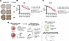 Figure 7
Higher RAD52 expression is associated with poor prognosis in high-grade serous ovarian cancer. (A) Representative immunohistochemical images of RAD52 staining in the tissue microarray (TMA). Scale bar: 50 μm. (B) Kaplan-Meier analysis of overall survival (OS) in patients with high-grade serous ovarian cancer represented in the TMA, dichotomized into high (H-score ≥ 168) and low (H-score < 168) RAD52 protein expression groups. The log-rank test was used to analyze survival at 4 years after diagnosis. (C) Kaplan-Meier analysis of OS in patients with serous ovarian cancer with TP53 mutations from The Cancer Genome Atlas (TCGA), stratified by high or low RAD52 mRNA expression. The log-rank test was used to analyze survival at 4 years after diagnosis. (D) Graphical overview summarizing the key findings in this study. Created in BioRender (https://BioRender.com/u905vy9).


