
Research ArticleInflammationOphthalmologyVascular biology
Open Access |  10.1172/jci.insight.203452
Mononuclear phagocyte–specific cGAS/STING targeting suppresses experimental choroidal neovascularization
Le Shi,1 Durgadas Cherukaraveedu,2 Hongkwan Cho,1 Kaoru Ri,1 Lingli Zhou,1 Yingxue Cao,1 Narendra Kale,2 Zhenhua Xu,1 Wathsala Liyanage,2 Rangaramanujam M. Kannan,2,3 and Elia J. Duh1
1Department of Ophthalmology, School of Medicine, Johns Hopkins University, Baltimore, Maryland, USA.
2Center for Nanomedicine at the Wilmer Eye Institute, Johns Hopkins University School of Medicine, Baltimore, Maryland, USA.
3Department of Chemical and Biomolecular Engineering, Johns Hopkins University, Baltimore, Maryland, USA.
Address correspondence to: Elia J. Duh, Johns Hopkins School of Medicine, 400 North Broadway, Smith Building Room 3011, Baltimore, Maryland, 21231, USA. Phone: 410.370.1917; Email: eduh@jhmi.edu. Or to: Rangaramanujam M. Kannan, Johns Hopkins School of Medicine, 400 North Broadway, Smith Building Room 6023, Baltimore, Maryland, 21231, USA. Phone: 443.287.8634; Email: krangar1@jhmi.edu.
Authorship note: LS and DC contributed equally to this work
Find articles by Shi, L. in: PubMed | Google Scholar
1Department of Ophthalmology, School of Medicine, Johns Hopkins University, Baltimore, Maryland, USA.
2Center for Nanomedicine at the Wilmer Eye Institute, Johns Hopkins University School of Medicine, Baltimore, Maryland, USA.
3Department of Chemical and Biomolecular Engineering, Johns Hopkins University, Baltimore, Maryland, USA.
Address correspondence to: Elia J. Duh, Johns Hopkins School of Medicine, 400 North Broadway, Smith Building Room 3011, Baltimore, Maryland, 21231, USA. Phone: 410.370.1917; Email: eduh@jhmi.edu. Or to: Rangaramanujam M. Kannan, Johns Hopkins School of Medicine, 400 North Broadway, Smith Building Room 6023, Baltimore, Maryland, 21231, USA. Phone: 443.287.8634; Email: krangar1@jhmi.edu.
Authorship note: LS and DC contributed equally to this work
Find articles by Cherukaraveedu, D. in: PubMed | Google Scholar
1Department of Ophthalmology, School of Medicine, Johns Hopkins University, Baltimore, Maryland, USA.
2Center for Nanomedicine at the Wilmer Eye Institute, Johns Hopkins University School of Medicine, Baltimore, Maryland, USA.
3Department of Chemical and Biomolecular Engineering, Johns Hopkins University, Baltimore, Maryland, USA.
Address correspondence to: Elia J. Duh, Johns Hopkins School of Medicine, 400 North Broadway, Smith Building Room 3011, Baltimore, Maryland, 21231, USA. Phone: 410.370.1917; Email: eduh@jhmi.edu. Or to: Rangaramanujam M. Kannan, Johns Hopkins School of Medicine, 400 North Broadway, Smith Building Room 6023, Baltimore, Maryland, 21231, USA. Phone: 443.287.8634; Email: krangar1@jhmi.edu.
Authorship note: LS and DC contributed equally to this work
Find articles by Cho, H. in: PubMed | Google Scholar
1Department of Ophthalmology, School of Medicine, Johns Hopkins University, Baltimore, Maryland, USA.
2Center for Nanomedicine at the Wilmer Eye Institute, Johns Hopkins University School of Medicine, Baltimore, Maryland, USA.
3Department of Chemical and Biomolecular Engineering, Johns Hopkins University, Baltimore, Maryland, USA.
Address correspondence to: Elia J. Duh, Johns Hopkins School of Medicine, 400 North Broadway, Smith Building Room 3011, Baltimore, Maryland, 21231, USA. Phone: 410.370.1917; Email: eduh@jhmi.edu. Or to: Rangaramanujam M. Kannan, Johns Hopkins School of Medicine, 400 North Broadway, Smith Building Room 6023, Baltimore, Maryland, 21231, USA. Phone: 443.287.8634; Email: krangar1@jhmi.edu.
Authorship note: LS and DC contributed equally to this work
Find articles by Ri, K. in: PubMed | Google Scholar
1Department of Ophthalmology, School of Medicine, Johns Hopkins University, Baltimore, Maryland, USA.
2Center for Nanomedicine at the Wilmer Eye Institute, Johns Hopkins University School of Medicine, Baltimore, Maryland, USA.
3Department of Chemical and Biomolecular Engineering, Johns Hopkins University, Baltimore, Maryland, USA.
Address correspondence to: Elia J. Duh, Johns Hopkins School of Medicine, 400 North Broadway, Smith Building Room 3011, Baltimore, Maryland, 21231, USA. Phone: 410.370.1917; Email: eduh@jhmi.edu. Or to: Rangaramanujam M. Kannan, Johns Hopkins School of Medicine, 400 North Broadway, Smith Building Room 6023, Baltimore, Maryland, 21231, USA. Phone: 443.287.8634; Email: krangar1@jhmi.edu.
Authorship note: LS and DC contributed equally to this work
Find articles by
Zhou, L.
in:
PubMed
|
Google Scholar
|

1Department of Ophthalmology, School of Medicine, Johns Hopkins University, Baltimore, Maryland, USA.
2Center for Nanomedicine at the Wilmer Eye Institute, Johns Hopkins University School of Medicine, Baltimore, Maryland, USA.
3Department of Chemical and Biomolecular Engineering, Johns Hopkins University, Baltimore, Maryland, USA.
Address correspondence to: Elia J. Duh, Johns Hopkins School of Medicine, 400 North Broadway, Smith Building Room 3011, Baltimore, Maryland, 21231, USA. Phone: 410.370.1917; Email: eduh@jhmi.edu. Or to: Rangaramanujam M. Kannan, Johns Hopkins School of Medicine, 400 North Broadway, Smith Building Room 6023, Baltimore, Maryland, 21231, USA. Phone: 443.287.8634; Email: krangar1@jhmi.edu.
Authorship note: LS and DC contributed equally to this work
Find articles by Cao, Y. in: PubMed | Google Scholar
1Department of Ophthalmology, School of Medicine, Johns Hopkins University, Baltimore, Maryland, USA.
2Center for Nanomedicine at the Wilmer Eye Institute, Johns Hopkins University School of Medicine, Baltimore, Maryland, USA.
3Department of Chemical and Biomolecular Engineering, Johns Hopkins University, Baltimore, Maryland, USA.
Address correspondence to: Elia J. Duh, Johns Hopkins School of Medicine, 400 North Broadway, Smith Building Room 3011, Baltimore, Maryland, 21231, USA. Phone: 410.370.1917; Email: eduh@jhmi.edu. Or to: Rangaramanujam M. Kannan, Johns Hopkins School of Medicine, 400 North Broadway, Smith Building Room 6023, Baltimore, Maryland, 21231, USA. Phone: 443.287.8634; Email: krangar1@jhmi.edu.
Authorship note: LS and DC contributed equally to this work
Find articles by Kale, N. in: PubMed | Google Scholar
1Department of Ophthalmology, School of Medicine, Johns Hopkins University, Baltimore, Maryland, USA.
2Center for Nanomedicine at the Wilmer Eye Institute, Johns Hopkins University School of Medicine, Baltimore, Maryland, USA.
3Department of Chemical and Biomolecular Engineering, Johns Hopkins University, Baltimore, Maryland, USA.
Address correspondence to: Elia J. Duh, Johns Hopkins School of Medicine, 400 North Broadway, Smith Building Room 3011, Baltimore, Maryland, 21231, USA. Phone: 410.370.1917; Email: eduh@jhmi.edu. Or to: Rangaramanujam M. Kannan, Johns Hopkins School of Medicine, 400 North Broadway, Smith Building Room 6023, Baltimore, Maryland, 21231, USA. Phone: 443.287.8634; Email: krangar1@jhmi.edu.
Authorship note: LS and DC contributed equally to this work
Find articles by Xu, Z. in: PubMed | Google Scholar
1Department of Ophthalmology, School of Medicine, Johns Hopkins University, Baltimore, Maryland, USA.
2Center for Nanomedicine at the Wilmer Eye Institute, Johns Hopkins University School of Medicine, Baltimore, Maryland, USA.
3Department of Chemical and Biomolecular Engineering, Johns Hopkins University, Baltimore, Maryland, USA.
Address correspondence to: Elia J. Duh, Johns Hopkins School of Medicine, 400 North Broadway, Smith Building Room 3011, Baltimore, Maryland, 21231, USA. Phone: 410.370.1917; Email: eduh@jhmi.edu. Or to: Rangaramanujam M. Kannan, Johns Hopkins School of Medicine, 400 North Broadway, Smith Building Room 6023, Baltimore, Maryland, 21231, USA. Phone: 443.287.8634; Email: krangar1@jhmi.edu.
Authorship note: LS and DC contributed equally to this work
Find articles by Liyanage, W. in: PubMed | Google Scholar
1Department of Ophthalmology, School of Medicine, Johns Hopkins University, Baltimore, Maryland, USA.
2Center for Nanomedicine at the Wilmer Eye Institute, Johns Hopkins University School of Medicine, Baltimore, Maryland, USA.
3Department of Chemical and Biomolecular Engineering, Johns Hopkins University, Baltimore, Maryland, USA.
Address correspondence to: Elia J. Duh, Johns Hopkins School of Medicine, 400 North Broadway, Smith Building Room 3011, Baltimore, Maryland, 21231, USA. Phone: 410.370.1917; Email: eduh@jhmi.edu. Or to: Rangaramanujam M. Kannan, Johns Hopkins School of Medicine, 400 North Broadway, Smith Building Room 6023, Baltimore, Maryland, 21231, USA. Phone: 443.287.8634; Email: krangar1@jhmi.edu.
Authorship note: LS and DC contributed equally to this work
Find articles by Kannan, R. in: PubMed | Google Scholar
1Department of Ophthalmology, School of Medicine, Johns Hopkins University, Baltimore, Maryland, USA.
2Center for Nanomedicine at the Wilmer Eye Institute, Johns Hopkins University School of Medicine, Baltimore, Maryland, USA.
3Department of Chemical and Biomolecular Engineering, Johns Hopkins University, Baltimore, Maryland, USA.
Address correspondence to: Elia J. Duh, Johns Hopkins School of Medicine, 400 North Broadway, Smith Building Room 3011, Baltimore, Maryland, 21231, USA. Phone: 410.370.1917; Email: eduh@jhmi.edu. Or to: Rangaramanujam M. Kannan, Johns Hopkins School of Medicine, 400 North Broadway, Smith Building Room 6023, Baltimore, Maryland, 21231, USA. Phone: 443.287.8634; Email: krangar1@jhmi.edu.
Authorship note: LS and DC contributed equally to this work
Find articles by Duh, E. in: PubMed | Google Scholar
Published August 10, 2026 - More info
JCI Insight. 2026;11(15):e203452. https://doi.org/10.1172/jci.insight.203452.
© 2026 Le Shi et al. This work is licensed under the Creative Commons Attribution 4.0 International License. To view a copy of this license, visit http://creativecommons.org/licenses/by/4.0/.
Received: December 12, 2025; Accepted: June 11, 2026


-
Results
The cGAS/STING pathway is active in MPs in both human and mouse nAMD. To determine whether the cGAS/STING pathway is active in nAMD and to identify the responsible cell type, we examined the temporal dynamics of cGAS/STING signaling in a laser-induced CNV mouse model. Consistent with previous reports (6), in retinal cross sections, IBA1+ MPs were present in the CNV lesion area as early as day 1 after laser injury, peaked on day 3, and gradually declined thereafter, whereas in controls, IBA1+ cells remained confined to the inner retina in cross section (Figure 1A). Notably, cGAS was highly colocalized with reactive IBA1+ MPs within the CNV lesions but absent in IBA1+ cells located outside the lesion, indicating that cGAS expression is primarily associated with lesion-resident MPs (Figure 1A). We also performed RPE/choroid flat-mount staining, which similarly showed that mild cGAS signal began to appear at day 1 after laser, without obvious cGAS+IBA1+ cells. At days 3 and 4, IBA1+ cells markedly accumulated within the lesion, and abundant colocalization signals of cGAS and IBA1 were observed; by days 5 and 7, the cGAS signal and colocalization gradually decreased (Supplemental Figure 1; supplemental material available online with this article; https://doi.org/10.1172/jci.insight.203452DS1). Western blot analysis further showed a marked increase in STING protein levels in the retina-choroid-RPE complex at day 3 after laser administration compared with controls (Figure 1, B and C). We were interested in deducing the human ocular cell types that endogenously express cGAS. For this purpose, we first analyzed single-cell transcriptomic data from the Human Protein Atlas eye database. The results showed that cGAS expression was particularly prominent in immune cell populations, including macrophages and lymphocytes, although neurons, glial cells, epithelial cells, and vascular-associated cells exhibited expression at lower levels (Figure 1D). We next analyzed single-cell RNA-sequencing (RNA-seq)data from human retinal samples obtained from nAMD, dry AMD, and control donors. This analysis further confirmed that cGAS expression was particularly prominent in immune cells, with much lower expression in non-immune cell populations (Figure 1E). Among immune subsets, inflammatory macrophages exhibited the highest cGAS expression. Moreover, cGAS expression in inflammatory macrophages was substantially upregulated in both nAMD and dry AMD samples compared with controls (Figure 1E). Collectively, these results demonstrate that the cGAS/STING pathway is strongly active in reactive MPs in both human nAMD and the experimental laser-induced CNV model, suggesting the potential involvement of this pathway in the regulation of CNV by reactive MPs.
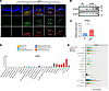 Figure 1
The cGAS/STING pathway is active in microglia/macrophages in human nAMD and laser-induced CNV mice. (A) Representative immunofluorescence images of ocular cross sections from control (Ctl) and experimental mice on day 1 (D1), D3, D4, D5, and D7 after laser induction of CNV. Sections were stained for IBA1 (green), cGAS (red), and Hoechst (blue). Arrowheads indicate IBA1+cGAS+ double-positive cells localized within or surrounding the CNV lesions. Scale bar: 100 μm. (B) Western blot analysis of STING in retina-RPE-choroid complexes from control and laser CNV eyes on D3 (n = 4–6 per group). β-Actin was used as a loading control. (C) Quantitation of STING protein levels in B. (D) cGAS expression levels across human ocular cell populations based on single-cell RNA-seq data obtained from the Human Protein Atlas. (E) Dot plot of single-cell RNA-seq expression of cGAS across retinal and choroidal cell populations from human donor eyes with AMD, nAMD, and controls. Individual dots represent single cells. Bar graphs depict the mean ± SEM. Note that the mean for most cell populations is close to zero. Statistical significance was assessed by 2-sided, unpaired t test. *P < 0.05.
Synthesis, characterization, and in vitro drug release profile of the HD-SN-011 conjugate. Our previous studies demonstrated that dendrimer-drug conjugates enable selective targeting of reactive MPs (28, 32, 34). To specifically modulate the cGAS/STING pathway in reactive phagocytes, we developed an HD conjugate of SN-011 (HD-SN-011) (Figure 2A). The intermediate compound HD-GABA-Boc (compound 3) was synthesized via ester coupling between HD and Boc-GABA-OH and characterized by proton nuclear magnetic resonance (¹H-NMR). The spectrum indicated approximately 4 GABA linkers conjugated to the HD backbone, based on the integration ratio between the tert-butyl protons of Boc-GABA-OH (1.35 ppm) and the amide protons of HD in the aromatic region. After Boc deprotection using 25% trifluoroacetic acid (TFA) in dichloromethane (DCM) under ice-cold conditions, the resulting HD-GABA-NH2 was obtained for eventual conjugation with modified SN-011. SN-011 was modified via an ester bond using succinic anhydride as a linker molecule. The final compound HD-SN-011 was synthesized by coupling the acid-functionalized SN-011 (SN-011 succinic acid linker) and HD-GABA-NH2 via an amide bond using EDC/NHS chemistry. The final HD-SN-011 conjugate was characterized by 1H-NMR and confirmed approximately 4 SN-011 molecules per dendrimer, with aromatic proton peaks (6.5–7.9 ppm) corresponding to SN-011 (Figure 2B). The ester linkage between SN-011 and the succinic acid spacer was designed to be hydrolyzable for intracellular drug release. Analytical high-performance liquid chromatography (HPLC) revealed a single dominant peak with greater than 98% purity for the HD-SN-011 conjugate (Figure 2C). Dynamic light scattering (DLS) showed that HD-SN-011 was monodisperse, with an average hydrodynamic diameter of 5.92 ± 0.88 nm and a ζ potential of –8.34 ± 0.21 mV, indicating excellent stability (Figure 2D). In vitro drug-release assays demonstrated that HD-SN-011 exhibited a sustained and esterase-responsive release profile (Figure 2E). Drug release was performed in 0.5 M citrate buffer (pH 5.5) containing porcine liver esterase, which mimics lysosomal conditions. The release of free SN-011 was quantified by HPLC at 260 nm based on the area under the curve (AUC). The enzyme was replenished every 48 hours to maintain catalytic activity, and samples were collected at predefined time points. Approximately 50% of SN-011 was released within 7 days and approximately 75% within 12 days, indicating controlled hydrolytic cleavage of the ester linkage under lysosome-like conditions. Collectively, these results demonstrate that HD-SN-011 is stable and “monodisperse,” exhibiting sustained and enzyme-responsive drug release under lysosome-mimicking conditions, thereby providing a basis for its further evaluation in ocular delivery applications.
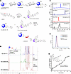 Figure 2
Synthesis and characterization of HD-SN-011 with improved solubility and physicochemical stability. (A) Schematic showing the multistep chemical synthesis of the HD-SN-011 conjugate. (B) 1H-NMR confirmed the formation of the HD-SN-011 conjugate via amide bonds on HD. (C) Analytical HPLC confirmed the conjugation of the GABA linker to HD at 210 nm detection (specific for HD), and the HD-drug conjugation at 260 nm detection. HD-SN-011 showed over 98% purity with a distinct retention time of approximately 17 minutes compared with the free drug at 26 minutes and HD-GABA-NH2 at 12.5 minutes. (D) DLS studies of the HD-SN-011 conjugate confirmed the monodisperse particle distribution with a size of approximately 5.9 nm. (E) The HD-SN-011 conjugate showed ester-mediated release of pure SN-011 under acidic pH (0.5 M citrate buffer). The analytical HPLC results show that nearly 75% of the drug was released over 11 days.
HD-SN-011 markedly reduces the in vitro and in vivo cytotoxicity of free SN-011. We next evaluated the biosafety of HD-SN-011 in multiple cell types, including murine macrophage-like RAW264.7 cells, human umbilical vein endothelial cells (HUVECs), and mouse photoreceptor-derived 661W cells. The results showed that free SN-011 induced significant cytotoxicity in all 3 cell types when the concentration exceeded 8 μg/mL, whereas cells treated with HD-SN-011 maintained more than 90% viability even at higher concentrations up to 32 μg/mL of conjugated SN-011, exhibiting negligible cytotoxicity (Figure 3, A–C). Notably, HUVECs were particularly sensitive to free SN-011, showing a marked decrease in viability at 0.125 μg/mL with a clear dose-dependent trend (Figure 3B). These findings indicate that conjugation to the HD effectively mitigates the concentration-dependent cytotoxicity of SN-011 (by greater than 250-fold) and improves its biocompatibility.
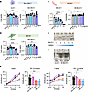 Figure 3
HD-SN-011 exhibits solubility, low cytotoxicity, and excellent ocular safety. (A–C) Viability assays in RAW264.7 cells (A), HUVECs (B), and 661W cells (C). Cells were treated with vehicle, free SN-011, or HD-SN-011 at the indicated concentrations for 16 hours, followed by measurement of cell viability using the CTG assay. A concentration of 32 μg/mL corresponds to 69 μM SN-011. (D) Representative images showing the solubility of free SN-011 in various solvent formulations. From left to right, the solvents are (1) 1% DMSO + 99% saline, (2) 3% DMSO + 10% PEG300 + 3% Tween 80 + 84% saline, (3) 5% DMSO + 40% PEG300 + 5% Tween 80 + 50% saline, (4) 8% DMSO + 40% PEG300 + 5% Tween 80 + 47% saline, and (5) 10% DMSO + 40% PEG300 + 5% Tween 80 + 45% saline. (E) Representative images showing the solubility of free SN-011 and HD-SN-011 at different concentrations when dissolved in saline. A concentration of 4 mM corresponds to 1.85 mg/mL SN-011, and 20 mM corresponds to 9.25 mg/mL SN-011. (F) In vivo retinal safety was assessed by ERG after intravitreal injection. Four experimental groups were included: vehicle control 1 (saline), HD-SN-011 dissolved in saline, vehicle control 2 (10% DMSO + 40% PEG300 + 5% Tween 80 + 45% saline), and free SN-011 dissolved in vehicle 2. Scotopic A- and B-wave amplitudes were recorded in response to increasing flash intensities at 7 days after injection. Data are represented as mean ± SEM. Statistical significance was assessed by 1-way ANOVA with Šídák’s multiple-comparison test. NS, not significant. *P < 0.05, **P < 0.01, ***P < 0.001, ****P < 0.0001.
Previous studies reported that intravitreal injection of SN-011 at 4 mM effectively reduced laser-induced CNV (20). Based on this, we evaluated the potential retinal toxicity of 4 mM HD-SN-011 and free SN-011 following intravitreal injection. Because free SN-011 is highly hydrophobic and poorly soluble in water, we first tested several solvent systems containing various ratios of DMSO, PEG300, Tween 80, and saline. Formulations with low DMSO content (1%–8%) or lacking PEG300 and Tween 80 appeared turbid or showed visible precipitation, whereas only the mixture containing 10% DMSO, 40% PEG300, 5% Tween 80, and 45% saline formed a clear solution (Figure 3D). Therefore, this formulation was used as the vehicle for free SN-011. In contrast, HD-SN-011, owing to the hydrophilic dendrimer carrier, was completely soluble in saline at both 4 mM and 20 mM without visible precipitation (Figure 3E).
To assess in vivo ocular safety, we performed electroretinography (ERG) at 7 days after intravitreal injection of different drug formulations in mice (Figure 3F). Four groups were tested: (a) saline control; (b) HD-SN-011 (4 mM) dissolved in saline; (c) free SN-011 dissolved in vehicle (10% DMSO, 40% PEG300, 5% Tween 80, and 45% saline); and (d) vehicle control. Mice injected with saline-dissolved HD-SN-011 exhibited normal scotopic A- and B-wave amplitudes comparable to those of saline controls, indicating preserved retinal function. In contrast, eyes injected with free SN-011 or its vehicle formulation showed markedly reduced ERG amplitudes, consistent with retinal toxicity induced by high DMSO concentrations and other organic cosolvents (Figure 3F and Supplemental Figure 2, A–D), indicating that high concentrations of DMSO and other organic cosolvents impair retinal function. In parallel, H&E staining and quantitative analysis of total retinal thickness performed 2 weeks after intravitreal injection revealed no significant differences among the 4 groups (Supplemental Figure 2E), indicating the absence of overt structural retinal damage under these conditions. Previous studies have also reported that intravitreal injection of DMSO-containing formulations can induce retinal ganglion cell apoptosis (38). Collectively, these findings demonstrate that HD-SN-011 exhibits excellent biocompatibility and substantially reduces the in vitro and in vivo cytotoxicity associated with free SN-011, supporting its potential as a safe and effective platform for intravitreal drug delivery.
HD-SN-011 is rapidly and selectively taken up by MPs and suppresses cGAS/STING pathway activation in vitro. To evaluate whether HDs can be selectively and efficiently internalized by MPs, we synthesized a Cy3-labeled HD (HD-Cy3) (Supplemental Figure 3) and monitored its cellular uptake over time. Confocal imaging revealed minimal background fluorescence in untreated controls, whereas RAW264.7 macrophages and BV2 microglia exhibited clear intracellular red fluorescence as early as 3 hours after addition of HD-Cy3. The fluorescence intensity further increased at 8 and 24 hours, indicating efficient and sustained internalization of HD-Cy3 by phagocytes (Figure 4, A–D). We next examined the regulatory effects of HD-SN-011 on cGAS/STING pathway activation in cultured RAW264.7 macrophages. Stimulation of macrophages with 2′3′-cGAMP robustly activated the STING signaling cascade, as evidenced by increased TBK1 phosphorylation, elevated IRF3 phosphorylation, and promoted IRF3 nuclear translocation (Figure 4, E–H). Pretreatment with HD-SN-011 markedly attenuated these responses, substantially reducing p-TBK1 levels and inhibiting IRF3 nuclear accumulation (Figure 4, F–H). Notably, free SN-011 exhibited a stronger inhibitory effect at the same mass concentration (Figure 4, F and G), which is likely attributable to its immediately bioavailable form, whereas HD-SN-011 requires intracellular ester-bond hydrolysis for active-drug release. On the other hand, HD-SN-011’s dendrimer-based design enables efficient, targeted uptake by MPs and intracellular release of SN-011 over time, which would be expected to provide a longer duration of treatment effect. Collectively, these in vitro findings demonstrate that HDs are rapidly and effectively internalized by MPs, and that HD-SN-011 effectively suppresses activation of the cGAS/STING pathway, supporting its therapeutic potential.
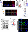 Figure 4
HD-Cy3 is efficiently taken up by microglia/macrophages and inhibits cGAS/STING activation in vitro. (A and C) Time-dependent uptake of HD-Cy3 in RAW264.7 macrophages (A) and BV2 microglia (C). Cells were incubated with HD-Cy3 (10 μg/mL) for 0, 3, 8, or 24 hours, then fixed and counterstained with Hoechst (blue). Representative images show a progressive increase in intracellular Cy3 signal (red). Yellow dashed boxes highlight regions of interest shown at higher magnification. Scale bars: 50 μm. (B and D) Quantification of mean fluorescence intensity (MFI) per cell demonstrates robust, time-dependent accumulation of HD-Cy3 in both RAW264.7 (B) and BV2 cells (D). (E) Schematic illustration of the cGAS/STING signaling cascade activated by 2′3′-cGAMP, leading to phosphorylation of TBK1 and IRF3 and induction of type I interferons. (F) Western blot analysis of STING pathway activation in RAW264.7 cells. Cells were pretreated with free SN-011 or HD-SN-011 (8 μg/mL) for 16 hours, followed by stimulation with 2′3′-cGAMP (25 nM) for 6 hours. β-Actin was used as a loading control. (G) Densitometric quantification of p-TBK1 normalized to total TBK1 in F (n = 3). (H) Immunofluorescent staining of IRF3 (green) in RAW264.7 cells. Cells were pretreated with free SN-011 or HD-SN-011 (8 μg/mL) for 16 hours, followed by stimulation with 2′3′-cGAMP (25 nM) for 6 hours. Nuclei were counterstained with Hoechst (blue). Scale bar: 10 μm. Statistical significance was assessed by 1-way ANOVA with Šídák’s multiple-comparison test. **P < 0.01; ***P < 0.001; ****P < 0.0001.
HD-Cy3 can be specifically taken up by reactive MPs in the laser-induced CNV model in vivo. To determine whether PAMAM HDs can selectively target reactive phagocytes in vivo, we took advantage of the well-defined temporal dynamics of inflammatory cell recruitment in the laser-induced CNV model; MPs begin to infiltrate the lesion at around day 1 after laser injury and reach a peak at approximately day 3. For dendrimer localization studies, HD-Cy3 was intravitreally injected at different time points after laser induction (immediately, day 1, day 2, day 3, or day 5), and all eyes were collected on day 7 for parallel analysis (Figure 5A). Because unengulfed dendrimers are rapidly cleared via renal filtration within approximately 24 hours (28), HD-Cy3 will be present in the retina on day 7 after laser only in cells that have internalized them. Confocal imaging revealed that when HD-Cy3 was injected immediately after laser injury, virtually no signal was detected in the CNV region on day 7, indicating rapid clearance in the absence of reactive MPs and further supporting the selectivity and ocular safety of the HD platform (Figure 5, B and C). In contrast, injections performed on day 1, day 2, day 3, or day 5 resulted in pronounced accumulation of HD-Cy3 within IBA1+ MPs inside the CNV lesion; quantitative analysis showed that more than 70% of IBA1+ cells were HD-Cy3+ across all 4 groups (Figure 5, B and C). Three-dimensional reconstruction further confirmed tight spatial colocalization between HD-Cy3 and IBA1+ microglia/macrophages (Figure 5F). To further assess the spatial precision of HD localization, we compared HD-Cy3 distribution inside versus outside the CNV lesion. More than 80% of the total HD-Cy3 signal was confined to the CNV core (Figure 5D), indicating lesion-specific enrichment. Individual cell analysis of the fluorescence signal additionally revealed that IBA1+ cells located within the lesion exhibited approximately 3- to 4-fold higher HD-Cy3 fluorescence intensity compared with IBA1+ cells outside the lesion (Figure 5E), demonstrating that HD uptake is not a universal property of all retinal phagocytes but is tightly associated with the lesion-recruited reactive population. In addition, we examined the colocalization of HD-Cy3 with RPE cells after intravitreal injection on day 1 or day 4 following laser injury. Limited colocalization of HD-Cy3 signal with RPE cells was observed, indicating that HD-Cy3 is also taken up by RPE cells (Supplemental Figure 4), although to a lesser extent compared with MPs. Finally, to evaluate long-term intracellular retention, we injected HD-Cy3 on day 1 after laser induction and examined CNV lesions on day 14. HD-Cy3 signal remained detectable in IBA1+ cells surrounding the regressed lesions, indicating stable and prolonged intracellular persistence of the conjugate (Figure 5G). In summary, HDs target and are preferentially taken up by reactive phagocytes in CNV lesions in vivo.
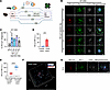 Figure 5
HD-Cy3 is specifically taken up by mononuclear phagocytes in laser-induced CNV in vivo. (A) Schematic of timeline of the experimental workflow. CNV was induced by laser photocoagulation on day 0 (D0). HD-Cy3 was intravitreally injected at different time points (D0, D1, D2, D3, or D5) after laser administration, and all eyes were collected on D7 after laser for flat-mount preparation and immunostaining. (B) Representative confocal images of CNV lesions from eyes injected with HD-Cy3 on D0, D1, D2, D3, or D5 and collected on D7. Flat-mounts were immunostained for IBA1 (green) and CD31 (gray), with nuclei counterstained with Hoechst (blue). Scale bar: 10 μm. (C) Quantification of the percentage of HD-Cy3+IBA1+ cells at D7 in eyes injected at the indicated time points after laser (n = 4 lesions). (D) Quantification of HD-Cy3 fluorescence intensity in B, comparing retinal quadrants inside and outside the CNV lesion (n = 4 lesions). (E) Fluorescence quantification of HD-Cy3+IBA1+ macrophages/microglia in B, comparing cells located inside the lesion (n = 44 IBA1+ cells) and outside the lesion (n = 60 IBA1+ cells). Cells were randomly selected from 4 lesion areas. (F) Three-dimensional reconstruction of the D7 CNV lesion demonstrating spatial colocalization of HD-Cy3 (red) with IBA1+ phagocytes (blue) within the neovascular core. (G) Representative confocal images showing HD-Cy3 uptake at D14 after HD-Cy3 injection on D1. Flat-mounts were immunostained for IBA1 (green) and CD31 (gray), with nuclei counterstained with Hoechst (blue). Scale bar: 10 μm. Data are represented as mean ± SEM. Statistical significance was assessed by 1-way ANOVA with Šídák’s multiple-comparison test (C) or 2-sided, unpaired t test (D and E). *P < 0.05; ***P < 0.001; ****P < 0.0001.
HD-SN-011 alleviates pathological neovascularization and vascular leakage in the laser-induced CNV model in vivo. To evaluate the therapeutic efficacy of HD-SN-011 in vivo, intravitreal injections of HD-SN-011 were performed 1 day and 3 days after laser induction, and the pathological features of CNV were analyzed on day 7 (Figure 6A). Fundus fluorescein angiography (FFA) showed no significant difference in vascular leakage between the saline and HD-only groups, whereas HD-SN-011 treatment markedly reduced vascular leakage. Quantitative analysis, calculated as the fluorescence intensity at 10 minutes minus that at 5 minutes, further confirmed that CNV leakage was reduced by approximately 50% following HD-SN-011 treatment (Figure 6, B and C). In addition, RPE-choroid flat-mount analysis demonstrated that HD-SN-011 treatment reduced CNV lesion size by approximately 60% compared with saline (55% compared with HD-only), indicating that HD-SN-011 effectively inhibits pathological neovascularization in vivo (Figure 6, D and E). Immunofluorescent staining showed strong IL-1β and TNF-α expression in IBA1+ MPs within CNV lesions in the saline group, whereas HD-SN-011 treatment markedly reduced the expression of both cytokines (Figure 6, F and G). Together, these findings demonstrate that HD-SN-011 significantly reduces vascular leakage and CNV lesion size in the laser-induced model, highlighting its potential as a therapeutic approach for nAMD through inhibition of both pathologic angiogenesis and CNV leakage.
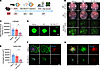 Figure 6
HD-SN-011 reduces CNV leakage, lesion size, and inflammatory signaling in the laser-induced CNV model. (A) Schematic timeline of the experiment. CNV was induced by laser photocoagulation on day 0 (D0), followed by intravitreal injection of saline or HD-SN-011 (4 mM, 1.85 mg/mL) on D1 or D3. On D7, fundus fluorescein angiography (FFA) and choroidal flat-mount analyses were performed. (B) Representative D0 fundus images after laser photocoagulation, D7 fundus images, and D7 FFA images acquired 5 and 10 minutes after intraperitoneal injection of sodium fluorescein in saline-, HD-, and HD-SN-011–treated mice. (C) Quantification of fluorescein leakage based on FFA images in B, calculated as the difference between fluorescence signal intensity at 10 minutes compared with 5 minutes after fluorescein injection (n = 7 mice). (D) Representative choroidal flat-mount images stained with IB4 lectin on D7 in saline- HD-, and HD-SN-011–treated eyes. Scale bar: 100 μm. (E) Quantification of CNV lesion area from choroidal flat-mounts in D (n = 7 mice). (F and G) Immunofluorescent staining of CNV lesions on D7. Flat-mounts were stained for IBA1 (green) together with IL-1β (F, red) or TNF-α (G, red), with nuclei counterstained using Hoechst (blue). Scale bars: 100 μm. Data are shown as mean ± SEM. Statistical analysis was performed using a 2-sided, unpaired t test. *P < 0.05.
HD-SN-011 attenuates MP cGAS/STING activation and downstream type I interferon–mediated inflammation. To confirm whether HD-SN-011 involves direct modulation of cGAS/STING signaling in MPs, we employed the Cx3cr1-CreER; Rpl22HA/HA (RiboTag) mouse model (39), which enables selective isolation of ribosome-bound mRNA from MPs following tamoxifen-induced Cre activation (40, 41) (Figure 7A and Supplemental Figure 5A). HD-SN-011 or saline was intravitreally injected on day 1 after laser induction, and MP-specific translatome profiling was performed on days 2 and 3 after laser. Successful enrichment of MP-restricted transcripts such as Cx3cr1 confirmed the effectiveness of the isolation strategy (Figure 7B). Quantitative PCR (qPCR) analysis revealed a time-dependent inflammatory response in saline-treated CNV eyes, in which Ccl2 and Tnf were markedly upregulated on day 2, whereas Ifnb1 and Cxcl10 were significantly induced on day 3. HD-SN-011 treatment significantly suppressed the induction of Ccl2 and Tnf on day 2 and markedly reduced Ifnb1 and Cxcl10 expression on day 3 (Figure 7, C–H, and Supplemental Figure 5, B and C). Collectively, these results demonstrate that HD-SN-011 suppresses cGAS/STING-associated inflammatory signaling and proinflammatory cytokine production in retinal MPs, thereby attenuating local inflammatory activation and contributing to its protective effect against pathological angiogenesis in AMD.
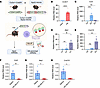 Figure 7
HD-SN-011 attenuates MP cGAS/STING activation and downstream type I interferon–related inflammatory gene expression. (A) Schematic of the Cx3cr1-CreER; Rpl22HA/HA mouse model and HA-RiboTag enrichment workflow. Cx3cr1-CreER mice were crossed with Rpl22HA/HA mice. Tamoxifen was administered to induce HA-tagged ribosome expression in Cx3cr1+ MPs. Retina-RPE-choroid complexes were collected to obtain input RNA and HA-immunoprecipitated (HA-IP) MP–enriched mRNAs. (B) qPCR validation of MP enrichment by comparing Cx3cr1 expression between input and HA-IP RNA fractions. (C–E) qPCR analysis of Ccl2 (C), Ifnb1 (D), and Cxcl10 (E) in HA-IP–enriched mRNAs at baseline (Ctl) and on day 2 (D2) or D3 after laser-induced CNV in saline-treated eyes. (F–H) Mice subjected to laser-induced CNV received intravitreal injection of saline or HD-SN-011 on D1, followed by qPCR analysis of Ccl2 on D2 (F), and Ifnb1 and Cxcl10 on D3. (G and H). The saline control shown in F corresponds to the same D2 control in C, whereas the saline controls shown in G and H correspond to the same D3 controls as in D and E, respectively. Data are represented as mean ± SEM. Statistical significance was assessed by 1-way ANOVA with Šídák’s multiple-comparison test (C–E) or 2-sided, unpaired t test (F–H). NS, not significant. *P < 0.05; **P < 0.01; ***P < 0.001; ****P < 0.0001.


