
Clinical Research and Public HealthImmunologyInfectious diseaseMicrobiology
Open Access |  10.1172/jci.insight.202134
Rare variable M. tuberculosis antigens induce predominant Th17 responses in human infection
Paul Ogongo,1,2 Liya Wassie,3 Anthony Tran,1 Devin Columbus,1 Julia Huffaker,1 Lisa Sharling,4 Gregory Ouma,5 Samuel Gurrion Ouma,5 Kidist Bobosha,3 Cecilia S. Lindestam Arlehamn,6,7 Neel R. Gandhi,4,8,9 Sara C. Auld,4,9,10 Jyothi Rengarajan,8,11 Cheryl L. Day,11,12 Artur Queiroz,13,14 Mariana Araújo-Pereira,13,14 Eduardo Fukutani,13,14 Bruno B. Andrade,13,14 John D. Altman,11 Henry M. Blumberg,4,8,9 Joel D. Ernst,1 and the TBRU ASTRa Study Group15
1Division of Experimental Medicine, UCSF, San Francisco, California, USA.
2Department of Tropical and Infectious Diseases, Kenya Institute of Primate Research, Nairobi, Kenya.
3Mycobacterial Disease Research Directorate, Armauer Hansen Research Institute, Addis Ababa, Ethiopia.
4Department of Epidemiology, Emory University Rollins School of Public Health, Atlanta, Georgia, USA.
5Center for Global Health Research, Kenya Medical Research Institute, Kisumu, Kenya.
6Center for Vaccine Innovation, La Jolla Institute for Immunology, La Jolla, California, USA.
7Department of Infectious Disease and Immunology, Center for Vaccine Research, Statens Serum Institut, Copenhagen, Denmark.
8Department of Medicine, Division of Infectious Diseases, Emory University School of Medicine, Atlanta, Georgia, USA.
9Department of Global Health, Emory University Rollins School of Public Health, Atlanta, Georgia, USA.
10Department of Medicine, Division of Pulmonary and Critical Care Medicine, Emory University School of Medicine, Atlanta, Georgia, USA.
11Emory Vaccine Center, Emory University, Atlanta, Georgia, USA.
12Department of Microbiology and Immunology, Emory University School of Medicine, Atlanta, Georgia, USA.
13Multinational Organization Network Sponsoring Translational and Epidemiological Research (MONSTER) Initiative, Salvador, Brazil.
14Laboratório de Pesquisa Clínica e Translacional, Instituto Gonçalo Moniz, Fundação Oswaldo Cruz, Salvador, Brazil.
15TBRU ASTRa Study Group is detailed in Supplemental Acknowledgments.
Address correspondence to: Paul Ogongo or Joel D. Ernst, UCSF, 2540 23rd Street, Pride Hall, Room 3724, San Francisco, California, 94110, USA. Phone: 415.476.1227; Email: paul.ogongo@ucsf.edu (PO). Email: joel.ernst@ucsf.edu (JDE).
Find articles by
Ogongo, P.
in:
PubMed
|
Google Scholar
|

1Division of Experimental Medicine, UCSF, San Francisco, California, USA.
2Department of Tropical and Infectious Diseases, Kenya Institute of Primate Research, Nairobi, Kenya.
3Mycobacterial Disease Research Directorate, Armauer Hansen Research Institute, Addis Ababa, Ethiopia.
4Department of Epidemiology, Emory University Rollins School of Public Health, Atlanta, Georgia, USA.
5Center for Global Health Research, Kenya Medical Research Institute, Kisumu, Kenya.
6Center for Vaccine Innovation, La Jolla Institute for Immunology, La Jolla, California, USA.
7Department of Infectious Disease and Immunology, Center for Vaccine Research, Statens Serum Institut, Copenhagen, Denmark.
8Department of Medicine, Division of Infectious Diseases, Emory University School of Medicine, Atlanta, Georgia, USA.
9Department of Global Health, Emory University Rollins School of Public Health, Atlanta, Georgia, USA.
10Department of Medicine, Division of Pulmonary and Critical Care Medicine, Emory University School of Medicine, Atlanta, Georgia, USA.
11Emory Vaccine Center, Emory University, Atlanta, Georgia, USA.
12Department of Microbiology and Immunology, Emory University School of Medicine, Atlanta, Georgia, USA.
13Multinational Organization Network Sponsoring Translational and Epidemiological Research (MONSTER) Initiative, Salvador, Brazil.
14Laboratório de Pesquisa Clínica e Translacional, Instituto Gonçalo Moniz, Fundação Oswaldo Cruz, Salvador, Brazil.
15TBRU ASTRa Study Group is detailed in Supplemental Acknowledgments.
Address correspondence to: Paul Ogongo or Joel D. Ernst, UCSF, 2540 23rd Street, Pride Hall, Room 3724, San Francisco, California, 94110, USA. Phone: 415.476.1227; Email: paul.ogongo@ucsf.edu (PO). Email: joel.ernst@ucsf.edu (JDE).
Find articles by Wassie, L. in: PubMed | Google Scholar
1Division of Experimental Medicine, UCSF, San Francisco, California, USA.
2Department of Tropical and Infectious Diseases, Kenya Institute of Primate Research, Nairobi, Kenya.
3Mycobacterial Disease Research Directorate, Armauer Hansen Research Institute, Addis Ababa, Ethiopia.
4Department of Epidemiology, Emory University Rollins School of Public Health, Atlanta, Georgia, USA.
5Center for Global Health Research, Kenya Medical Research Institute, Kisumu, Kenya.
6Center for Vaccine Innovation, La Jolla Institute for Immunology, La Jolla, California, USA.
7Department of Infectious Disease and Immunology, Center for Vaccine Research, Statens Serum Institut, Copenhagen, Denmark.
8Department of Medicine, Division of Infectious Diseases, Emory University School of Medicine, Atlanta, Georgia, USA.
9Department of Global Health, Emory University Rollins School of Public Health, Atlanta, Georgia, USA.
10Department of Medicine, Division of Pulmonary and Critical Care Medicine, Emory University School of Medicine, Atlanta, Georgia, USA.
11Emory Vaccine Center, Emory University, Atlanta, Georgia, USA.
12Department of Microbiology and Immunology, Emory University School of Medicine, Atlanta, Georgia, USA.
13Multinational Organization Network Sponsoring Translational and Epidemiological Research (MONSTER) Initiative, Salvador, Brazil.
14Laboratório de Pesquisa Clínica e Translacional, Instituto Gonçalo Moniz, Fundação Oswaldo Cruz, Salvador, Brazil.
15TBRU ASTRa Study Group is detailed in Supplemental Acknowledgments.
Address correspondence to: Paul Ogongo or Joel D. Ernst, UCSF, 2540 23rd Street, Pride Hall, Room 3724, San Francisco, California, 94110, USA. Phone: 415.476.1227; Email: paul.ogongo@ucsf.edu (PO). Email: joel.ernst@ucsf.edu (JDE).
Find articles by Tran, A. in: PubMed | Google Scholar
1Division of Experimental Medicine, UCSF, San Francisco, California, USA.
2Department of Tropical and Infectious Diseases, Kenya Institute of Primate Research, Nairobi, Kenya.
3Mycobacterial Disease Research Directorate, Armauer Hansen Research Institute, Addis Ababa, Ethiopia.
4Department of Epidemiology, Emory University Rollins School of Public Health, Atlanta, Georgia, USA.
5Center for Global Health Research, Kenya Medical Research Institute, Kisumu, Kenya.
6Center for Vaccine Innovation, La Jolla Institute for Immunology, La Jolla, California, USA.
7Department of Infectious Disease and Immunology, Center for Vaccine Research, Statens Serum Institut, Copenhagen, Denmark.
8Department of Medicine, Division of Infectious Diseases, Emory University School of Medicine, Atlanta, Georgia, USA.
9Department of Global Health, Emory University Rollins School of Public Health, Atlanta, Georgia, USA.
10Department of Medicine, Division of Pulmonary and Critical Care Medicine, Emory University School of Medicine, Atlanta, Georgia, USA.
11Emory Vaccine Center, Emory University, Atlanta, Georgia, USA.
12Department of Microbiology and Immunology, Emory University School of Medicine, Atlanta, Georgia, USA.
13Multinational Organization Network Sponsoring Translational and Epidemiological Research (MONSTER) Initiative, Salvador, Brazil.
14Laboratório de Pesquisa Clínica e Translacional, Instituto Gonçalo Moniz, Fundação Oswaldo Cruz, Salvador, Brazil.
15TBRU ASTRa Study Group is detailed in Supplemental Acknowledgments.
Address correspondence to: Paul Ogongo or Joel D. Ernst, UCSF, 2540 23rd Street, Pride Hall, Room 3724, San Francisco, California, 94110, USA. Phone: 415.476.1227; Email: paul.ogongo@ucsf.edu (PO). Email: joel.ernst@ucsf.edu (JDE).
Find articles by Columbus, D. in: PubMed | Google Scholar
1Division of Experimental Medicine, UCSF, San Francisco, California, USA.
2Department of Tropical and Infectious Diseases, Kenya Institute of Primate Research, Nairobi, Kenya.
3Mycobacterial Disease Research Directorate, Armauer Hansen Research Institute, Addis Ababa, Ethiopia.
4Department of Epidemiology, Emory University Rollins School of Public Health, Atlanta, Georgia, USA.
5Center for Global Health Research, Kenya Medical Research Institute, Kisumu, Kenya.
6Center for Vaccine Innovation, La Jolla Institute for Immunology, La Jolla, California, USA.
7Department of Infectious Disease and Immunology, Center for Vaccine Research, Statens Serum Institut, Copenhagen, Denmark.
8Department of Medicine, Division of Infectious Diseases, Emory University School of Medicine, Atlanta, Georgia, USA.
9Department of Global Health, Emory University Rollins School of Public Health, Atlanta, Georgia, USA.
10Department of Medicine, Division of Pulmonary and Critical Care Medicine, Emory University School of Medicine, Atlanta, Georgia, USA.
11Emory Vaccine Center, Emory University, Atlanta, Georgia, USA.
12Department of Microbiology and Immunology, Emory University School of Medicine, Atlanta, Georgia, USA.
13Multinational Organization Network Sponsoring Translational and Epidemiological Research (MONSTER) Initiative, Salvador, Brazil.
14Laboratório de Pesquisa Clínica e Translacional, Instituto Gonçalo Moniz, Fundação Oswaldo Cruz, Salvador, Brazil.
15TBRU ASTRa Study Group is detailed in Supplemental Acknowledgments.
Address correspondence to: Paul Ogongo or Joel D. Ernst, UCSF, 2540 23rd Street, Pride Hall, Room 3724, San Francisco, California, 94110, USA. Phone: 415.476.1227; Email: paul.ogongo@ucsf.edu (PO). Email: joel.ernst@ucsf.edu (JDE).
Find articles by Huffaker, J. in: PubMed | Google Scholar
1Division of Experimental Medicine, UCSF, San Francisco, California, USA.
2Department of Tropical and Infectious Diseases, Kenya Institute of Primate Research, Nairobi, Kenya.
3Mycobacterial Disease Research Directorate, Armauer Hansen Research Institute, Addis Ababa, Ethiopia.
4Department of Epidemiology, Emory University Rollins School of Public Health, Atlanta, Georgia, USA.
5Center for Global Health Research, Kenya Medical Research Institute, Kisumu, Kenya.
6Center for Vaccine Innovation, La Jolla Institute for Immunology, La Jolla, California, USA.
7Department of Infectious Disease and Immunology, Center for Vaccine Research, Statens Serum Institut, Copenhagen, Denmark.
8Department of Medicine, Division of Infectious Diseases, Emory University School of Medicine, Atlanta, Georgia, USA.
9Department of Global Health, Emory University Rollins School of Public Health, Atlanta, Georgia, USA.
10Department of Medicine, Division of Pulmonary and Critical Care Medicine, Emory University School of Medicine, Atlanta, Georgia, USA.
11Emory Vaccine Center, Emory University, Atlanta, Georgia, USA.
12Department of Microbiology and Immunology, Emory University School of Medicine, Atlanta, Georgia, USA.
13Multinational Organization Network Sponsoring Translational and Epidemiological Research (MONSTER) Initiative, Salvador, Brazil.
14Laboratório de Pesquisa Clínica e Translacional, Instituto Gonçalo Moniz, Fundação Oswaldo Cruz, Salvador, Brazil.
15TBRU ASTRa Study Group is detailed in Supplemental Acknowledgments.
Address correspondence to: Paul Ogongo or Joel D. Ernst, UCSF, 2540 23rd Street, Pride Hall, Room 3724, San Francisco, California, 94110, USA. Phone: 415.476.1227; Email: paul.ogongo@ucsf.edu (PO). Email: joel.ernst@ucsf.edu (JDE).
Find articles by Sharling, L. in: PubMed | Google Scholar
1Division of Experimental Medicine, UCSF, San Francisco, California, USA.
2Department of Tropical and Infectious Diseases, Kenya Institute of Primate Research, Nairobi, Kenya.
3Mycobacterial Disease Research Directorate, Armauer Hansen Research Institute, Addis Ababa, Ethiopia.
4Department of Epidemiology, Emory University Rollins School of Public Health, Atlanta, Georgia, USA.
5Center for Global Health Research, Kenya Medical Research Institute, Kisumu, Kenya.
6Center for Vaccine Innovation, La Jolla Institute for Immunology, La Jolla, California, USA.
7Department of Infectious Disease and Immunology, Center for Vaccine Research, Statens Serum Institut, Copenhagen, Denmark.
8Department of Medicine, Division of Infectious Diseases, Emory University School of Medicine, Atlanta, Georgia, USA.
9Department of Global Health, Emory University Rollins School of Public Health, Atlanta, Georgia, USA.
10Department of Medicine, Division of Pulmonary and Critical Care Medicine, Emory University School of Medicine, Atlanta, Georgia, USA.
11Emory Vaccine Center, Emory University, Atlanta, Georgia, USA.
12Department of Microbiology and Immunology, Emory University School of Medicine, Atlanta, Georgia, USA.
13Multinational Organization Network Sponsoring Translational and Epidemiological Research (MONSTER) Initiative, Salvador, Brazil.
14Laboratório de Pesquisa Clínica e Translacional, Instituto Gonçalo Moniz, Fundação Oswaldo Cruz, Salvador, Brazil.
15TBRU ASTRa Study Group is detailed in Supplemental Acknowledgments.
Address correspondence to: Paul Ogongo or Joel D. Ernst, UCSF, 2540 23rd Street, Pride Hall, Room 3724, San Francisco, California, 94110, USA. Phone: 415.476.1227; Email: paul.ogongo@ucsf.edu (PO). Email: joel.ernst@ucsf.edu (JDE).
Find articles by Ouma, G. in: PubMed | Google Scholar
1Division of Experimental Medicine, UCSF, San Francisco, California, USA.
2Department of Tropical and Infectious Diseases, Kenya Institute of Primate Research, Nairobi, Kenya.
3Mycobacterial Disease Research Directorate, Armauer Hansen Research Institute, Addis Ababa, Ethiopia.
4Department of Epidemiology, Emory University Rollins School of Public Health, Atlanta, Georgia, USA.
5Center for Global Health Research, Kenya Medical Research Institute, Kisumu, Kenya.
6Center for Vaccine Innovation, La Jolla Institute for Immunology, La Jolla, California, USA.
7Department of Infectious Disease and Immunology, Center for Vaccine Research, Statens Serum Institut, Copenhagen, Denmark.
8Department of Medicine, Division of Infectious Diseases, Emory University School of Medicine, Atlanta, Georgia, USA.
9Department of Global Health, Emory University Rollins School of Public Health, Atlanta, Georgia, USA.
10Department of Medicine, Division of Pulmonary and Critical Care Medicine, Emory University School of Medicine, Atlanta, Georgia, USA.
11Emory Vaccine Center, Emory University, Atlanta, Georgia, USA.
12Department of Microbiology and Immunology, Emory University School of Medicine, Atlanta, Georgia, USA.
13Multinational Organization Network Sponsoring Translational and Epidemiological Research (MONSTER) Initiative, Salvador, Brazil.
14Laboratório de Pesquisa Clínica e Translacional, Instituto Gonçalo Moniz, Fundação Oswaldo Cruz, Salvador, Brazil.
15TBRU ASTRa Study Group is detailed in Supplemental Acknowledgments.
Address correspondence to: Paul Ogongo or Joel D. Ernst, UCSF, 2540 23rd Street, Pride Hall, Room 3724, San Francisco, California, 94110, USA. Phone: 415.476.1227; Email: paul.ogongo@ucsf.edu (PO). Email: joel.ernst@ucsf.edu (JDE).
Find articles by Ouma, S. in: PubMed | Google Scholar
1Division of Experimental Medicine, UCSF, San Francisco, California, USA.
2Department of Tropical and Infectious Diseases, Kenya Institute of Primate Research, Nairobi, Kenya.
3Mycobacterial Disease Research Directorate, Armauer Hansen Research Institute, Addis Ababa, Ethiopia.
4Department of Epidemiology, Emory University Rollins School of Public Health, Atlanta, Georgia, USA.
5Center for Global Health Research, Kenya Medical Research Institute, Kisumu, Kenya.
6Center for Vaccine Innovation, La Jolla Institute for Immunology, La Jolla, California, USA.
7Department of Infectious Disease and Immunology, Center for Vaccine Research, Statens Serum Institut, Copenhagen, Denmark.
8Department of Medicine, Division of Infectious Diseases, Emory University School of Medicine, Atlanta, Georgia, USA.
9Department of Global Health, Emory University Rollins School of Public Health, Atlanta, Georgia, USA.
10Department of Medicine, Division of Pulmonary and Critical Care Medicine, Emory University School of Medicine, Atlanta, Georgia, USA.
11Emory Vaccine Center, Emory University, Atlanta, Georgia, USA.
12Department of Microbiology and Immunology, Emory University School of Medicine, Atlanta, Georgia, USA.
13Multinational Organization Network Sponsoring Translational and Epidemiological Research (MONSTER) Initiative, Salvador, Brazil.
14Laboratório de Pesquisa Clínica e Translacional, Instituto Gonçalo Moniz, Fundação Oswaldo Cruz, Salvador, Brazil.
15TBRU ASTRa Study Group is detailed in Supplemental Acknowledgments.
Address correspondence to: Paul Ogongo or Joel D. Ernst, UCSF, 2540 23rd Street, Pride Hall, Room 3724, San Francisco, California, 94110, USA. Phone: 415.476.1227; Email: paul.ogongo@ucsf.edu (PO). Email: joel.ernst@ucsf.edu (JDE).
Find articles by Bobosha, K. in: PubMed | Google Scholar
1Division of Experimental Medicine, UCSF, San Francisco, California, USA.
2Department of Tropical and Infectious Diseases, Kenya Institute of Primate Research, Nairobi, Kenya.
3Mycobacterial Disease Research Directorate, Armauer Hansen Research Institute, Addis Ababa, Ethiopia.
4Department of Epidemiology, Emory University Rollins School of Public Health, Atlanta, Georgia, USA.
5Center for Global Health Research, Kenya Medical Research Institute, Kisumu, Kenya.
6Center for Vaccine Innovation, La Jolla Institute for Immunology, La Jolla, California, USA.
7Department of Infectious Disease and Immunology, Center for Vaccine Research, Statens Serum Institut, Copenhagen, Denmark.
8Department of Medicine, Division of Infectious Diseases, Emory University School of Medicine, Atlanta, Georgia, USA.
9Department of Global Health, Emory University Rollins School of Public Health, Atlanta, Georgia, USA.
10Department of Medicine, Division of Pulmonary and Critical Care Medicine, Emory University School of Medicine, Atlanta, Georgia, USA.
11Emory Vaccine Center, Emory University, Atlanta, Georgia, USA.
12Department of Microbiology and Immunology, Emory University School of Medicine, Atlanta, Georgia, USA.
13Multinational Organization Network Sponsoring Translational and Epidemiological Research (MONSTER) Initiative, Salvador, Brazil.
14Laboratório de Pesquisa Clínica e Translacional, Instituto Gonçalo Moniz, Fundação Oswaldo Cruz, Salvador, Brazil.
15TBRU ASTRa Study Group is detailed in Supplemental Acknowledgments.
Address correspondence to: Paul Ogongo or Joel D. Ernst, UCSF, 2540 23rd Street, Pride Hall, Room 3724, San Francisco, California, 94110, USA. Phone: 415.476.1227; Email: paul.ogongo@ucsf.edu (PO). Email: joel.ernst@ucsf.edu (JDE).
Find articles by Lindestam Arlehamn, C. in: PubMed | Google Scholar
1Division of Experimental Medicine, UCSF, San Francisco, California, USA.
2Department of Tropical and Infectious Diseases, Kenya Institute of Primate Research, Nairobi, Kenya.
3Mycobacterial Disease Research Directorate, Armauer Hansen Research Institute, Addis Ababa, Ethiopia.
4Department of Epidemiology, Emory University Rollins School of Public Health, Atlanta, Georgia, USA.
5Center for Global Health Research, Kenya Medical Research Institute, Kisumu, Kenya.
6Center for Vaccine Innovation, La Jolla Institute for Immunology, La Jolla, California, USA.
7Department of Infectious Disease and Immunology, Center for Vaccine Research, Statens Serum Institut, Copenhagen, Denmark.
8Department of Medicine, Division of Infectious Diseases, Emory University School of Medicine, Atlanta, Georgia, USA.
9Department of Global Health, Emory University Rollins School of Public Health, Atlanta, Georgia, USA.
10Department of Medicine, Division of Pulmonary and Critical Care Medicine, Emory University School of Medicine, Atlanta, Georgia, USA.
11Emory Vaccine Center, Emory University, Atlanta, Georgia, USA.
12Department of Microbiology and Immunology, Emory University School of Medicine, Atlanta, Georgia, USA.
13Multinational Organization Network Sponsoring Translational and Epidemiological Research (MONSTER) Initiative, Salvador, Brazil.
14Laboratório de Pesquisa Clínica e Translacional, Instituto Gonçalo Moniz, Fundação Oswaldo Cruz, Salvador, Brazil.
15TBRU ASTRa Study Group is detailed in Supplemental Acknowledgments.
Address correspondence to: Paul Ogongo or Joel D. Ernst, UCSF, 2540 23rd Street, Pride Hall, Room 3724, San Francisco, California, 94110, USA. Phone: 415.476.1227; Email: paul.ogongo@ucsf.edu (PO). Email: joel.ernst@ucsf.edu (JDE).
Find articles by
Gandhi, N.
in:
PubMed
|
Google Scholar
|
1Division of Experimental Medicine, UCSF, San Francisco, California, USA.
2Department of Tropical and Infectious Diseases, Kenya Institute of Primate Research, Nairobi, Kenya.
3Mycobacterial Disease Research Directorate, Armauer Hansen Research Institute, Addis Ababa, Ethiopia.
4Department of Epidemiology, Emory University Rollins School of Public Health, Atlanta, Georgia, USA.
5Center for Global Health Research, Kenya Medical Research Institute, Kisumu, Kenya.
6Center for Vaccine Innovation, La Jolla Institute for Immunology, La Jolla, California, USA.
7Department of Infectious Disease and Immunology, Center for Vaccine Research, Statens Serum Institut, Copenhagen, Denmark.
8Department of Medicine, Division of Infectious Diseases, Emory University School of Medicine, Atlanta, Georgia, USA.
9Department of Global Health, Emory University Rollins School of Public Health, Atlanta, Georgia, USA.
10Department of Medicine, Division of Pulmonary and Critical Care Medicine, Emory University School of Medicine, Atlanta, Georgia, USA.
11Emory Vaccine Center, Emory University, Atlanta, Georgia, USA.
12Department of Microbiology and Immunology, Emory University School of Medicine, Atlanta, Georgia, USA.
13Multinational Organization Network Sponsoring Translational and Epidemiological Research (MONSTER) Initiative, Salvador, Brazil.
14Laboratório de Pesquisa Clínica e Translacional, Instituto Gonçalo Moniz, Fundação Oswaldo Cruz, Salvador, Brazil.
15TBRU ASTRa Study Group is detailed in Supplemental Acknowledgments.
Address correspondence to: Paul Ogongo or Joel D. Ernst, UCSF, 2540 23rd Street, Pride Hall, Room 3724, San Francisco, California, 94110, USA. Phone: 415.476.1227; Email: paul.ogongo@ucsf.edu (PO). Email: joel.ernst@ucsf.edu (JDE).
Find articles by Auld, S. in: PubMed | Google Scholar
1Division of Experimental Medicine, UCSF, San Francisco, California, USA.
2Department of Tropical and Infectious Diseases, Kenya Institute of Primate Research, Nairobi, Kenya.
3Mycobacterial Disease Research Directorate, Armauer Hansen Research Institute, Addis Ababa, Ethiopia.
4Department of Epidemiology, Emory University Rollins School of Public Health, Atlanta, Georgia, USA.
5Center for Global Health Research, Kenya Medical Research Institute, Kisumu, Kenya.
6Center for Vaccine Innovation, La Jolla Institute for Immunology, La Jolla, California, USA.
7Department of Infectious Disease and Immunology, Center for Vaccine Research, Statens Serum Institut, Copenhagen, Denmark.
8Department of Medicine, Division of Infectious Diseases, Emory University School of Medicine, Atlanta, Georgia, USA.
9Department of Global Health, Emory University Rollins School of Public Health, Atlanta, Georgia, USA.
10Department of Medicine, Division of Pulmonary and Critical Care Medicine, Emory University School of Medicine, Atlanta, Georgia, USA.
11Emory Vaccine Center, Emory University, Atlanta, Georgia, USA.
12Department of Microbiology and Immunology, Emory University School of Medicine, Atlanta, Georgia, USA.
13Multinational Organization Network Sponsoring Translational and Epidemiological Research (MONSTER) Initiative, Salvador, Brazil.
14Laboratório de Pesquisa Clínica e Translacional, Instituto Gonçalo Moniz, Fundação Oswaldo Cruz, Salvador, Brazil.
15TBRU ASTRa Study Group is detailed in Supplemental Acknowledgments.
Address correspondence to: Paul Ogongo or Joel D. Ernst, UCSF, 2540 23rd Street, Pride Hall, Room 3724, San Francisco, California, 94110, USA. Phone: 415.476.1227; Email: paul.ogongo@ucsf.edu (PO). Email: joel.ernst@ucsf.edu (JDE).
Find articles by
Rengarajan, J.
in:
PubMed
|
Google Scholar
|
1Division of Experimental Medicine, UCSF, San Francisco, California, USA.
2Department of Tropical and Infectious Diseases, Kenya Institute of Primate Research, Nairobi, Kenya.
3Mycobacterial Disease Research Directorate, Armauer Hansen Research Institute, Addis Ababa, Ethiopia.
4Department of Epidemiology, Emory University Rollins School of Public Health, Atlanta, Georgia, USA.
5Center for Global Health Research, Kenya Medical Research Institute, Kisumu, Kenya.
6Center for Vaccine Innovation, La Jolla Institute for Immunology, La Jolla, California, USA.
7Department of Infectious Disease and Immunology, Center for Vaccine Research, Statens Serum Institut, Copenhagen, Denmark.
8Department of Medicine, Division of Infectious Diseases, Emory University School of Medicine, Atlanta, Georgia, USA.
9Department of Global Health, Emory University Rollins School of Public Health, Atlanta, Georgia, USA.
10Department of Medicine, Division of Pulmonary and Critical Care Medicine, Emory University School of Medicine, Atlanta, Georgia, USA.
11Emory Vaccine Center, Emory University, Atlanta, Georgia, USA.
12Department of Microbiology and Immunology, Emory University School of Medicine, Atlanta, Georgia, USA.
13Multinational Organization Network Sponsoring Translational and Epidemiological Research (MONSTER) Initiative, Salvador, Brazil.
14Laboratório de Pesquisa Clínica e Translacional, Instituto Gonçalo Moniz, Fundação Oswaldo Cruz, Salvador, Brazil.
15TBRU ASTRa Study Group is detailed in Supplemental Acknowledgments.
Address correspondence to: Paul Ogongo or Joel D. Ernst, UCSF, 2540 23rd Street, Pride Hall, Room 3724, San Francisco, California, 94110, USA. Phone: 415.476.1227; Email: paul.ogongo@ucsf.edu (PO). Email: joel.ernst@ucsf.edu (JDE).
Find articles by
Day, C.
in:
PubMed
|
Google Scholar
|
1Division of Experimental Medicine, UCSF, San Francisco, California, USA.
2Department of Tropical and Infectious Diseases, Kenya Institute of Primate Research, Nairobi, Kenya.
3Mycobacterial Disease Research Directorate, Armauer Hansen Research Institute, Addis Ababa, Ethiopia.
4Department of Epidemiology, Emory University Rollins School of Public Health, Atlanta, Georgia, USA.
5Center for Global Health Research, Kenya Medical Research Institute, Kisumu, Kenya.
6Center for Vaccine Innovation, La Jolla Institute for Immunology, La Jolla, California, USA.
7Department of Infectious Disease and Immunology, Center for Vaccine Research, Statens Serum Institut, Copenhagen, Denmark.
8Department of Medicine, Division of Infectious Diseases, Emory University School of Medicine, Atlanta, Georgia, USA.
9Department of Global Health, Emory University Rollins School of Public Health, Atlanta, Georgia, USA.
10Department of Medicine, Division of Pulmonary and Critical Care Medicine, Emory University School of Medicine, Atlanta, Georgia, USA.
11Emory Vaccine Center, Emory University, Atlanta, Georgia, USA.
12Department of Microbiology and Immunology, Emory University School of Medicine, Atlanta, Georgia, USA.
13Multinational Organization Network Sponsoring Translational and Epidemiological Research (MONSTER) Initiative, Salvador, Brazil.
14Laboratório de Pesquisa Clínica e Translacional, Instituto Gonçalo Moniz, Fundação Oswaldo Cruz, Salvador, Brazil.
15TBRU ASTRa Study Group is detailed in Supplemental Acknowledgments.
Address correspondence to: Paul Ogongo or Joel D. Ernst, UCSF, 2540 23rd Street, Pride Hall, Room 3724, San Francisco, California, 94110, USA. Phone: 415.476.1227; Email: paul.ogongo@ucsf.edu (PO). Email: joel.ernst@ucsf.edu (JDE).
Find articles by Queiroz, A. in: PubMed | Google Scholar
1Division of Experimental Medicine, UCSF, San Francisco, California, USA.
2Department of Tropical and Infectious Diseases, Kenya Institute of Primate Research, Nairobi, Kenya.
3Mycobacterial Disease Research Directorate, Armauer Hansen Research Institute, Addis Ababa, Ethiopia.
4Department of Epidemiology, Emory University Rollins School of Public Health, Atlanta, Georgia, USA.
5Center for Global Health Research, Kenya Medical Research Institute, Kisumu, Kenya.
6Center for Vaccine Innovation, La Jolla Institute for Immunology, La Jolla, California, USA.
7Department of Infectious Disease and Immunology, Center for Vaccine Research, Statens Serum Institut, Copenhagen, Denmark.
8Department of Medicine, Division of Infectious Diseases, Emory University School of Medicine, Atlanta, Georgia, USA.
9Department of Global Health, Emory University Rollins School of Public Health, Atlanta, Georgia, USA.
10Department of Medicine, Division of Pulmonary and Critical Care Medicine, Emory University School of Medicine, Atlanta, Georgia, USA.
11Emory Vaccine Center, Emory University, Atlanta, Georgia, USA.
12Department of Microbiology and Immunology, Emory University School of Medicine, Atlanta, Georgia, USA.
13Multinational Organization Network Sponsoring Translational and Epidemiological Research (MONSTER) Initiative, Salvador, Brazil.
14Laboratório de Pesquisa Clínica e Translacional, Instituto Gonçalo Moniz, Fundação Oswaldo Cruz, Salvador, Brazil.
15TBRU ASTRa Study Group is detailed in Supplemental Acknowledgments.
Address correspondence to: Paul Ogongo or Joel D. Ernst, UCSF, 2540 23rd Street, Pride Hall, Room 3724, San Francisco, California, 94110, USA. Phone: 415.476.1227; Email: paul.ogongo@ucsf.edu (PO). Email: joel.ernst@ucsf.edu (JDE).
Find articles by Araújo-Pereira, M. in: PubMed | Google Scholar
1Division of Experimental Medicine, UCSF, San Francisco, California, USA.
2Department of Tropical and Infectious Diseases, Kenya Institute of Primate Research, Nairobi, Kenya.
3Mycobacterial Disease Research Directorate, Armauer Hansen Research Institute, Addis Ababa, Ethiopia.
4Department of Epidemiology, Emory University Rollins School of Public Health, Atlanta, Georgia, USA.
5Center for Global Health Research, Kenya Medical Research Institute, Kisumu, Kenya.
6Center for Vaccine Innovation, La Jolla Institute for Immunology, La Jolla, California, USA.
7Department of Infectious Disease and Immunology, Center for Vaccine Research, Statens Serum Institut, Copenhagen, Denmark.
8Department of Medicine, Division of Infectious Diseases, Emory University School of Medicine, Atlanta, Georgia, USA.
9Department of Global Health, Emory University Rollins School of Public Health, Atlanta, Georgia, USA.
10Department of Medicine, Division of Pulmonary and Critical Care Medicine, Emory University School of Medicine, Atlanta, Georgia, USA.
11Emory Vaccine Center, Emory University, Atlanta, Georgia, USA.
12Department of Microbiology and Immunology, Emory University School of Medicine, Atlanta, Georgia, USA.
13Multinational Organization Network Sponsoring Translational and Epidemiological Research (MONSTER) Initiative, Salvador, Brazil.
14Laboratório de Pesquisa Clínica e Translacional, Instituto Gonçalo Moniz, Fundação Oswaldo Cruz, Salvador, Brazil.
15TBRU ASTRa Study Group is detailed in Supplemental Acknowledgments.
Address correspondence to: Paul Ogongo or Joel D. Ernst, UCSF, 2540 23rd Street, Pride Hall, Room 3724, San Francisco, California, 94110, USA. Phone: 415.476.1227; Email: paul.ogongo@ucsf.edu (PO). Email: joel.ernst@ucsf.edu (JDE).
Find articles by Fukutani, E. in: PubMed | Google Scholar
1Division of Experimental Medicine, UCSF, San Francisco, California, USA.
2Department of Tropical and Infectious Diseases, Kenya Institute of Primate Research, Nairobi, Kenya.
3Mycobacterial Disease Research Directorate, Armauer Hansen Research Institute, Addis Ababa, Ethiopia.
4Department of Epidemiology, Emory University Rollins School of Public Health, Atlanta, Georgia, USA.
5Center for Global Health Research, Kenya Medical Research Institute, Kisumu, Kenya.
6Center for Vaccine Innovation, La Jolla Institute for Immunology, La Jolla, California, USA.
7Department of Infectious Disease and Immunology, Center for Vaccine Research, Statens Serum Institut, Copenhagen, Denmark.
8Department of Medicine, Division of Infectious Diseases, Emory University School of Medicine, Atlanta, Georgia, USA.
9Department of Global Health, Emory University Rollins School of Public Health, Atlanta, Georgia, USA.
10Department of Medicine, Division of Pulmonary and Critical Care Medicine, Emory University School of Medicine, Atlanta, Georgia, USA.
11Emory Vaccine Center, Emory University, Atlanta, Georgia, USA.
12Department of Microbiology and Immunology, Emory University School of Medicine, Atlanta, Georgia, USA.
13Multinational Organization Network Sponsoring Translational and Epidemiological Research (MONSTER) Initiative, Salvador, Brazil.
14Laboratório de Pesquisa Clínica e Translacional, Instituto Gonçalo Moniz, Fundação Oswaldo Cruz, Salvador, Brazil.
15TBRU ASTRa Study Group is detailed in Supplemental Acknowledgments.
Address correspondence to: Paul Ogongo or Joel D. Ernst, UCSF, 2540 23rd Street, Pride Hall, Room 3724, San Francisco, California, 94110, USA. Phone: 415.476.1227; Email: paul.ogongo@ucsf.edu (PO). Email: joel.ernst@ucsf.edu (JDE).
Find articles by
Andrade, B.
in:
PubMed
|
Google Scholar
|
1Division of Experimental Medicine, UCSF, San Francisco, California, USA.
2Department of Tropical and Infectious Diseases, Kenya Institute of Primate Research, Nairobi, Kenya.
3Mycobacterial Disease Research Directorate, Armauer Hansen Research Institute, Addis Ababa, Ethiopia.
4Department of Epidemiology, Emory University Rollins School of Public Health, Atlanta, Georgia, USA.
5Center for Global Health Research, Kenya Medical Research Institute, Kisumu, Kenya.
6Center for Vaccine Innovation, La Jolla Institute for Immunology, La Jolla, California, USA.
7Department of Infectious Disease and Immunology, Center for Vaccine Research, Statens Serum Institut, Copenhagen, Denmark.
8Department of Medicine, Division of Infectious Diseases, Emory University School of Medicine, Atlanta, Georgia, USA.
9Department of Global Health, Emory University Rollins School of Public Health, Atlanta, Georgia, USA.
10Department of Medicine, Division of Pulmonary and Critical Care Medicine, Emory University School of Medicine, Atlanta, Georgia, USA.
11Emory Vaccine Center, Emory University, Atlanta, Georgia, USA.
12Department of Microbiology and Immunology, Emory University School of Medicine, Atlanta, Georgia, USA.
13Multinational Organization Network Sponsoring Translational and Epidemiological Research (MONSTER) Initiative, Salvador, Brazil.
14Laboratório de Pesquisa Clínica e Translacional, Instituto Gonçalo Moniz, Fundação Oswaldo Cruz, Salvador, Brazil.
15TBRU ASTRa Study Group is detailed in Supplemental Acknowledgments.
Address correspondence to: Paul Ogongo or Joel D. Ernst, UCSF, 2540 23rd Street, Pride Hall, Room 3724, San Francisco, California, 94110, USA. Phone: 415.476.1227; Email: paul.ogongo@ucsf.edu (PO). Email: joel.ernst@ucsf.edu (JDE).
Find articles by
Altman, J.
in:
PubMed
|
Google Scholar
|
1Division of Experimental Medicine, UCSF, San Francisco, California, USA.
2Department of Tropical and Infectious Diseases, Kenya Institute of Primate Research, Nairobi, Kenya.
3Mycobacterial Disease Research Directorate, Armauer Hansen Research Institute, Addis Ababa, Ethiopia.
4Department of Epidemiology, Emory University Rollins School of Public Health, Atlanta, Georgia, USA.
5Center for Global Health Research, Kenya Medical Research Institute, Kisumu, Kenya.
6Center for Vaccine Innovation, La Jolla Institute for Immunology, La Jolla, California, USA.
7Department of Infectious Disease and Immunology, Center for Vaccine Research, Statens Serum Institut, Copenhagen, Denmark.
8Department of Medicine, Division of Infectious Diseases, Emory University School of Medicine, Atlanta, Georgia, USA.
9Department of Global Health, Emory University Rollins School of Public Health, Atlanta, Georgia, USA.
10Department of Medicine, Division of Pulmonary and Critical Care Medicine, Emory University School of Medicine, Atlanta, Georgia, USA.
11Emory Vaccine Center, Emory University, Atlanta, Georgia, USA.
12Department of Microbiology and Immunology, Emory University School of Medicine, Atlanta, Georgia, USA.
13Multinational Organization Network Sponsoring Translational and Epidemiological Research (MONSTER) Initiative, Salvador, Brazil.
14Laboratório de Pesquisa Clínica e Translacional, Instituto Gonçalo Moniz, Fundação Oswaldo Cruz, Salvador, Brazil.
15TBRU ASTRa Study Group is detailed in Supplemental Acknowledgments.
Address correspondence to: Paul Ogongo or Joel D. Ernst, UCSF, 2540 23rd Street, Pride Hall, Room 3724, San Francisco, California, 94110, USA. Phone: 415.476.1227; Email: paul.ogongo@ucsf.edu (PO). Email: joel.ernst@ucsf.edu (JDE).
Find articles by
Blumberg, H.
in:
PubMed
|
Google Scholar
|
1Division of Experimental Medicine, UCSF, San Francisco, California, USA.
2Department of Tropical and Infectious Diseases, Kenya Institute of Primate Research, Nairobi, Kenya.
3Mycobacterial Disease Research Directorate, Armauer Hansen Research Institute, Addis Ababa, Ethiopia.
4Department of Epidemiology, Emory University Rollins School of Public Health, Atlanta, Georgia, USA.
5Center for Global Health Research, Kenya Medical Research Institute, Kisumu, Kenya.
6Center for Vaccine Innovation, La Jolla Institute for Immunology, La Jolla, California, USA.
7Department of Infectious Disease and Immunology, Center for Vaccine Research, Statens Serum Institut, Copenhagen, Denmark.
8Department of Medicine, Division of Infectious Diseases, Emory University School of Medicine, Atlanta, Georgia, USA.
9Department of Global Health, Emory University Rollins School of Public Health, Atlanta, Georgia, USA.
10Department of Medicine, Division of Pulmonary and Critical Care Medicine, Emory University School of Medicine, Atlanta, Georgia, USA.
11Emory Vaccine Center, Emory University, Atlanta, Georgia, USA.
12Department of Microbiology and Immunology, Emory University School of Medicine, Atlanta, Georgia, USA.
13Multinational Organization Network Sponsoring Translational and Epidemiological Research (MONSTER) Initiative, Salvador, Brazil.
14Laboratório de Pesquisa Clínica e Translacional, Instituto Gonçalo Moniz, Fundação Oswaldo Cruz, Salvador, Brazil.
15TBRU ASTRa Study Group is detailed in Supplemental Acknowledgments.
Address correspondence to: Paul Ogongo or Joel D. Ernst, UCSF, 2540 23rd Street, Pride Hall, Room 3724, San Francisco, California, 94110, USA. Phone: 415.476.1227; Email: paul.ogongo@ucsf.edu (PO). Email: joel.ernst@ucsf.edu (JDE).
Find articles by
Ernst, J.
in:
PubMed
|
Google Scholar
|
1Division of Experimental Medicine, UCSF, San Francisco, California, USA.
2Department of Tropical and Infectious Diseases, Kenya Institute of Primate Research, Nairobi, Kenya.
3Mycobacterial Disease Research Directorate, Armauer Hansen Research Institute, Addis Ababa, Ethiopia.
4Department of Epidemiology, Emory University Rollins School of Public Health, Atlanta, Georgia, USA.
5Center for Global Health Research, Kenya Medical Research Institute, Kisumu, Kenya.
6Center for Vaccine Innovation, La Jolla Institute for Immunology, La Jolla, California, USA.
7Department of Infectious Disease and Immunology, Center for Vaccine Research, Statens Serum Institut, Copenhagen, Denmark.
8Department of Medicine, Division of Infectious Diseases, Emory University School of Medicine, Atlanta, Georgia, USA.
9Department of Global Health, Emory University Rollins School of Public Health, Atlanta, Georgia, USA.
10Department of Medicine, Division of Pulmonary and Critical Care Medicine, Emory University School of Medicine, Atlanta, Georgia, USA.
11Emory Vaccine Center, Emory University, Atlanta, Georgia, USA.
12Department of Microbiology and Immunology, Emory University School of Medicine, Atlanta, Georgia, USA.
13Multinational Organization Network Sponsoring Translational and Epidemiological Research (MONSTER) Initiative, Salvador, Brazil.
14Laboratório de Pesquisa Clínica e Translacional, Instituto Gonçalo Moniz, Fundação Oswaldo Cruz, Salvador, Brazil.
15TBRU ASTRa Study Group is detailed in Supplemental Acknowledgments.
Address correspondence to: Paul Ogongo or Joel D. Ernst, UCSF, 2540 23rd Street, Pride Hall, Room 3724, San Francisco, California, 94110, USA. Phone: 415.476.1227; Email: paul.ogongo@ucsf.edu (PO). Email: joel.ernst@ucsf.edu (JDE).
Published January 27, 2026 - More info
JCI Insight. 2026;11(6):e202134. https://doi.org/10.1172/jci.insight.202134.
© 2026 Ogongo et al. This work is licensed under the Creative Commons Attribution 4.0 International License. To view a copy of this license, visit http://creativecommons.org/licenses/by/4.0/.
Received: November 6, 2025; Accepted: January 22, 2026


-
Results
Individual Mtb antigens elicit IFN-γ responses that vary in frequency and magnitude
To characterize the frequency (the percentage of participants whose cells responded) and magnitude (the quantitative response in a given sample) of responses to 60 individual Mtb antigens, we assayed samples from 71 QFT+HIV– adults after recent (≤3 months) household exposure to active pulmonary TB from western Kenya (Cohort 1). After quality control and subtraction of background, we found considerable diversity in IFN-γ secretion by antigen (columns) and by participant (rows) (Figure 1A). By arranging the antigens by the position of their genes on the Mtb chromosome, the results reveal apparent “antigenic islands” and “antigenic deserts” reflected in the frequency (percentage of samples with responses) and magnitude (amount of IFN-γ produced) of responses. The antigenic islands largely comprise known Mtb classical antigens, including EsxH (Tb10.4), PE13, PPE18, Ag85B, PPE46, Ag85A, CFP-10, and ESAT-6 (median values, 50–279 pg/mL), consistent with other reports that these antigens induce IFN-γ responses. In some samples, responses to these antigens exceeded the upper limit of the IFN-γ assay (1,000 pg/mL), contributing to potential underestimation of the response magnitudes.
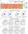 Figure 1
Distinct Mtb antigens elicit T cell responses with different functional properties (Cohort 1). (A) Whole blood samples from QFT+HIV– participants in Cohort 1 (KEMRI, Kisumu, Kenya) were stimulated with 60 individual Mtb antigens as overlapping peptides (1 μg/mL) for 7 days, and supernatants were harvested to quantitate IFN-γ by ELISA. The antigens are arranged left to right (antigens 1–60) according to their positions on the Mtb chromosome, and individual participant samples are arranged in rows. Data presented are results after subtraction of the average of 6 unstimulated wells as the background response. IFN-γ levels are color coded according to magnitude, with red for the lowest and green for the highest responses. Responses greater than the highest standard were assigned the value of the highest standard (1,000 pg/mL). Selected antigens are highlighted at the top of the heatmap, with arrows indicating 6 of the 7 RVMA. The identity of all of the antigens ordered in the same fashion is available in ref. 37. (B) Whole blood IFN-γ response magnitude of selected classical antigens (Tb10.4 [EsxH], PE13, PPE18, Ag85B, PPE46, Ag85A, CFP-10, ESAT-6, and EspI) compared with the 6 RVMA (Rv0010c, Rv0012, RimJ, LldD2, Rv2719c, and TB7.3); the red horizontal line represents the median response. Statistical significance was determined by the Mann-Whitney U test. (C) Representative flow cytometry plots comparing CD4 T cells producing IFN-γ or IL-17 in response to stimulation with one of the RVMA (Rv0012); background response for each cytokine is shown for comparison. (D and E) Cryopreserved peripheral blood mononuclear cells (PBMC) were stimulated with indicated antigens (2 mg/mL) for a total of 20 hours in the presence of Golgi Stop and Golgi Plug as well as costimulatory antibodies anti-CD28 and anti-CD49d; cytokine production by CD4+ T cells was determined by intracellular cytokine staining and flow cytometry. Data are values after subtraction of unstimulated cells, with values lower than the unstimulated control cells indicated below the dotted line (cut-off of positive response). The frequency of participants with a detectable response is shown at the top of each antigen plot. Each symbol is a distinct participant; the horizontal blue line indicates the median cytokine response. Results for RVMA are shown in D, and results for the classical antigens are shown in E.
In contrast to the findings in ref. 33, our assay platform and participant population did not show evidence for an antigenic island involving the ESX-3 locus. While EsxH (also known as Tb10.4) induced responses of modest magnitudes (median, 102 pg/mL), the other components of the Esx-3 locus (EsxG, EspG3, EccD3, MycP3, EccE3, and Rv0293c) induced minimal IFN-γ responses (medians, 4–14 pg/mL).
Compared with responses to the known classical antigens, we found lower magnitude IFN-γ responses (0–19 pg/mL; red color) to other Mtb antigens. The antigens that induced low magnitude IFN-γ responses include Rv0010c, Rv0012, RimJ, LldD2, Rv2719c, and TB7.3 (Rv3221c), which we previously identified as showing evidence of evolutionary diversifying selection (shown in arrows) and termed RVMA (30). While the overall IFN-γ responses to RVMA were low, some participants had higher magnitude IFN-γ responses (>100 pg/mL) to certain RVMA, although these were still lower than the magnitude of responses to the classical antigens (Figure 1B).
We also observed variation in responses to each of the 60 antigens at the level of individual participants (Figure 1A, rows), emphasizing that responses to a single Mtb antigen are not representative of the response to other Mtb antigens within an individual. Thus, we confirmed the diversity in responses to Mtb infection in humans in agreement with a previous study (38) and found that classical Mtb antigens elicit a robust IFN-γ response in whole blood of individuals with LTBI, while other antigens, including the RVMA, elicit lower frequency and lower magnitude IFN-γ responses.
Mtb antigens induce diverse cytokine responses that vary by antigen class
Response frequencies. Since RVMA are characterized by evidence of diversifying evolutionary selection and T cell epitope sequence variation, we hypothesized that they induce CD4 T cell responses distinct from those induced by the commonly studied classical Mtb antigens. We therefore asked whether RVMA induce CD4 T cells that express cytokines other than IFN-γ. For this analysis (and guided by the cell input requirement), we first randomly selected 4 RVMA that induced low frequency and low magnitude IFN-γ responses in the 60-antigen whole blood assay and compared the responses to 4 classical Mtb antigens that induced high frequency and high-magnitude IFN-γ responses. We stimulated PBMC from participants in Cohort 1 (Supplemental Table 1; supplemental material available online with this article; https://doi.org/10.1172/jci.insight.202134DS1) with the individual antigens and quantitated CD4 T cells expressing TNF-α, IFN-γ, IL-17, and/or GM-CSF by ICS and flow cytometry (Figure 1C and Supplemental Figure 1).
Consistent with the results of the whole blood assay, the 4 RVMA (Rv0010c, Rv0012, RimJ, and LldD2) induced IFN-γ–producing CD4 T cells in only a modest fraction of participants (38%–71%, depending on the individual antigen). In contrast, CD4 T cells responding to individual RVMA produced IL-17 in a high proportion (72%–85%) of participants. The frequency of participants whose CD4 T cells produced TNF-α or GM-CSF in response to the RVMA varied widely (45%–92%, depending on the individual antigen) (Figure 1D and Table 1).
At the level of individual antigens, all 4 RVMA induced IL-17–producing CD4 T cells in > 70% of the participants, while other cytokine responses varied widely by antigen. Of the RVMA, Rv0010c induced IFN-γ and GM-CSF at the lowest frequency (38% and 50%, respectively). Notably, Rv0012 induced GM-CSF–producing CD4 T cells in a high frequency (92%) of participants, a rate higher than any of the other RVMA or any of the classical antigens. RimJ induced TNF-α in only 54% and induced IFN-γ and GM-CSF with frequencies intermediate between those of IL-17 and TNF-α. LldD2 induced the lowest frequencies of TNF-α–, IFN-γ–, or GM-CSF–producing CD4 T cells (45%, 45%, and 55%, respectively) but induced IL-17–producing cells in 85% (Figure 1D and Table 1).
For comparison, we studied CD4 T cell production of the same 4 cytokines in response to 4 IFN-γ–dominant classical antigens. This revealed considerable variation in the frequency of individuals who responded with CD4 T cell production of TNF-α, IFN-γ, IL-17, and GM-CSF after stimulation with individual classical antigens (PPE18 [Rv1196], PPE46 [Rv3018c], ESAT-6 [Rv3875], and EspI [Rv3876]) (Figure 1E and Table 1). TNF-α responses to individual classical antigens were observed in 70%–87% of samples, with the highest frequency of TNF-α responses to PPE46. IFN-γ responses to the 4 classical antigens were observed in 57%–75% of samples, with little variation in the frequency of responses to PPE18, PPE46, and ESAT-6 and fewer responders to EspI. By comparison, the 4 classical antigens induced IL-17 (45%–68%) and GM-CSF (45%–75%) depending on the antigen.
When we compared the frequency of responders (that is, participants whose CD4 T cells expressed a given cytokine in response to Mtb antigen stimulation) by antigen class (i.e., classical versus RVMA), IL-17 responses were more frequent for RVMA than for classical Mtb antigens, while TNF-α, IFN-γ, and GM-CSF responses did not differ significantly (Table 2).
These data indicate that Mtb antigens vary in their induction of CD4 T cells that express different cytokines. The data also demonstrate that RVMA induce IL-17–producing CD4 T cells more frequently than IFN-γ– or TNF-α–producing T cells, and they induce IL-17 responses more frequently than do the classical antigens in a cohort of recently exposed household contacts (HHCs) that have controlled Mtb infection.
Response magnitudes. In addition to variation in the proportion (frequency) of participants whose CD4 T cells responded to individual antigens with expression of different cytokines, we found variation in the magnitude of the CD4 T cell responses, defined as the percent of CD4 T cells that produced the cytokine of interest in response to individual antigens.
For each of the individual RVMA, the magnitude of IL-17 responses was greater than for TNF-α, IFN-γ, or GM-CSF, with the exception that Rv0012 induced GM-CSF responses that exceeded those of each of the other 3 cytokines (Figure 1D and Table 3). As expected for the individual classical antigens, the magnitudes of TNF-α and IFN-γ response magnitudes were highest, with the exception that PPE46 induced GM-CSF responses that exceeded the magnitude of IFN-γ responses to Espl. IL-17 responses were present with the lowest magnitudes for all 4 of the classical antigens.
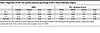 Table 3
Magnitudes of CD4 T cell cytokine responses (percentage of CD4 T cells) to individual antigens
For the RVMA as a group, the highest magnitude responses were for CD4 T cells that produce IL-17 (median, 0.46% of CD4 T cells), while the lowest magnitude responses were for IFN-γ (median 0.01%) (Figure 1D and Table 4). GM-CSF– and TNF-α–producing CD4 T cells were found with intermediate magnitudes (medians of 0.20% and 0.15%, respectively). In contrast, for the classical antigens as a group, the highest magnitude responses were for TNF-α–producing CD4 T cells (median, 0.19% of CD4 T cells) with slightly lower magnitude responses for IFN-γ (0.08%), IL-17 (0.05%), and GM-CSF (0.09%). When comparing cytokine responses by antigen group, differences were significant for IL-17 (RVMA > classical) and IFN-γ (classical > RVMA) (Table 4). These analyses revealed that, consistent with the frequency of responders, the magnitude of IL-17–producing CD4 T cells is higher for RVMA than for classical antigens.
To determine whether the IL-17 bias of CD4 T cell responses to RVMA extends to another human population, we used the same experimental procedures to study samples from participants recruited in the same manner and with the same criteria as for Cohort 1, in an independent cohort in Addis Ababa, Ethiopia (Cohort 2). The 2 cohorts were comparable in their distribution of age, sex, body mass index (BMI), and hemoglobin A1c (Supplemental Table 1). The distribution of sex was comparable between the cohorts (P = 0.2611, Fisher’s exact test), and when we considered sex as a biological variable, we found no systematic differences in the results within or between cohorts according to sex. Results of QuantiFERON testing differed between the cohorts: Cohort 1 had significantly higher magnitude responses for TB Antigen minus Nil, while Cohort 2 had higher responses for Mitogen minus Nil (Supplemental Table 1).
In Cohort 2, the frequency of participants whose cells responded to RVMA was lower than for classical antigens; the difference was significant for IFN-γ and for IL-17 but not for TNF-α or GM-CSF (Figure 2, A and B, and Supplemental Tables 2 and 3). Likewise, the magnitudes of cytokine-producing CD4 T cells in Cohort 2 were lower in response to RVMA compared with classical antigens; the differences in magnitudes between RVMA and classical antigens were significant for TNF-α, IFN-γ, and IL-17 but not for GM-CSF (Supplemental Table 4). Because of the lower-magnitude responses, the IL-17 response magnitudes to the RVMA did not exceed the IL-17 responses to classical antigens. Despite the overall lower-magnitude responses, IL-17 responses were higher than other cytokine responses to RVMA (Figure 2A and Supplemental Table 4). When we analyzed IL-17 and IFN-γ responses to individual RVMA in Cohort 2, we found that the frequencies of IL-17 responders exceeded those for IFN-γ for all 4 of the RVMA (Supplemental Table 5). Likewise, the magnitudes of IL-17 responses exceeded those of IFN-γ responses for all 4 of the RVMA; the differences were significant for Rv0012 and LldD2. Comparison of the cytokine response magnitudes to all of the RVMA together revealed that IL-17 responses were significantly greater than TNF-α or IFN-γ (P = 0.0378 and 0.0018, respectively) but not GM-CSF (Friedman test with Dunn’s correction for multiple comparisons). Despite the differences in the overall magnitudes of CD4 T cell responses to RVMA in Cohort 2 compared with Cohort 1, the results confirmed the findings in Cohort 1 that RVMA predominantly induce CD4 T cells that produce IL-17.
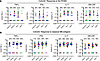 Figure 2
Distinct Mtb antigens elicit T cell responses with different functional properties (Cohort 2). Procedures and analyses were as described in Figure 1; the samples were obtained from participants in Cohort 2 (AHRI, Addis Ababa, Ethiopia). (A and B) Results for RVMA are shown in A; results for classical Mtb antigens are shown in B.
In additional experiments, we assessed the same cytokine responses to the remaining 3 RVMA (Rv0990c, Rv2719c, and Rv3221c) (30), and 4 additional antigens that are conserved and nonsecreted proteins and that exhibited low IFN-γ responses by ELISA (Rv1366, Rv1957, Rv2022c, and Rv2031c) — the LICA — using PBMC obtained from Cohort 2 participants. Consistent with findings from the first RVMA set, these antigens predominantly induced IL-17 responses, both in response frequencies and magnitudes (Supplemental Figure 2 and Supplemental Table 5), with the hierarchy of responses being IL-17 > GM-CSF > IFN-γ > TNF-α. IL-17 responses were significantly greater than TNF-α or IFN-γ (P = 0.0005 and 0.0478, respectively) but not GM-CSF (Friedman test with Dunn’s correction for multiple comparisons). Rv3221c elicited the highest frequency and magnitude responses (57%–87%; 0.04%–0.51% of CD4 T cells) for any of the cytokines (Supplemental Figure 2 and Supplemental Table 5). Grouped analysis of RVMA revealed significantly higher responses for IL-17 than IFN-γ (Supplemental Table 6).
The LICA induced strong IL-17 and GM-CSF responses (>71% prevalence for each antigen) with comparable magnitudes across antigens (Supplemental Figure 2). IFN-γ responses were lower (54%–71% prevalence; median magnitude 0.04%–0.19 % of CD4 T cells), whereas TNF-α responses were the lowest — e.g., Rv1366 responses were detectable in only 21% of donors with a median magnitude below the limit of detection. When grouped, the LICA showed significantly higher IL-17 than IFN-γ responses (Supplemental Table 6). Collectively, these findings reinforce the preferential induction of IL-17 by RVMA and underscore heterogeneity in IFN-γ responses to classical Mtb antigens.
In Cohort 1, RVMA and IFN-γ–dominant classical antigens induced similar magnitudes of TNF+IL-17+ and IFN-γ+IL-17+ cells, while RVMA induced lower magnitudes of TNF+IFN-γ+ cells than did classical antigens. In Cohort 2, RVMA induced lower magnitudes of cells expressing each of the 3 dual cytokine combinations compared with those induced by IFN-γ–dominant classical antigens (Supplemental Figure 3).
RVMA-responsive CD4 T cells include bona fide Th17 cells
To further investigate CD4 T cells that respond to RVMA, we extended our studies to include additional markers associated with Th17 cells. Since peptide:HLA multimers for the Cohort 1 and Cohort 2 participants’ HLA alleles are not yet available, we used a T cell activation induced marker (AIM) assay (39–41) based on surface expression of CD154 after brief antigen stimulation in the absence of protein transport inhibitors, and we then assayed expression of RORγT and CCR6 (Th17) or T-bet and CXCR3 (Th1) on the Mtb antigen–activated CD154+ cells (Figure 3A). Stimulation with RVMA induced lower frequencies of CD154+ CD4 T cells in PBMC than did classical antigens (Figure 3B), consistent with immunodominance of classical antigens in the latter group. When we analyzed lineage-defining transcription factor (TF) expression on antigen-responsive (CD4+CD154+) cells, we found a significantly higher fraction of RORγT+T-bet– cells in RVMA-responsive cells than in cells that responded to classical antigens (Figure 3C). In turn, we observed a significantly higher fraction of RORγT–T-bet+ cells responding to classical antigens than to RVMA. These results are in accord with the cytokine data (Figures 1 and 2) and indicate that, beyond cytokine production, RVMA-responsive CD4 T cells exhibit other characteristics of Th17 cells while confirming that classical antigen-responsive T cells are typical Th1 cells. We found no difference in double-positive (RORγT+T-bet+) cells according to antigen category (Figure 3C).
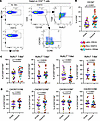 Figure 3
Antigen-activated cells express markers of Th17 and Th1 cells upon stimulation with RVMA and classical antigens, respectively. Cryopreserved PBMC from participants in Cohort 1 were stimulated with antigens for a total of 20 hours in the presence of costimulatory antibodies anti-CD28 and anti-CD49d and anti-CD40 blocking antibody without protein transport inhibitors, and antigen-activated cells were identified by CD154 surface expression on CD4+ T cells. (A) Representative flow cytometry plots. (B) CD154 surface expression on CD4+ T cells; CD154+ was used to identify antigen-activated T cells. (C and D) Expression of Th cell lineage-defining transcription factors (RORγT = Th17 and T-bet = Th1) (C) and expression of chemokine receptors (CCR6 = Th17 marker) and (CXCR3 = Th1 marker) on antigen-activated T cells (D). Wilcoxon matched pairs test was used.
Since certain chemokine receptors (CRs) are reported to be associated with specific CD4 T cell subsets, we also examined the expression of CCR6 (characteristic of Th17 cells) and CXCR3 (characteristic of Th1 cells). This revealed a significantly higher incidence of CXCR3+CCR6– (Th1) and CXCR3+CCR6+ (Th1*) on antigen-activated (CD4+CD154+) cells responding to classical antigens than to the RVMA (Figure 3D), consistent with the previously reported Mtb-responsive human Th1* responses to Mtb peptide pools (33, 42–44). In contrast, the incidence of CD4+CD154+ cells that were CXCR3–CCR6+ (Figure 3D) did not differ by antigen class. Notably, T cells that responded (CD4+CD154+) to RVMA exhibit a higher incidence of being CXCR3–CCR6– than are classical antigen-activated CD4+ cells, suggesting that some RVMA-responsive cells do not adhere to a conventional pattern of CR expression.
RVMA are enriched for nonclassical Th1 or Th17 responses
The ICS data reveal prevalent detection of GM-CSF (50%–92%, depending on antigen, with Rv0012 particularly robust in inducing GM-CSF production) in both Cohort 1 and Cohort 2 (Figure 1D, Figure 2A, and Supplemental Figure 2A), with a relative magnitude of 0.1% of CD4+ T cells. The cytokine GM-CSF has been little studied in TB, and its role in the outcome of Mtb infection remains unclear, although studies in mice indicate that T cell–derived GM-CSF can contribute to restricting infection (45). We also observed enrichment of CD154+CCR6–CXCR3– and CD154+T-bet–RORγT– RVMA-responsive CD4 T cells compared with classical antigens (Figure 3). To comprehensively investigate whether RVMA induce T cell responses with phenotypes other than classical Th17 (and in some cases low-level Th1), we selected participants in whom cytokines, TFs, and CRs were measured and antigens for which this data was complete. This yielded 41 samples in Cohort 1 and 29 samples in Cohort 2 with 3 RVMA (Rv0012, RimJ, and LldD2) and 3 classical antigens (PPE18, PPE46, and ESAT-6).
To identify features capable of distinguishing between classical and RVMA antigen responses, we applied a Random Forest classification algorithm using the caret package in R and performed the analysis on Cohort 1 and Cohort 2 (see computational analysis methods).
In Cohort 1, the top marker combination distinguishing RVMA from classical antigens was CD154+CCR6–CXCR3–, followed by CD154+T-bet–RORγT– and GM-CSF (Figure 4A). The abundance of CD154+CCR6–CXCR3– and CD154+T-bet–RORγT– cells was significantly higher for RVMA compared with classical antigens (Figure 4B). Among RVMA antigens, the strongest discriminant was GM-CSF, followed by CD154+CCR6–CXCR3– and CD154+T-bet–RORγT– (Figure 4C), though their abundance did not differ significantly across individual antigens (Figure 4D).
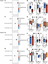 Figure 4
RVMA are enriched for nonclassical Th1 or Th17 responses. (A–H) Comparison of RVMA and classical antigen-specific antigens marker profile in Cohort 1 (A–D) and Cohort 2 (E–H). A, C, E, and G show random forest feature selection analysis results. The y axis shows the variable names ordered by the Mean Decrease Gini. The x axis is displaying the Mean Decrease Gini values, while the color represents the Mean decrease in Accuracy. B, D, F, and H show box and whisker plots from the most informative variables identified by the random forest algorithm. The y axis shows the variable abundance value transformed in log10, while the x axis and colors display the antigen groups or antigen identity. Group comparison was performed with the Wilcoxon test, and P values were adjusted by FDR.
In Cohort 2, the key discriminant between RVMA and classical antigens was CD154+CCR6–CXCR3– followed by CD154+T-bet–RORγT– and GM-CSF (Figure 4E), with no significant differences in their abundance (Figure 4F). When comparing individual RVMA antigens, CD154+T-bet–RORγT– emerged as the strongest discriminator, followed by CD154+CCR6–CXCR3– and GM-CSF (Figure 4G). Notably, CD154+T-bet–RORγT– cells were significantly more abundant in LldD2 compared with RimJ and Rv0012 responsive cells, while GM-CSF and CD154+CCR6–CXCR3– levels did not differ among the antigens (Figure 4H). Together, these findings highlight the heterogeneity of CD4 T cell responses induced by RVMA antigens, including nonclassical Th17 and Th1 phenotypes, and underscore the diversity of immune responses in human populations.
RVMA-responsive CD4 T cells have fewer intermarker interactions than classical antigens
Finally, we examined associations between cytokines, TFs, and CRs expressed by antigen-responsive CD4 T cells that responded to RVMA (Rv0012, RimJ, and LldD2) or to classical antigens (ESAT-6, PPE18, and PPE46). Analyses were restricted to participants with complete data for all 3 variables. To investigate correlations among cytokines (blue), TFs (purple), and CRs (yellow), and by Th1 (orange) or Th17 (green) subset, we applied Spearman’s correlation separately for each cohort and antigen group (RVMA and classical). Only statistically significant correlations, defined as having an FDR-adjusted P < 0.05 and an absolute Spearman’s rho > 0.4, were included. These significant relationships were visualized using chord diagrams (Figure 5, A–D) to highlight the most relevant interactions.
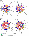 Figure 5
RVMA-responsive CD4 T cells exhibit fewer intermarker interactions. Correlations identified in Cohort 1 (top) and Cohort 2 (bottom), in both RVMA (Rv0012, RimJ, and LldD2) and classical (PPE18, PPE46, and ESAT-6) antigen–stimulated samples. Chord diagrams depicting the correlation identified in RVMA (A and C) and classical (B and D) antigens, using Spearman’s correlation analysis. Only correlations with |rho| > 0.4 and FDR < 0.05 were considered. Positive and negative correlations are represented by the red and blue lines, respectively. Cytokines, TFs, and CRs are highlighted in the outer ring as blue, purple, and yellow areas, respectively. Th1 and Th17 variables are highlighted in the inner ring as Orange and Green, respectively.
CD4 T cells that responded to RVMA antigens exhibited fewer intermarker network interactions than those responsive to classical antigens. While there were more likely interactions between TFs and CRs, the cytokine interactions were mostly restricted to within the cytokine variable. Consistent with the expected biology, CD154+RORγT–T-bet– correlated positively with CD154+CCR6–CXCR3–, suggesting Mtb-antigen responsive CD4 T cells that are neither Th17 nor Th1, and CD154+CXCR3+CCR6– correlated positively with CD154+RORγT–T-bet+, indicating a Th1 subset, whereas CD154+RORγT+T-bet– inversely correlated with TNF-α+IFN-γ+. Notably, GM-CSF strongly correlated with IL-17, particularly in Cohort 1. These results indicate that Mtb antigens influence CD4 T cell differentiation through distinct transcription factor and chemokine receptor expression profiles and highlight a functional link between GM-CSF– and IL-17–producing CD4 T cells.


