
Research ArticleCell biologyPulmonology
Open Access |  10.1172/jci.insight.201695
αTAT1 links microtubules to stiffness sensing, fibroblast activation, and fibrosis
Alba Santos,1,2,3 Tamanna Islam,4,5 Maria-Anna Chrysovergi,1,2,3 Paula E. Grasberger,1,2,3 Vera Auernheimer,6 Taslim A. Al-Hilal,4,5 Santiago Lamas,7 and David Lagares1,2,3
1Department of Medicine;
2Fibrosis Research Center; and
3Center for Immunology and Inflammatory Diseases; Massachusetts General Hospital, Harvard Medical School, Boston, Massachusetts, USA.
4Department of Molecular Pharmaceutics, College of Pharmacy; and
5Department of Biomedical Engineering, John and Marcia Price College of Engineering; University of Utah, Salt Lake City, Utah, USA.
6Friedrich-Alexander University of Erlangen-Nuremberg, Erlangen, Germany.
7Centro de Biología Molecular “Severo Ochoa” (CSIC-UAM), Madrid, Spain.
Address for correspondence. David Lagares, Center for Immunology and Inflammatory Diseases, Massachusetts General Hospital, Harvard Medical School, 149 13th Street, Charlestown, Massachusetts 02129, USA. Phone: 857.264.8078; Email: david.lagares@sloan.mit.edu or dlagares@mgh.harvard.edu.
Find articles by Santos, A. in: PubMed | Google Scholar
1Department of Medicine;
2Fibrosis Research Center; and
3Center for Immunology and Inflammatory Diseases; Massachusetts General Hospital, Harvard Medical School, Boston, Massachusetts, USA.
4Department of Molecular Pharmaceutics, College of Pharmacy; and
5Department of Biomedical Engineering, John and Marcia Price College of Engineering; University of Utah, Salt Lake City, Utah, USA.
6Friedrich-Alexander University of Erlangen-Nuremberg, Erlangen, Germany.
7Centro de Biología Molecular “Severo Ochoa” (CSIC-UAM), Madrid, Spain.
Address for correspondence. David Lagares, Center for Immunology and Inflammatory Diseases, Massachusetts General Hospital, Harvard Medical School, 149 13th Street, Charlestown, Massachusetts 02129, USA. Phone: 857.264.8078; Email: david.lagares@sloan.mit.edu or dlagares@mgh.harvard.edu.
Find articles by Islam, T. in: PubMed | Google Scholar
1Department of Medicine;
2Fibrosis Research Center; and
3Center for Immunology and Inflammatory Diseases; Massachusetts General Hospital, Harvard Medical School, Boston, Massachusetts, USA.
4Department of Molecular Pharmaceutics, College of Pharmacy; and
5Department of Biomedical Engineering, John and Marcia Price College of Engineering; University of Utah, Salt Lake City, Utah, USA.
6Friedrich-Alexander University of Erlangen-Nuremberg, Erlangen, Germany.
7Centro de Biología Molecular “Severo Ochoa” (CSIC-UAM), Madrid, Spain.
Address for correspondence. David Lagares, Center for Immunology and Inflammatory Diseases, Massachusetts General Hospital, Harvard Medical School, 149 13th Street, Charlestown, Massachusetts 02129, USA. Phone: 857.264.8078; Email: david.lagares@sloan.mit.edu or dlagares@mgh.harvard.edu.
Find articles by
Chrysovergi, M.
in:
PubMed
|
Google Scholar
|

1Department of Medicine;
2Fibrosis Research Center; and
3Center for Immunology and Inflammatory Diseases; Massachusetts General Hospital, Harvard Medical School, Boston, Massachusetts, USA.
4Department of Molecular Pharmaceutics, College of Pharmacy; and
5Department of Biomedical Engineering, John and Marcia Price College of Engineering; University of Utah, Salt Lake City, Utah, USA.
6Friedrich-Alexander University of Erlangen-Nuremberg, Erlangen, Germany.
7Centro de Biología Molecular “Severo Ochoa” (CSIC-UAM), Madrid, Spain.
Address for correspondence. David Lagares, Center for Immunology and Inflammatory Diseases, Massachusetts General Hospital, Harvard Medical School, 149 13th Street, Charlestown, Massachusetts 02129, USA. Phone: 857.264.8078; Email: david.lagares@sloan.mit.edu or dlagares@mgh.harvard.edu.
Find articles by Grasberger, P. in: PubMed | Google Scholar
1Department of Medicine;
2Fibrosis Research Center; and
3Center for Immunology and Inflammatory Diseases; Massachusetts General Hospital, Harvard Medical School, Boston, Massachusetts, USA.
4Department of Molecular Pharmaceutics, College of Pharmacy; and
5Department of Biomedical Engineering, John and Marcia Price College of Engineering; University of Utah, Salt Lake City, Utah, USA.
6Friedrich-Alexander University of Erlangen-Nuremberg, Erlangen, Germany.
7Centro de Biología Molecular “Severo Ochoa” (CSIC-UAM), Madrid, Spain.
Address for correspondence. David Lagares, Center for Immunology and Inflammatory Diseases, Massachusetts General Hospital, Harvard Medical School, 149 13th Street, Charlestown, Massachusetts 02129, USA. Phone: 857.264.8078; Email: david.lagares@sloan.mit.edu or dlagares@mgh.harvard.edu.
Find articles by Auernheimer, V. in: PubMed | Google Scholar
1Department of Medicine;
2Fibrosis Research Center; and
3Center for Immunology and Inflammatory Diseases; Massachusetts General Hospital, Harvard Medical School, Boston, Massachusetts, USA.
4Department of Molecular Pharmaceutics, College of Pharmacy; and
5Department of Biomedical Engineering, John and Marcia Price College of Engineering; University of Utah, Salt Lake City, Utah, USA.
6Friedrich-Alexander University of Erlangen-Nuremberg, Erlangen, Germany.
7Centro de Biología Molecular “Severo Ochoa” (CSIC-UAM), Madrid, Spain.
Address for correspondence. David Lagares, Center for Immunology and Inflammatory Diseases, Massachusetts General Hospital, Harvard Medical School, 149 13th Street, Charlestown, Massachusetts 02129, USA. Phone: 857.264.8078; Email: david.lagares@sloan.mit.edu or dlagares@mgh.harvard.edu.
Find articles by Al-Hilal, T. in: PubMed | Google Scholar
1Department of Medicine;
2Fibrosis Research Center; and
3Center for Immunology and Inflammatory Diseases; Massachusetts General Hospital, Harvard Medical School, Boston, Massachusetts, USA.
4Department of Molecular Pharmaceutics, College of Pharmacy; and
5Department of Biomedical Engineering, John and Marcia Price College of Engineering; University of Utah, Salt Lake City, Utah, USA.
6Friedrich-Alexander University of Erlangen-Nuremberg, Erlangen, Germany.
7Centro de Biología Molecular “Severo Ochoa” (CSIC-UAM), Madrid, Spain.
Address for correspondence. David Lagares, Center for Immunology and Inflammatory Diseases, Massachusetts General Hospital, Harvard Medical School, 149 13th Street, Charlestown, Massachusetts 02129, USA. Phone: 857.264.8078; Email: david.lagares@sloan.mit.edu or dlagares@mgh.harvard.edu.
Find articles by
Lamas, S.
in:
PubMed
|
Google Scholar
|
1Department of Medicine;
2Fibrosis Research Center; and
3Center for Immunology and Inflammatory Diseases; Massachusetts General Hospital, Harvard Medical School, Boston, Massachusetts, USA.
4Department of Molecular Pharmaceutics, College of Pharmacy; and
5Department of Biomedical Engineering, John and Marcia Price College of Engineering; University of Utah, Salt Lake City, Utah, USA.
6Friedrich-Alexander University of Erlangen-Nuremberg, Erlangen, Germany.
7Centro de Biología Molecular “Severo Ochoa” (CSIC-UAM), Madrid, Spain.
Address for correspondence. David Lagares, Center for Immunology and Inflammatory Diseases, Massachusetts General Hospital, Harvard Medical School, 149 13th Street, Charlestown, Massachusetts 02129, USA. Phone: 857.264.8078; Email: david.lagares@sloan.mit.edu or dlagares@mgh.harvard.edu.
Find articles by
Lagares, D.
in:
PubMed
|
Google Scholar
|
Published November 9, 2026 - More info
JCI Insight. 2026;11(21):e201695. https://doi.org/10.1172/jci.insight.201695.
© 2026 Santos et al. This work is licensed under the Creative Commons Attribution 4.0 International License. To view a copy of this license, visit http://creativecommons.org/licenses/by/4.0/.
Received: October 27, 2025; Accepted: September 21, 2026

-
Results
Microtubule acetylation underlies exaggerated durotaxis in IPF fibroblasts. To investigate the role of microtubules in stiffness sensing and fibroblast durotaxis, we established in vitro stiffness-gradient models using primary human lung fibroblasts from patients with IPF and healthy controls. Using microfluidic-engineered hydrogels spanning a stiffness range of 4–40 kPa over 1 mm (8), we performed long-term live-cell imaging to track single-cell migration in real time. On these substrates, fibroblasts exhibited durotactic responses and clear front-rear polarization oriented toward stiffer regions (Figure 1, A and B). Notably, IPF fibroblasts exhibited enhanced durotactic responses and a more narrowly focused migratory trajectory along the stiffness gradient compared with healthy fibroblasts. Morphological analysis revealed that durotaxing IPF fibroblasts formed a higher number of leading-edge protrusions aligned with the direction of increasing stiffness (Figure 1, B and C). These protrusions were highly dynamic, undergoing rapid cycles of microtubule polymerization and depolymerization, known as dynamic instability, which enables cells to explore their mechanical environment (24). Costaining for F-actin and α-tubulin showed that while actin networks organized in polarized stress fibers, the protrusive structures in durotaxing IPF fibroblasts were enriched in microtubules (Figure 1C). These observations suggest that leading-edge microtubule dynamics facilitate cellular probing and alignment along stiffness gradients. Since microtubule dynamics are finely tuned by posttranslational modifications, we next examined α-tubulin acetylation at K40, a modification previously described to promote directional persistence (25). Importantly, fibroblasts derived from IPF patients exhibited elevated basal α-tubulin K40 acetylation compared with healthy controls (Figure 1D). Moreover, Western blot analyses revealed a stiffness-dependent increase in K40 α-tubulin acetylation in healthy control fibroblasts, with higher levels in fibroblasts cultured on stiff (64 kPa) versus soft (0.5 kPa) matrices (Figure 1E). Immunofluorescence staining confirmed increased acetylated α-tubulin in healthy fibroblasts cultured on stiff matrices compared with soft substrates (Figure 1F). Together, these findings link leading-edge microtubule organization and K40 acetylation to stiffness sensing and the exaggerated durotactic response of IPF fibroblasts.
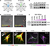 Figure 1
Microtubule acetylation underlies exaggerated durotaxis in IPF fibroblasts. (A) Rose diagrams showing migration directionality of primary human lung fibroblasts from healthy donors and patients with idiopathic pulmonary fibrosis (IPF) seeded on stiffness-gradient hydrogels. IPF fibroblasts exhibit enhanced directional persistence toward stiffer regions. Radial length indicates the number of events in each trajectory (n = 4 control, n = 4 IPF). (B) Representative phase-contrast images of fibroblasts migrating along a stiffness gradient. IPF fibroblasts display more polarized protrusions aligned with the stiffness gradient than control fibroblasts (red arrows) (n = 4 control, n = 4 IPF). Scale bar: 50 μm. (C) Confocal images of control and IPF fibroblasts stained for F-actin (red), α-tubulin (green), and nuclei (DAPI, blue). IPF fibroblasts show enhanced microtubule-based protrusions and cytoskeletal polarization toward stiffer substrates (n = 4). Scale bar: 50 μm. (D) Western blot analysis of acetylated α-tubulin and α-smooth muscle actin (αSMA) in fibroblasts from healthy donors (n = 3, N1–N3) and IPF patients (n = 3, IPF1–IPF3) cultured on regular tissue culture plastic, showing elevated α-tubulin K40 acetylation in IPF cells. GAPDH, loading control. (E) Matrix stiffness–dependent induction of α-tubulin K40 acetylation in normal lung fibroblasts cultured on polyacrylamide substrates of increasing stiffness (0.2–64 kPa). α-Tubulin was used as a loading control (n = 3). (F) Immunofluorescence images showing increased acetylated α-tubulin (magenta) in healthy control fibroblasts cultured on stiff (64 kPa) compared with soft (0.5 kPa) matrices. Total α-tubulin (green) marks the microtubule network (n = 4). Scale bar: 50 μm.
αTAT1-mediated α-tubulin K40 acetylation controls fibroblast durotaxis in vitro. Given the enrichment of α-tubulin K40 acetylation at the leading edge of durotaxing fibroblasts, we next investigated the molecular mechanisms governing this modification. α-Tubulin K40 acetylation is catalyzed by αTAT1, which localizes to clathrin-coated pits at the leading edge, spatially polarizing microtubule acetylation to promote directional migration (25). In contrast, histone deacetylase 6 (HDAC6) and sirtuin 2 (SIRT2) mediate deacetylation of the same residue, thereby regulating the dynamic balance of this posttranslational modification (26, 27) (Figure 2A). We found that αTAT1 expression was significantly upregulated in fibroblasts isolated from fibrotic lungs of patients with IPF compared with healthy controls, whereas HDAC6 and SIRT2 levels remained unchanged (Figure 2B), suggesting selective upregulation of the αTAT1-dependent acetylation pathway during fibrotic remodeling. To determine whether αTAT1-dependent acetylation is required for durotaxis, we silenced ATAT1 mRNA using siRNA. ATAT1 knockdown markedly impaired durotaxis on step-gradient durotaxis hydrogels in control fibroblasts. Extending these findings to human disease fibroblasts, both IPF and scleroderma fibroblasts displayed pronounced hyperdurotaxis compared with healthy controls, and ATAT1 knockdown attenuated their directional migration along stiffness gradients (Figure 2C). Importantly, loss of ATAT1 did not alter chemotaxis toward 20% serum in control or diseased fibroblasts, indicating that αTAT1 specifically controls stiffness-directed migration rather than general motility (Figure 2D). Together, these results identify αTAT1-mediated α-tubulin K40 acetylation as a key molecular driver of fibroblast durotaxis in fibrotic disease.
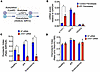 Figure 2
αTAT1 regulates fibroblast durotaxis through α-tubulin acetylation. (A) Schematic illustrating reversible α-tubulin acetylation catalyzed by αTAT1 at lysine-40 (K40) and deacetylation by HDAC6 and SIRT2. (B) mRNA expression levels of αTAT1, SIRT2, and HDAC6 in fibroblasts derived from healthy donors and patients with IPF, normalized to GAPDH (n = 2 control, n = 2 IPF). (C) Quantification of durotactic migration in fibroblasts from healthy donors and patients with IPF and patients with scleroderma transfected with non-targeting (NT) or ATAT1 siRNA. ATAT1 knockdown significantly reduced stiffness-directed migration across all fibroblast types (n = 3 per group). (D) Quantification of chemotaxis toward 20% FBS. Loss of ATAT1 does not affect chemotactic migration, indicating that αTAT1 specifically regulates stiffness-directed (durotactic) migration rather than soluble factor–driven migration (n = 3 per group). Data are presented as mean ± SD. Statistical significance was determined by 2-tailed unpaired (B) and paired (C and D) Student’s t test. *P < 0.05, **P < 0.01.
Atat1-deficient mice are protected from bleomycin-induced lung fibrosis. Building on our in vitro studies, we next examined how αTAT1-dependent acetylation influences fibroblast biology in vivo and contributes to tissue fibrosis. To begin investigating the role of microtubule acetylation in vivo, we first assessed α-tubulin K40 acetylation in lung tissues from mice subjected to bleomycin-induced lung fibrosis (28, 29) (Figure 3A). Immunofluorescence costaining of lung sections for αSMA, a marker of myofibroblast activation, and acetylated α-tubulin revealed that α-tubulin K40 acetylation was increased in α-smooth muscle actin–positive (αSMA+) myofibroblasts in fibrotic lungs compared with saline-treated controls (Figure 3, A and B). In control mice, K40-acetylated α-tubulin was restricted primarily to bronchial epithelial cells, consistent with previous observations. To determine whether this increase was also evident in fibroblasts isolated from fibrotic lungs, we isolated primary lung fibroblasts from bleomycin- or PBS-treated mice. Western blot analysis confirmed that fibroblasts from fibrotic lungs exhibited elevated α-tubulin K40 acetylation compared with controls (Figure 3C). We next examined the functional role of αTAT1 in vivo using Atat1-deficient mice (Atat1–/–) from the Knockout Mouse Project. As expected, primary lung fibroblasts isolated from Atat1–/– mice lacked detectable α-tubulin K40 acetylation by Western blot compared with fibroblasts from wild-type (WT) Atat1+/+ mice (Figure 3, D and E). In functional in vitro studies, Atat1–/– lung fibroblasts exhibited reduced durotactic migration compared with Atat1+/+ control lung fibroblasts (Figure 3F). To determine whether αTAT1 activity contributes to lung fibrosis development in vivo, we examined the impact of global Atat1 deletion in the bleomycin-induced lung fibrosis model. Atat1–/– mice were markedly protected from bleomycin-induced fibrosis compared with WT Atat1+/+ controls, as demonstrated by reduced fibrosis in Masson’s trichrome–stained lung sections at day 14 after bleomycin challenge (Figure 3G). Consistent with these observations, biochemical quantification of total collagen content by hydroxyproline assay confirmed significantly lower levels of fibrosis in Atat1-deficient lungs (Figure 3H). To define the mechanism underlying this protection, we assessed inflammation and vascular permeability at day 7 following bleomycin exposure. Vascular permeability measured by total protein in bronchoalveolar lavage (BAL) fluid was indistinguishable between genotypes (Figure 3I), suggesting that αTAT1 does not influence vascular leak or acute epithelial-endothelial barrier disruption. Flow cytometric analysis of BAL cells revealed comparable numbers of total leukocytes (Figure 3J), including neutrophils, T cells, and monocytes, in Atat1–/– and Atat1+/+ mice (data not shown), indicating that αTAT1 does not regulate the inflammatory phase of lung injury. We next examined whether αTAT1 contributes to myofibroblast activation during fibrosis. Immunofluorescence revealed increased α-tubulin K40 acetylation in αSMA+ myofibroblasts of bleomycin-treated Atat1+/+ lungs, whereas Atat1-deficient mice exhibited markedly fewer αSMA+ cells within fibrotic foci (Figure 3, K and L). Taken together, our results identify αTAT1 as a key regulator of fibroblast mechanosensing and myofibroblast activation in vivo. Loss of Atat1 attenuates lung fibrosis without affecting inflammation or vascular injury, supporting a direct role for αTAT1 in driving fibrotic remodeling.
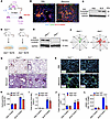 Figure 3
αTAT1 deficiency protects mice from bleomycin-induced lung fibrosis. (A) Schematic of the bleomycin-induced (BLM-induced) lung fibrosis model. Mice received a single intratracheal (i.t.) instillation of BLM (1.2 U/kg), and lungs were harvested 14 days later. (B) Immunofluorescence staining of lung sections from PBS- and BLM-treated mice showing increased α-tubulin K40 acetylation (Ac α-tubulin, red) in αSMA+ myofibroblasts (green). Nuclei: DAPI (blue). (n = 4 mice per group.) Scale bar: 100 μm. (C) Immunoblot analysis of α-tubulin K40 acetylation in fibroblasts isolated from fibrotic versus control mouse lungs. GAPDH, loading control. (n = 3 mice per group.) (D) Schematic of lung fibroblast isolation from wild-type (WT; Atat1+/+) and Atat1-deficient (Atat1–/–) mice. (E) Western blot showing loss of α-tubulin K40 acetylation in Atat1–/– fibroblasts compared with controls (n = 4 per group). (F) Quantification of fibroblast durotaxis on stiffness-gradient hydrogels, showing impaired directional migration in Atat1–/– cells (n = 3 mice per group.) (G) Representative Masson’s trichrome–stained lung sections from Atat1+/+ and Atat1–/– mice after PBS or BLM challenge, showing reduced collagen deposition in Atat1–/– lungs (n = 6 mice per group). Scale bar: 100 μm. (H) Quantification of lung collagen content by hydroxyproline (n = 6 mice per group). (I) Bronchoalveolar lavage (BAL) fluid protein concentration (n = 6 mice per group). (J) Flow cytometric quantification of immune cells in BAL at day 7 (n = 4 mice per group). (K) Immunofluorescence staining showing reduced αSMA+ myofibroblast accumulation (green) in BLM-treated Atat1–/– mice compared with Atat1+/+ controls. Nuclei: DAPI (blue). (n = 4 mice per group.) Scale bar: 100 μm. (L) Quantification of αSMA+ myofibroblasts per field (n = 4 mice per group). Data are presented as mean ± SD. For animal experiments, statistical significance was determined by 1-way ANOVA followed by Šidák’s multiple-comparison test. ****P < 0.0001.
Atat1-deficient mice are protected from bleomycin-induced dermal fibrosis. To determine whether the protective effects of genetic ablation of Atat1 extend beyond the lung, we next examined dermal fibrosis in Atat1-deficient mice subjected to daily bleomycin injections for 28 days (Figure 4A). Histological analysis of Masson’s trichrome–stained sections revealed a striking reduction in both dermal thickening and collagen deposition in Atat1–/– mice compared with WT controls (Figure 4, A–D). Biochemical quantification confirmed a similar decrease in total collagen content, as measured by hydroxyproline assay (Figure 4C). Importantly, Atat1–/– mice exhibited markedly fewer αSMA+ myofibroblasts following bleomycin challenge compared with WT controls (Figure 4, E and F), consistent with impaired fibroblast recruitment and/or activation. Together, these findings demonstrate that αTAT1-mediated α-tubulin acetylation promotes fibroblast activation and myofibroblast accumulation in fibrotic skin, paralleling its role in the lung. To directly determine the role of αTAT1 in fibroblast-driven fibrosis in vivo, we generated mice with conditional fibroblast-specific Atat1 deficiency by crossing Atat1fl/fl mice with Pdgfra-CreERT2 mice, which express a tamoxifen-inducible Cre recombinase in mesenchymal lineages including dermal fibroblasts (Figure 4G). Tamoxifen administration to Pdgfra-CreERT2 Atat1fl/fl mice resulted in efficient genetic ablation of Atat1 and complete loss of acetylated α-tubulin in primary dermal fibroblasts, as demonstrated by Western blot analysis (Figure 4H). Consistent with our findings in global-knockout mice, fibroblast-specific Atat1 deletion led to a significant reduction in dermal thickness and collagen deposition in the bleomycin-induced skin fibrosis model (Figure 4, I and J). Together, these data demonstrate that fibroblast-specific loss of Atat1 is sufficient to attenuate fibrotic remodeling, supporting a fibroblast-intrinsic role for αTAT1-dependent mechanotransduction in vivo.
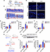 Figure 4
αTAT1 deficiency protects mice from bleomycin-induced skin fibrosis. (A) Schematic of the mouse model of bleomycin-induced (BLM-induced) skin fibrosis. Mice received daily BLM injections for 28 days before skin harvest. (B) Representative Masson’s trichrome–stained skin sections from WT (Atat1+/+) and Atat1-deficient (Atat1–/–) mice 28 days after PBS or BLM challenge, showing reduced collagen deposition in Atat1–/– mice (n = 6 mice per group). Scale bar: 100 μm. (C) Quantification of skin collagen content by hydroxyproline assay in PBS- and BLM-treated Atat1+/+ and Atat1–/– mice (n = 6 mice per group). (D) Quantification of dermal thickness in PBS- and BLM-treated Atat1+/+ and Atat1–/– mice (n = 6 mice per group). (E) Immunofluorescence staining of skin sections showing reduced αSMA+ myofibroblast accumulation (green) in BLM-treated Atat1–/– mice compared with Atat1+/+ controls. Nuclei are stained with DAPI (blue). Scale bar: 100 μm. (F) Quantification of αSMA+ myofibroblasts per field showing reduced myofibroblast accumulation in Atat1–/– mice (n = 4 mice per group). (G) Generation of mice with conditional, fibroblast-specific Atat1 deficiency by crossing Atat1-floxed mice (INFRAFRONTIER European biorepository) with Pdgfra-CreERT2 mice (The Jackson Laboratory), which express a tamoxifen-inducible Cre recombinase in mesenchymal lineages including fibroblasts. (H) Tamoxifen administration to Pdgfra-CreERT2 Atat1fl/fl mice resulted in efficient Atat1 deletion and a marked reduction of acetylated α-tubulin in primary dermal fibroblasts, as confirmed by Western blotting. (I) Quantification of dermal thickness in fibroblast-specific Atat1-deficient mice after PBS or BLM treatment (n = 6 mice per group). (J) Quantification of skin collagen content by hydroxyproline assay in fibroblast-specific Atat1-deficient mice showing reduced collagen deposition after BLM treatment (n = 6 mice per group). For all animal experiments, data are presented as mean ± SD. Statistical significance was determined by 1-way ANOVA followed by Šidák’s multiple-comparison test. ****P < 0.0001.
Atat1-deficient mice are protected from kidney fibrosis in the unilateral ureteral obstruction model. To assess whether genetic loss of Atat1 confers multi-organ protection from fibrosis, we subjected Atat1–/– mice to the unilateral ureteral obstruction model of renal fibrosis and evaluated fibrotic remodeling and myofibroblast accumulation 14 days after ligation (Figure 5A). Histopathological examination revealed extensive peritubular collagen accumulation in WT kidneys, as evidenced by Picrosirius red staining, whereas Atat1-deficient mice exhibited markedly reduced collagen deposition and preservation of normal parenchymal architecture (Figure 5, B and C). Consistent with the reduction in fibrosis, immunostaining demonstrated a pronounced reduction in αSMA+ myofibroblasts within the interstitial compartment of Atat1–/– kidneys compared with Atat1+/+ controls (Figure 5D), further supporting αTAT1-dependent microtubule acetylation as a shared mechanotransductive pathway driving fibroblast activation across fibrotic tissues.
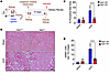 Figure 5
αTAT1 deficiency protects mice from kidney fibrosis in the unilateral ureteral obstruction model. (A) Schematic of the unilateral ureteral obstruction mouse model of kidney fibrosis. (B) Picrosirius red–stained kidney sections from WT (Atat1+/+) and αTAT1-deficient (Atat1–/–) mice 14 days after ligation, showing reduced collagen deposition in Atat1–/– kidneys (n = 4 per group). Scale bar: 100 μm. (C) Kidney collagen content by Picrosirius red staining in Atat1+/+ and Atat1–/– mice (n = 4 mice per group). (D) Quantification of αSMA+ myofibroblasts per field, showing reduced myofibroblast accumulation in Atat1-deficient kidneys (n = 4 mice per group). For animal experiments, data are presented as mean ± SD. Statistical significance was determined by 1-way ANOVA followed by Šidák’s multiple-comparison test. ****P < 0.0001.
αTAT1 controls fibroblast durotaxis and activation via the FAK/YAP mechanosensitive axis. To elucidate how αTAT1 orchestrates fibroblast durotaxis and mechano-activation, we investigated whether its catalytic activity toward α-tubulin K40 acetylation governs cytoskeletal polarization and stiffness-dependent signaling. αTAT1 catalyzes the acetylation of α-tubulin at K40, a posttranslational modification enriched in long-lived microtubules that enhances their flexibility and resilience under mechanical stress. We reasoned that this enzymatic activity might be required for establishing the microtubule alignment necessary for directional migration along stiffness gradients. To test this, we generated fibroblasts overexpressing either WT αTAT1 or a catalytically inactive mutant, αTAT1-D157N, which carries a single amino acid substitution that abolishes acetyltransferase activity (Figure 6A). When plated on hydrogels containing continuous stiffness gradients, WT αTAT1 fibroblasts exhibited pronounced front-rear polarity and oriented their microtubule network toward regions of increasing stiffness. In contrast, fibroblasts expressing αTAT1-D157N failed to establish this polarized architecture, displaying disorganized and isotropic microtubule arrays (Figure 6B). Quantitative analysis confirmed a marked reduction in directional migration, with decreased durotaxis and loss of directional bias in D157N-expressing cells (Figure 6C), demonstrating that αTAT1 catalytic activity is required for stiffness-guided migration. We next assessed whether loss of αTAT1 function affects fibroblast profibrotic responses using step-gradient hydrogels, which enable simultaneous evaluation of durotaxis and mechano-activation upon reaching stiff regions. Consistent with impaired directional migration (Figure 6, D and E), D157N-expressing fibroblasts exhibited reduced secretion of collagen type I (Figure 6F), indicating defective stiffness-dependent mechano-activation. Given the central role of focal adhesions in mechanosensing, we examined focal adhesion dynamics and signaling. Loss of αTAT1 activity resulted in reduced phosphorylated FAK (p-FAK) (Tyr397) levels and decreased focal adhesion size, consistent with impaired focal adhesion maturation and force transmission (Figure 6, G–I). We then evaluated downstream mechanotransduction through YAP, a key effector of stiffness sensing. WT fibroblasts exhibited robust YAP nuclear accumulation in stiff regions, whereas αTAT1-D157N cells displayed reduced nuclear YAP and increased cytoplasmic retention (Figure 6, J and K), indicating defective coupling of mechanical cues to transcriptional activation. Finally, we assessed whether αTAT1 directly regulates myofibroblast differentiation. Genetic deletion or siRNA-mediated knockdown of ATAT1 attenuated TGF-β–induced αSMA expression in both mouse and human lung fibroblasts (Figure 6, L and M), and reduced activation of profibrotic FAK signaling without altering canonical p-SMAD2 signaling (Figure 6N). Together, these findings demonstrate that αTAT1 catalytic activity is required for microtubule polarization, durotaxis, and focal adhesion–dependent mechanotransduction. These data define an αTAT1-dependent microtubule mechanosensing axis that operates through focal adhesions and FAK/YAP signaling to link stiffness sensing with fibroblast activation and profibrotic responses.
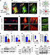 Figure 6
αTAT1 controls fibroblast durotaxis and activation via the FAK/YAP mechanosensitive axis. (A) Structural model of αTAT1 showing the acetyl-CoA ligand and catalytic residues D157 and C120. (B) Immunofluorescence images of human lung fibroblasts expressing WT αTAT1 or catalytically inactive αTAT1-D157N on continuous-stiffness-gradient hydrogels. α-Tubulin (green), F-actin (red), and nuclei (DAPI, blue). (n = 4.) Scale bars: 10 μm. (C) Rose plots showing reduced durotaxis in D157N-expressing fibroblasts compared with WT (n = 4). (D) Representative images of fibroblast durotaxis on step-gradient hydrogels. F-actin (red) and nuclei (DAPI, blue). (n = 5.) Scale bars: 50 μm. (E) Quantification of durotaxis index from D, demonstrating reduced directional migration in D157N-expressing fibroblasts (n = 5). (F) Quantification of secreted collagen type I from WT and D157N-expressing fibroblasts on stiffness gradients (n = 4). (G) Immunofluorescence images of p-FAK and focal adhesions (n = 4). Scale bars: 50 μm (left) and 10 μm (right). (H) Quantification of p-FAK–positive cells (n = 4). (I) Quantification of focal adhesion area (60 focal adhesions per condition, n = 4). (J) Immunofluorescence images of YAP localization in fibroblasts cultured on stiffness gradients (n = 4). Scale bars: 50 μm. (K) Quantification of nuclear YAP localization (n = 4). (L) Western blot analysis of α-SMA expression in mouse lung fibroblasts isolated from Atat1+/+ and Atat1–/– mice following TGF-β stimulation. GAPDH, loading control. (n = 3.) (M) Western blot analysis of αSMA expression in human lung fibroblasts transfected with Atat1 siRNA or non-targeting (NT) control following TGF-β stimulation. GAPDH, loading control. (n = 3.) (N) Western blot analysis of p-FAK and p-SMAD2 following TGF-β stimulation. GAPDH, loading control. (n = 3.) Data are presented as mean ± SD. Statistical significance was determined by 2-tailed paired Student’s t test. *P < 0.05, **P < 0.01.
-
Methods
Sex as a biological variable. This study used male and female animals in mouse model studies, with no significant differences between sexes. Data presented include results from both sexes.
Animal experiments. Pathogen-free C57BL/6N (6- to 8-week-old) mice purchased from the National Cancer Institute Frederick Mouse Repository (Frederick, Maryland, USA) were used for established mouse models of skin, lung, and kidney fibrosis (28). Data distribution was assumed to be normal. Data collection and analysis were not performed blind to the conditions of the experiments.
Mouse model of lung fibrosis. Lung fibrosis in 6- to 8-week-old mice was induced by intratracheal administration of bleomycin (50 μL at 1.2 U/kg body weight), as previously described (8, 29, 35). Sterile saline was used as control. Lungs were collected for histological, flow cytometry, molecular, and biochemical studies as well as hydroxyproline analysis.
Mouse model of skin fibrosis. Skin fibrosis in 6- to 8-week-old mice was induced by daily subcutaneous injection of bleomycin (100 μL from 10 μg/mL stock) for 28 days, as previously described (30). Sterile saline was used as the control. At the conclusion of experiments, mice were euthanized, and full-thickness 6-mm punch biopsies were obtained for histological, molecular, and biochemical studies as well as hydroxyproline analysis.
Mouse model of kidney fibrosis. Renal fibrosis in 6- to 8-week-old mice was induced by unilateral ureteral obstruction surgery, as previously described (8). In brief, the left ureter was ligated using a suture, leading to an obstruction of the kidney outflow tract on the ligated side so that the urine could not drain anymore, causing hydronephrosis with tubular dilation and kidney fibrosis at day 14. Kidneys were collected for histological, flow cytometry, molecular, and biochemical studies as well as hydroxyproline analysis.
Atat1-knockout mice. Atat1tm1(KOMP)Vlcg heterozygous embryonic stem cells were obtained from the Knockout Mouse Project (KOMP) repository affiliated with the University of California, Davis (www.komp.org). These embryonic stem cells were then injected into C57BL/6 blastocysts and implanted in receiving females at the Transgenic Research Center at the University of California, Davis. Atat1tm1(KOMP)Vlcg heterozygous C57BL/6 male mice were obtained and crossed with C57BL/6 females purchased from Charles River Laboratories to achieve germline transmission. Finally, heterozygous males and females were mated to obtain Atat1–/– and Atat1+/+ mice.
Genotyping protocol. Genotyping was performed using the Phire Animal Tissue Direct PCR Kit (Thermo Fisher Scientific), according to the manufacturer’s instructions. At the time of weaning, every mouse was ear-punched for identification purposes, and a small piece of tail tissue was collected in an Eppendorf tube. Then, genomic DNA from the tail was extracted by addition of 20 μL of dilution buffer and 0.5 μL of DNA release enzyme to each tube. The samples were centrifuged at 13,000 x g for 5 minutes at room temperature, followed by incubation at 98°C for 2 minutes. The extract was then ready for use in PCR, as shown in Table 1.
Agarose gel electrophoresis. Agarose gels (1.5% w/v) were prepared by dissolving 3 g of agarose (Sigma-Aldrich) in 200 mL of 1× Tris-Acetate-EDTA (TAE) buffer (Fisher Bioreagents), supplemented with 10 μL of ethidium bromide (10 mg/mL) (Sigma-Aldrich). After gel polymerization, 20 μL of PCR product was mixed with 2 μL of 10× DNA loading dye (Roche) and loaded into the gel wells. Electrophoresis was performed for 40 minutes at 120–150 V using a horizontal electrophoresis system (Bio-Rad) containing 1× TAE buffer. Gel images were obtained using a UV light system (Bio-Rad), and the amplification bands were analyzed to determine genotype. The expected amplicon sizes were 321 bp for WT (Atat1+/+) mice and 210 bp for Atat1-/- mice.
Generation of fibroblast-specific Atat1-deficient mice. We generated mice with conditional fibroblast-specific Atat1 deficiency by crossing Atat1-floxed mice (obtained from the European biorepository INFRAFRONTIER EMMA) to Pdgfrα-CreERT2 mice (The Jackson Laboratory), which express a tamoxifen-inducible Cre recombinase in mesenchymal cells including lung fibroblasts. Tamoxifen administration to Pdgfra-CreERT2 Atat1fl/fl mice resulted in Atat1 deletion and markedly reduced α-tubulin acetylation in primary fibroblasts, as demonstrated by Western blot analysis. Atat1 was conditionally ablated by the administration of 2 mg of tamoxifen injected intraperitoneally for 5 consecutive days starting at 4 weeks of age, according to our standard protocols. Atat1fl/fl mice treated with tamoxifen and untreated Pdgfra-CreERT2 Atat1fl/fl mice were used as controls.
Histopathology and immunohistochemistry analysis. Once the mice were completely anesthetized, the right ventricle was flushed with 50 mL of PBS, and lungs were inflated at 25 cm H2O. Lungs were subsequently excised from the thoracic cavity and fixed in 10% formalin before being embedded in paraffin. Histopathological analysis of 5 μm sections of paraffin-embedded lungs was performed on lung sections stained with hematoxylin and eosin and Masson’s trichrome using standard procedures. Immunohistochemistry stainings were performed with the following primary antibodies at a 1:100 dilution: mouse monoclonal αSMA (clone 1A4, Sigma-Aldrich), rabbit polyclonal α-tubulin (2144, Cell Signaling Technology), and rabbit polyclonal acetyl-α-tubulin Lys40 (5335, Cell Signaling Technology). For secondary antibodies, goat anti-mouse Alexa Fluor 488 (A32723, Thermo Fisher Scientific) and goat anti-rabbit Alexa Fluor 555 (A-21428, Thermo Fisher Scientific) were used at a 1:100 dilution.
Collection of bronchoalveolar lavage. In order to analyze the total protein cellular content in the lungs, bronchoalveolar lavage (BAL) was performed in anesthetized mice as previously described (8). Briefly, BAL fluid was obtained by intubation of the trachea using a 3-way catheter containing the tubing for the trachea, a collection syringe, and a perfusion syringe with 5 mL of PBS. With this system, 1 mL of PBS was flushed at a time and then collected with the collection syringe. A total volume of 5 mL was obtained from each mouse, and cells in the BAL fluid were centrifuged at 500g for 10 minutes at 4°C. Afterward, the supernatant was transferred to a new tube and stored at –80°C until used, and the cellular pellet was then resuspended and counted for later experiments.
Hydroxyproline assay. In order to determine the collagen content of the lungs, lungs were minced and homogenized in 2 mL of PBS. Then, 1 mL was transferred to a new Eppendorf tube, desiccated, and hydrolyzed in 6N HCl at 110°C overnight. Twenty-five microliters of the resultant lysate was incubated for 20 minutes at room temperature with 1 mL solution of 1.4% chloramine T (Sigma-Aldrich), 10% n-propanol, and 0.5 M sodium acetate, pH 6.0. After the incubation step, 1 mL of Ehrlich’s solution (1 M p-dimethylaminobenzaldehyde [Sigma-Aldrich] in 70% n-propanol/20% perchloric acid) was added. The final mix was incubated for 15 minutes at 65°C. Absorbance was measured at 550 nm, and the amount of hydroxyproline was determined against a standard curve.
Flow cytometry. Single cells from digested lungs or BAL (100,000 cells) were incubated with antibodies against specific cell surface antigens for 45 minutes on ice and protected from light. Unstained cells, fluorescence minus one (FMO) controls, and single-antibody-stain controls were used. Before incubation with specific surface antigens, red blood cells were lysed by incubation of the samples on ice for 3 minutes with 1 mL of Hybri-Max red blood cell lysis buffer (R7757, Sigma-Aldrich). Immediately after, 10 mL of PBS was added to the tube, and cells were centrifuged at 450g for 5 minutes and filtered through a 40 μm strainer. Cells were pelleted by centrifugation at 450g for 5 minutes and finally resuspended in 100–200 μL of FACS buffer (PBS plus 2.5% fetal bovine serum [FBS]). After incubation of the cell samples, the FMO controls, and the single-antibody stains with the corresponding antibodies, the tubes were centrifuged at 450g for 5 minutes, washed once with FACS buffer, and resuspended in 300 μL of FACS buffer. At this point, the tubes were kept on ice until subsequent flow cytometry analysis using CytoFLEX S (Beckman Coulter) and FlowJo analysis software. A list of antibodies used for FACS is provided in Table 2.
Human lung fibroblasts. Primary human lung fibroblasts from healthy controls and IPF patients were obtained from Eric White, University of Michigan (Ann Arbor, Michigan, USA). Healthy lung fibroblasts were isolated from lung sections from patients without IPF who underwent lung transplantation, and then cultured to 80% confluence in complete medium with 10% FBS, 100 U/mL penicillin, and 100 μg/mL streptomycin. Cells were passaged when reaching confluence and used for experiments at passage 3 or 6.
Mouse lung fibroblasts. Primary mouse lung fibroblasts were isolated from C57BL/6 mice and cultured in complete medium supplemented with 15% FBS, 100 U/mL penicillin, and 100 μg/mL streptomycin. Cells were grown up to approximately 80% confluence. To isolate fibroblasts from lung tissue, lung lobes were first excised from terminally anesthetized mice, minced into small pieces, and digested with 0.28 U/mL Liberase Blendzyme 3 (Roche) and 60 U/mL DNase I in RPMI for 45 minutes at 37°C on a belly shaker. Then, the digested lungs were passed through a 70 μm filter, centrifuged at 540g at 4°C, and plated in tissue culture flasks in complete medium. Cells were passaged when reaching confluence and used for experiments at passage 3 or 4.
Durotaxis assay on steep stiffness gradients. As previously described (7, 8, 36), fibroblasts were seeded at a density of 10,000 cells per 20-mm hydrogel in serum-free medium on stripe-patterned polyacrylamide substrates (Zebraxis). Four hours after plating, cells were evenly distributed between soft and stiff regions (baseline durotactic index = 1). After 24 hours, cultures were fixed and stained with DAPI, and durotaxis was quantified as the ratio of cells localized on stiff versus soft stripes. For each hydrogel, 5 random fields were imaged at ×10 magnification, and cell counts were averaged across triplicate samples.
Generation of shallow-stiffness-gradient hydrogels via microfluidics. Gradient hydrogels were fabricated using a microfluidic system as previously reported (8, 36). Briefly, channels were perfused with acrylamide polymer solutions (10% acrylamide with 0.05% or 0.5% bis-acrylamide in deionized water) at 30 μL/min using a syringe pump (KD Scientific). Solutions were introduced sequentially through 3 inlets arranged low-high-low, producing a continuous stiffness gradient upon recombination. Polymerization was triggered by UV illumination for 6 minutes beneath the outlet region of the microchannel, generating stable polyacrylamide hydrogels with shallow elastic gradients.
Quantitative analysis of durotaxis by time-lapse microscopy. Cells were seeded at a density of 15,000 per hydrogel in serum-free medium and imaged for 24 hours using live-cell microscopy. Images were captured every 5 minutes with a ×10, 0.3 NA Plan Fluor objective. Single-cell trajectories (n = 75 cells per field) were manually tracked using the Manual Tracking plug-in in ImageJ (NIH). Migration parameters, including velocity and forward migration index, were calculated from the recorded trajectories to assess directional persistence along the stiffness gradient. All experiments were performed in biological triplicates.
Polyacrylamide hydrogel preparation. Polyacrylamide hydrogels were fabricated following established protocols (30, 37). Briefly, 18 mm glass coverslips (Fisher Scientific) were activated by incubation in 0.4% (vol/vol) 3-methacryloxypropyltrimethoxysilane (Sigma-Aldrich) diluted in acetone for 20 minutes, rinsed once with fresh acetone, and air-dried. Acrylamide and bis-acrylamide (Bio-Rad) were mixed at defined ratios to generate gels of distinct stiffness: 3%/0.11% for 0.5 kPa (soft) and 20%/0.24% for 64 kPa (stiff). After polymerization, gels were functionalized with collagen I (PureCol, Advanced BioMatrix) by incubation in PBS containing 0.05 mg/mL protein for 30 minutes under sterile conditions to promote cell adhesion.
Chemotaxis assays. Chemotactic migration toward soluble cues was evaluated using a 96-well FluoroBlok insert system (Corning; 8 μm pore size). Inserts were precoated with fibronectin (10 μg/mL in PBS; Sigma-Aldrich) for 4 hours at 37°C to promote cell adhesion. Before seeding, fibroblasts were labeled with DiIC12 fluorescent dye (1:1,000 dilution) for 1 hour, washed, and resuspended in serum-free medium. A total of 5 × 10⁴ cells in 50 μL were added to the upper chamber of each insert. The lower (apical) chamber was filled with 150 μL of either complete medium containing 20% FBS or serum-free medium supplemented with specific chemoattractants, including lysophosphatidic acid (Avanti Polar Lipids), PDGF-BB (R&D Systems), or fibronectin (Thermo Fisher Scientific). After a 24-hour incubation at 37°C in 5% CO2, the extent of cell migration was quantified by measurement of fluorescence (excitation/emission: 544/590 nm) from the bottom of the plate using a Fluoroskan Ascent FL reader (Thermo Fisher Scientific). Experiments were performed in triplicate, and the chemotactic index was calculated as (total absorbance toward any chemoattractant – chemoattractant background absorbance) / (total absorbance toward serum-free medium – serum-free medium background absorbance).
siRNA transfection. Human lung fibroblasts were transfected with siRNA targeting ATAT1 (L-014510-02-0005, Dharmacon) or a non-targeting control siRNA lacking homology to known mammalian genes (D-001206-14-05, Dharmacon). Transfections were carried out using HiPerfect Transfection Reagent (QIAGEN), which contains a proprietary lipid mixture that facilitates siRNA encapsulation and efficient intracellular delivery. For reverse transfection, 10 μL of 20 μM siRNA was diluted in 100 μL of Opti-MEM (Gibco, Life Technologies) and combined with 6 μL of HiPerfect reagent. After vortexing and a 10-minute incubation at room temperature to allow complex formation, the transfection mixture was added to each well of a 6-well plate. Subsequently, 1.0 × 10⁵ to 1.5 × 10⁵ fibroblasts were seeded per well in 2–3 mL of complete medium lacking antibiotics. Gene silencing efficiency was assessed 48 hours after transfection by Western blot analysis.
RNA extraction and quantification. Total RNA was extracted using TRIzol Reagent (15596026, Thermo Fisher Scientific) according to the manufacturer’s protocol. Briefly, cells were washed once with PBS and lysed in 1 mL of TRIzol per well. Lysates were transferred to microcentrifuge tubes and incubated for 5 minutes at room temperature to ensure complete dissociation of nucleoprotein complexes. Chloroform (200 μL; Sigma-Aldrich) was added, and samples were vortexed for 15 seconds, incubated for 2–3 minutes, and centrifuged at 12,000g for 20 minutes at 4°C. The aqueous phase was collected and mixed with isopropanol (500 μL; Sigma-Aldrich) and GlycoBlue Coprecipitant (30 μg/mL; AM9515, Invitrogen), followed by overnight precipitation at –20°C. The next day, RNA was pelleted by centrifugation (12,000g, 20 minutes, 4°C), washed 3 times with 1 mL of 75% ethanol, air-dried, and resuspended in 30–50 μL of RNase-free water. RNA concentration and purity were assessed using a NanoDrop spectrophotometer (Thermo Fisher Scientific).
cDNA synthesis. Complementary DNA (cDNA) was synthesized from 250–500 ng of total RNA using the iScript cDNA Synthesis Kit (Bio-Rad) following the manufacturer’s instructions. Each 20 μL reaction contained 4 μL of 5× iScript Supermix (including MMLV reverse transcriptase, dNTPs, oligo-dT and random primers, MgCl2, and RNase inhibitor) and nuclease-free water. Reactions were performed in a thermal cycler under the following conditions: (a) priming: 5 minutes at 25°C; (b) reverse transcription: 20 minutes at 46°C; (c) enzyme inactivation: 1 minute at 95°C; and (d) hold: 4°C.
Quantitative PCR. Quantitative PCR (qPCR) was performed using SYBR Green chemistry on an Mx4000 Multiplex Quantitative PCR System (Stratagene). Each 25 μL reaction contained 12.5 μL of SYBR Green qPCR SuperMix (Invitrogen; containing SYBR Green I dye, Taq DNA polymerase, MgCl2, dNTPs, UDG, and stabilizers), 1 μL of cDNA (≈250 ng/μL), and 11.5 μL of nuclease-free water. Gene-specific primers were designed from RefSeq mRNA sequences (NCBI) using Primer3 software (Table 3). Cycling conditions were as follows: initial denaturation: 95°C for 10 minutes; 40 amplification cycles: 95°C for 15 seconds, 60°C for 20 seconds, 72°C for 60 seconds (fluorescence measured each cycle); melt curve analysis: 95°C for 60 seconds, 55°C for 30 seconds, 95°C for 30 seconds with continuous fluorescence acquisition; final hold: 40°C.
Relative gene expression was normalized to GAPDH using the ΔCt method (ΔCt = Ct_target − Ct_GAPDH) and expressed as 2−ΔCt. Fold changes relative to controls were calculated using the ΔΔCt method (ΔΔCt = ΔCt_sample − ΔCt_control) and expressed as 2−ΔΔCt.
Western blotting. Cells were lysed in RIPA buffer (Thermo Fisher Scientific) supplemented with Halt Protease and Phosphatase Inhibitor Cocktail (1:100). Lysates were centrifuged at 6,000g for 10 minutes at 4°C, and protein concentrations were determined using a BCA Protein Assay Kit (Pierce). Equal amounts of protein were denatured for 10 minutes at 95°C in 4× LDS Sample Buffer (NP0008, Thermo Fisher Scientific) containing 10% β-mercaptoethanol (Sigma-Aldrich). Samples were resolved by 8%–12% SDS-PAGE and transferred to PVDF membranes at 27 V overnight at 4°C using NuPAGE electrophoresis and transfer systems (Life Technologies). Membranes were blocked with 5% BSA in TBS-T for 2 hours at room temperature and incubated overnight at 4°C with primary antibodies diluted in blocking buffer. After 3 washes in TBS-T (10 minutes each), membranes were incubated with HRP-conjugated secondary antibodies (Cell Signaling Technology) (Table 4) for 1 hour at room temperature. Blots were developed using Amersham ECL substrate (GE Healthcare), and band intensities were quantified by densitometry using ImageJ software (NIH).
Immunocytochemistry. Five thousand and 10,000 cells were seeded on fibronectin-coated coverslips (10 μg/mL; Sigma-Aldrich) in 24-well plates and cultured at 37°C in 5% CO2. After 24 hours, cells were washed with PBS and fixed in 4% paraformaldehyde for 10 minutes at room temperature. After fixation, cells were rinsed with PBS and stored in PBS at 4°C until further use. Permeabilization was performed using 0.1% Triton X-100 (Sigma-Aldrich) in blocking buffer composed of 3% BSA and 10% goat serum in TBS-T for 1 hour at room temperature. Cells were then washed with TBS-T and incubated with fresh blocking buffer for an additional hour to prevent nonspecific binding. Primary antibodies (Table 5) were diluted in blocking buffer and incubated overnight at 4°C. The next day, cells were washed 3 times with TBS-T and incubated with fluorescent secondary antibodies (Table 5) for 1 hour at room temperature in the dark. After final washes, coverslips were mounted cell-side-down on microscope slides using mounting medium containing DAPI (Sigma-Aldrich) for nuclear staining. Fluorescence images were acquired using a Zeiss wide-field fluorescence microscope (×10 and ×20 objectives), and image processing was performed with ZEN 2009 Light Edition software.
Statistics. Statistical analyses were performed using GraphPad Prism 7 (GraphPad Software). Data are presented as mean ± SD unless otherwise indicated. The independent biological replicate was used as the experimental unit. For measurements obtained from multiple cells, fields, or focal adhesions within the same experiment, these measurements were treated as subsamples and were not considered independent biological replicates. Comparisons between 2 independent groups were performed using a 2-tailed unpaired Student’s t test. Comparisons between matched conditions within the same biological replicate, including paired siRNA or WT-versus-mutant experiments, were performed using a 2-tailed paired Student’s t test. Animal experiments containing multiple independent treatment and genotype groups were analyzed by 1-way ANOVA followed by Šidák’s multiple-comparison test for prespecified comparisons. Statistical tests and sample sizes for individual experiments are specified in the corresponding figure legends. All tests were 2-sided, and statistical significance was defined as P < 0.05. Significance is indicated as *P < 0.05, **P < 0.01, ***P < 0.001, and ****P < 0.0001.
Study approval. All mouse experiments were performed in accordance with National Institutes of Health (NIH) guidelines, protocols were approved by the Massachusetts General Hospital Subcommittee on Research Animal Care (protocol 2019N000148), and all mice were maintained in a specific pathogen–free environment certified by the Association for Assessment and Accreditation of Laboratory Animal Care International.
Data availability. All data are available upon request. Supporting data values are provided in the Supporting Data Values Excel file; supplemental material available online with this article; https://doi.org/10.1172/jci.insight.201695DS1


