
Research ArticleCardiologyVascular biology
Open Access |

10.1172/jci.insight.201535
1Center for Cardiovascular Research, Division of Cardiology, Department of Medicine, and
2Department of Genetics, Washington University School of Medicine, St. Louis, Missouri, USA.
3John Cochran Veterans Affairs Medical Center, St. Louis, Missouri, USA.
Address correspondence to: In-Hyuk Jung or Nathan O. Stitziel, 660 S Euclid Ave., Campus Box 8086, St. Louis, Missouri, 63110, USA. Phone: 314.747.6267; Email: in-hyuk.jung@wustl.edu (IHJ). Phone: 314.747.8394; Email: nstitziel@wustl.edu (NOS).
Find articles by Jung, I. in: PubMed | Google Scholar
1Center for Cardiovascular Research, Division of Cardiology, Department of Medicine, and
2Department of Genetics, Washington University School of Medicine, St. Louis, Missouri, USA.
3John Cochran Veterans Affairs Medical Center, St. Louis, Missouri, USA.
Address correspondence to: In-Hyuk Jung or Nathan O. Stitziel, 660 S Euclid Ave., Campus Box 8086, St. Louis, Missouri, 63110, USA. Phone: 314.747.6267; Email: in-hyuk.jung@wustl.edu (IHJ). Phone: 314.747.8394; Email: nstitziel@wustl.edu (NOS).
Find articles by Amrute, J. in: PubMed | Google Scholar
1Center for Cardiovascular Research, Division of Cardiology, Department of Medicine, and
2Department of Genetics, Washington University School of Medicine, St. Louis, Missouri, USA.
3John Cochran Veterans Affairs Medical Center, St. Louis, Missouri, USA.
Address correspondence to: In-Hyuk Jung or Nathan O. Stitziel, 660 S Euclid Ave., Campus Box 8086, St. Louis, Missouri, 63110, USA. Phone: 314.747.6267; Email: in-hyuk.jung@wustl.edu (IHJ). Phone: 314.747.8394; Email: nstitziel@wustl.edu (NOS).
Find articles by Luna, S. in: PubMed | Google Scholar
1Center for Cardiovascular Research, Division of Cardiology, Department of Medicine, and
2Department of Genetics, Washington University School of Medicine, St. Louis, Missouri, USA.
3John Cochran Veterans Affairs Medical Center, St. Louis, Missouri, USA.
Address correspondence to: In-Hyuk Jung or Nathan O. Stitziel, 660 S Euclid Ave., Campus Box 8086, St. Louis, Missouri, 63110, USA. Phone: 314.747.6267; Email: in-hyuk.jung@wustl.edu (IHJ). Phone: 314.747.8394; Email: nstitziel@wustl.edu (NOS).
Find articles by Wagoner, R. in: PubMed | Google Scholar
1Center for Cardiovascular Research, Division of Cardiology, Department of Medicine, and
2Department of Genetics, Washington University School of Medicine, St. Louis, Missouri, USA.
3John Cochran Veterans Affairs Medical Center, St. Louis, Missouri, USA.
Address correspondence to: In-Hyuk Jung or Nathan O. Stitziel, 660 S Euclid Ave., Campus Box 8086, St. Louis, Missouri, 63110, USA. Phone: 314.747.6267; Email: in-hyuk.jung@wustl.edu (IHJ). Phone: 314.747.8394; Email: nstitziel@wustl.edu (NOS).
Find articles by Alisio, A. in: PubMed | Google Scholar
1Center for Cardiovascular Research, Division of Cardiology, Department of Medicine, and
2Department of Genetics, Washington University School of Medicine, St. Louis, Missouri, USA.
3John Cochran Veterans Affairs Medical Center, St. Louis, Missouri, USA.
Address correspondence to: In-Hyuk Jung or Nathan O. Stitziel, 660 S Euclid Ave., Campus Box 8086, St. Louis, Missouri, 63110, USA. Phone: 314.747.6267; Email: in-hyuk.jung@wustl.edu (IHJ). Phone: 314.747.8394; Email: nstitziel@wustl.edu (NOS).
Find articles by Lee, P. in: PubMed | Google Scholar
1Center for Cardiovascular Research, Division of Cardiology, Department of Medicine, and
2Department of Genetics, Washington University School of Medicine, St. Louis, Missouri, USA.
3John Cochran Veterans Affairs Medical Center, St. Louis, Missouri, USA.
Address correspondence to: In-Hyuk Jung or Nathan O. Stitziel, 660 S Euclid Ave., Campus Box 8086, St. Louis, Missouri, 63110, USA. Phone: 314.747.6267; Email: in-hyuk.jung@wustl.edu (IHJ). Phone: 314.747.8394; Email: nstitziel@wustl.edu (NOS).
Find articles by Burks, K. in: PubMed | Google Scholar
1Center for Cardiovascular Research, Division of Cardiology, Department of Medicine, and
2Department of Genetics, Washington University School of Medicine, St. Louis, Missouri, USA.
3John Cochran Veterans Affairs Medical Center, St. Louis, Missouri, USA.
Address correspondence to: In-Hyuk Jung or Nathan O. Stitziel, 660 S Euclid Ave., Campus Box 8086, St. Louis, Missouri, 63110, USA. Phone: 314.747.6267; Email: in-hyuk.jung@wustl.edu (IHJ). Phone: 314.747.8394; Email: nstitziel@wustl.edu (NOS).
Find articles by Oh, J. in: PubMed | Google Scholar
1Center for Cardiovascular Research, Division of Cardiology, Department of Medicine, and
2Department of Genetics, Washington University School of Medicine, St. Louis, Missouri, USA.
3John Cochran Veterans Affairs Medical Center, St. Louis, Missouri, USA.
Address correspondence to: In-Hyuk Jung or Nathan O. Stitziel, 660 S Euclid Ave., Campus Box 8086, St. Louis, Missouri, 63110, USA. Phone: 314.747.6267; Email: in-hyuk.jung@wustl.edu (IHJ). Phone: 314.747.8394; Email: nstitziel@wustl.edu (NOS).
Find articles by Plunkett Paletta, H. in: PubMed | Google Scholar
1Center for Cardiovascular Research, Division of Cardiology, Department of Medicine, and
2Department of Genetics, Washington University School of Medicine, St. Louis, Missouri, USA.
3John Cochran Veterans Affairs Medical Center, St. Louis, Missouri, USA.
Address correspondence to: In-Hyuk Jung or Nathan O. Stitziel, 660 S Euclid Ave., Campus Box 8086, St. Louis, Missouri, 63110, USA. Phone: 314.747.6267; Email: in-hyuk.jung@wustl.edu (IHJ). Phone: 314.747.8394; Email: nstitziel@wustl.edu (NOS).
Find articles by Kang, C. in: PubMed | Google Scholar
1Center for Cardiovascular Research, Division of Cardiology, Department of Medicine, and
2Department of Genetics, Washington University School of Medicine, St. Louis, Missouri, USA.
3John Cochran Veterans Affairs Medical Center, St. Louis, Missouri, USA.
Address correspondence to: In-Hyuk Jung or Nathan O. Stitziel, 660 S Euclid Ave., Campus Box 8086, St. Louis, Missouri, 63110, USA. Phone: 314.747.6267; Email: in-hyuk.jung@wustl.edu (IHJ). Phone: 314.747.8394; Email: nstitziel@wustl.edu (NOS).
Find articles by
Stitziel, N.
in:
PubMed
|
Google Scholar
|
Published July 22, 2026 - More info
A single-nucleotide missense polymorphism (rs1800449, R158Q) in the propeptide domain of lysyl oxidase (LOX-PP) is associated with increased risk of coronary artery disease (CAD) independent of changes in plasma lipid levels. Although the enzymatic function of LOX has an essential role for the cross-linking of extracellular matrix proteins in connective tissues, whether and how LOX-PP R158Q contributes to the development of atherosclerosis has not been clearly established. Here, hypercholesterolemia was induced in mice that were WT or homozygous for the LOX-PP R158Q polymorphism by adeno-associated virus-8–mediated overexpression of Pcsk9 followed by high-fat diet feeding for 16 weeks. We found that the R158Q polymorphism promoted atherosclerosis and induced proliferation of macrophages and vascular smooth muscle cells without altering LOX enzymatic activity. Using single-cell RNA sequencing, we found the transcriptional program of atherosclerotic plaques from mice harboring R158Q was strongly enriched for proliferation- and calcification-related genes in a regionally distinct manner. Together, these results establish an enzymatically independent proatherogenic role for the LOX-PP and suggest its potential as a novel therapeutic target.
The extracellular matrix (ECM) serves an important role in the development and maintenance of the arterial wall; altered ECM homeostasis and its remodeling have been implicated in a variety of diseases, including atherosclerosis. Comprised of a complex network of glycoproteins and proteoglycans, the ECM provides structural integrity to the vascular wall while actively influencing cell behavior. Major structural components of the arterial ECM include elastin and collagen, where elastin contributes to elastic recoil and collagen is responsible for maintaining the tensile strength of tissues (1). Lysyl oxidase (LOX) and 4 LOX-like isoenzymes (LOXL1–4) comprise a family of copper-dependent oxido-deaminases that catalyze the oxidation of lysine and hydroxylysine side chains on these structural proteins, forming elastic lamellae and collagen fibers for ECM homeostasis and remodeling (2). LOX is synthesized and secreted as a 50-kDa inactive proenzyme (Pro-LOX), which is cleaved by bone morphogenic protein-1 (BMP-1) to generate a mature active 32-kDa enzyme and 18-kDa propeptide (LOX-PP).
The LOX enzyme promotes cancer cell proliferation, metastasis formation, and angiogenesis (3, 4). However, LOX also has tumor suppressor function that is inactivated by methylation and loss of heterozygosity (5), and recent studies have demonstrated that this is due to enzymatically independent functions of the LOX-PP (6, 7), suggesting that the LOX enzyme and its propeptide play opposite roles in tumorigenesis. Studies in both human and animal models have shown that dysregulated LOX expression is also involved in the development of cardiovascular diseases (8–11), although all of these studies have investigated altered levels of full-length LOX, which includes both its propeptide and enzymatic domain.
A single-nucleotide polymorphism (SNP) in the propeptide of LOX leading to the substitution of glutamine (Q) for arginine (R) at codon 158 (hereafter referred to as R158Q) has been shown to impair the endogenous tumor suppressor function of LOX-PP (6). LOX-knockin mice harboring R158Q (homologous to R152Q in mice) exhibited more carcinogen-induced tumor development without a change in LOX enzyme activity (12), suggesting that LOX-PP tumor suppressor function is independent from its enzymatic activity. Notably, the LOX R158Q polymorphism is associated with increased risk of coronary artery disease (CAD) at a level of genome-wide significance (13). This variant is not significantly associated with traditional risk factors for CAD, such as plasma lipids, and therefore has the potential to help uncover new mechanisms leading to CAD.
The findings in recent studies of LOX along with the known biology of the protein led us to hypothesize that the R158Q LOX polymorphism could contribute to CAD risk through a mechanism linked to the ECM. Here, using a murine model of atherosclerosis in mice harboring R152Q (homologous to R158Q in humans), we address the impact of R158Q polymorphism in LOX-PP on the development of atherosclerosis and how it alters the transcriptional programs of cells within the developing plaque.
LOX R158Q polymorphism promotes atherosclerosis in the aortic root and induces cellular proliferation in mice. To address the effect of LOX R158Q polymorphism on the development of atherosclerosis, we generated mice harboring this polymorphism, which was previously shown to result in loss of the tumor suppressor effect of the LOX-PP without impacting LOX enzymatic function (6). Using clustered regularly interspaced short palindromic repeats (CRISPR) and CRISPR-associated protein-9 nuclease (Cas9) genome engineering tools, we introduced the arginine to glutamine substitution in the homologous site within the mouse LOX-PP (murine codon 152, corresponding to human R158; Supplemental Figure 1A; supplemental material available online with this article; https://doi.org/10.1172/jci.insight.201535DS1) and bred these animals to generate experimental groups of mice that were homozygous for glutamine (hereafter referred to as LoxR152Q) or arginine (hereafter referred to as LoxWT) as littermate controls. We then induced hypercholesterolemia in both groups of mice by adeno-associated virus-8–mediated overexpression of Pcsk9 (AAV8-Pcsk9) followed by high-fat diet (HFD) feeding for 16 weeks. AAV8-Pcsk9 and HFD efficiently elevated plasma cholesterol levels in both genotypes (Figure 1B) without inducing any genotype-specific differences in body weight, plasma total cholesterol, triglycerides, and glucose (Figure 1, A, C, and D). Although no significant differences in atherosclerotic plaque development were observed between LoxR152Q and LoxWT mice in the aortic arch or the entire aorta after HFD feeding (Figure 1, E and F), a significant increase in plaque burden was detected within the aortic root of LoxR152Q mice at multiple levels throughout the aortic sinus, as measured at defined distances from the root (Figure 1, G and H). Consistently, the mean plaque size within the aortic root was significantly larger in LoxR152Q mice than in control mice (Figure 1I). In addition, atheromas from LoxR152Q mice were more complex, containing significantly greater vascular smooth muscle cell (VSMC) staining (Supplemental Figure 1B), and modestly higher amounts of macrophage staining (Supplemental Figure 1C) as well as necrotic core size (Figure 1, J and K).
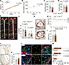
Figure 1R158Q polymorphism in LOX-PP promotes atherosclerosis and proliferation of VSMCs and macrophages. (A) Body weight of LoxWT and LoxR152Q male mice during HFD feeding (n = 9–12/group). (B) Plasma total cholesterol at 2, 8, and 16 weeks of HFD feeding (n = 9–12/group), (C) triglyceride (n = 9–10/group), and (D) glucose (n = 9–12/group) after 16 weeks of HFD feeding. (E) En face Oil Red O–stained aortas. (F) Quantification of Oil Red O–stained area in each aortic arch and whole aorta (n = 9–12/group). (G) Oil Red O–stained aortic root cross sections. (H) Quantification of Oil Red O–stained atherosclerotic plaque area from 8 sections taken at 5, 20, 35, 100, 115, 130, 195, 210 μm (section number 1 through 8) from the aortic root toward the top (n = 9–11/group). (I) Average plaque area was calculated from the values measured across the 8 sections in H. (J) Necrotic core of aortic roots outlined by H&E staining. Red arrows indicate necrotic core in plaque from each genotype. (K) Quantification of necrotic core as a percentage of plaque area (n = 7–10/group). (L) EdU staining in VSMCs and macrophages of the aortic root from LoxWT and LoxR152Q mice after 16 weeks of HFD feeding. Tissues were costained for SMα-actin and Mac3. Outlined areas indicate the regions magnified in the next panels. Yellow circles indicate EdU+ cells. (M) Quantification of EdU+ in SMα-actin+, Mac3+, and all DAPI+ cells (n = 6–7/group). Scale bars: 500 μm (G), 200 μm (J), and 50 μm (L). Data were analyzed with 2-way ANOVA test (A, B, and H) or unpaired nonparametric Mann-Whitney U test (C, D, F, I, K, and M) and are shown as the mean ± SEM. P, plaque; L, lumen; M, media.
To further characterize differences in plaque phenotypes induced by R158Q, we also performed in situ zymography in which gelatin was used as a substrate to assess gelatinase activity (as a surrogate for MMP-2 and MMP-9 activity) (14–17) in aortic root plaques, although we did not detect a significant difference between genotypes (Supplemental Figure 1, D and E). To address whether macrophage polarization was altered, we performed immunofluorescent staining for iNOS and CD206 to assess inflammatory and reparative macrophages, respectively. Although we did not observe a significant difference in iNOS-positive inflammatory macrophages (Supplemental Figure 1, F and G), we found that LoxR152Q mice had a significant reduction in CD206-positive reparative macrophages within the aortic root plaque compared with controls (Supplemental Figure 1, H and I). Cellular proliferation of VSMCs and macrophages within the arterial wall is a key contributor to plaque development (18–20). To assess whether the LOX-PP R158Q polymorphism might be mediating the proliferation of neointimal cells, we labeled proliferating cells by administering 5-ethynyl-2′-deoxyuridine (EdU) intraperitoneally to mice 96 hours before euthanasia. EdU labeling demonstrated that LoxR152Q mice had a significant increase in the number of proliferating cells, including VSMCs and macrophages, in the plaque when compared with controls (Figure 1, L and M). To further examine whether the proliferative phenotype associated with the R158Q polymorphism could be recapitulated in human VSMCs, we treated primary human coronary artery smooth muscle cells (CASMCs) and aortic smooth muscle cells (AoSMCs) with recombinant WT or R158Q LOX-PP in vitro. WT LOX-PP significantly inhibited proliferation of both CASMCs (Supplemental Figure 1, J and K) and AoSMCs (Supplemental Figure 1, L and M), whereas R158Q LOX-PP showed reduced inhibitory activity. This observation is consistent with previous work demonstrating that LOX-PP inhibits proliferation of primary rat AoSMCs (21) and supports an interpretation that impaired LOX-PP function can directly influence proliferative responses in VSMCs, although the mechanisms underlying increased macrophage proliferation in vivo remain to be further defined. Together, these findings suggest that the LOX R158Q polymorphism promotes atherosclerotic plaque formation and cellular changes that may compromise plaque stability, including increased cellular proliferation, increased plaque complexity, and impaired reparative macrophage polarization.
LOX R158Q polymorphism does not change LOX activity or expression. Given the location of the R158Q polymorphism in the PP region of the protein, we sought to determine the relative mechanisms by which the R158Q polymorphism could be increasing risk of CAD. Since LOX-PP is required for normal LOX protein processing (22, 23), it is possible that normal enzymatic function could be altered due to the location of the polymorphism within the propeptide. To address this, we determined the enzymatic activity within atherosclerotic plaque tissues from LoxR152Q and LoxWT mice. However, we did not observe any significant difference in the enzymatic activity from aortic root plaque tissues of LoxR152Q and LoxWT mice (Supplemental Figure 2A). We then assessed the number of elastic lamellae breaks as a surrogate of LOX function. Imaging of elastin autofluorescence in the aortic tissues of LoxR152Q and LoxWT mice did not demonstrate any difference in the number of medial VSMC layers and elastic lamellae breaks between genotypes (Supplemental Figure 2, B and C). We also did not detect a significant difference in collagen content between groups (Supplemental Figure 2, D and E). Together, these findings suggest that the LOX R158Q polymorphism did not alter LOX enzymatic activity or impact overall ECM composition.
Next, we tested whether the polymorphism could be affecting the production or secretion of the protein to the ECM. Expression data from the Genotype-Tissue Expression (GTEx) project indicated that the R158Q risk variant was not associated with changes in LOX mRNA expression in the aorta, coronary artery, or tibial artery (Supplemental Figure 2, F and G). We then performed in vitro studies of LOX WT and LOX R158Q to assess for differences in LOX expression and production. To determine this, human embryonic kidney cells transformed with large T antigen (HEK-239T) cells were transfected with cDNA constructs for c-Myc–tagged LOX WT, c-Myc–tagged LOX R158Q, or empty vector as a control. After confirming the correct sequence change for LOX R158Q (Supplemental Figure 2H), conditioned media were collected. The R158Q polymorphism did not alter protein production or its secretion (Supplemental Figure 2I), consistent with previous findings that this variant does not affect LOX protein processing or expression (12). Immunocytochemistry for Myc confirmed that both the amount and expression pattern of LOX were similar between the WT and R158Q constructs (Supplemental Figure 2J).
LOX is expressed by VSMCs in human and mice. It is well established that LOX is mainly expressed by VSMCs (24–26) and upregulated in atherosclerotic plaque lesions (27). To further confirm the expression of LOX during the development of atherosclerosis in this study, we initially searched for disease-relevant tissues and cell types that express LOX. Expression data from the GTEx project showed that human arterial tissues such as the aorta, tibial artery, and coronary artery express LOX (Supplemental Figure 3A). A reanalysis of single-cell RNA sequencing in human coronary arteries from explanted hearts of transplant recipients (GSE131780) (28) found that fibroblasts and modulated smooth muscle cells (modSMCs) were the main cell types expressing high levels of LOX (Supplemental Figure 3, B–D). We then performed RNA in situ hybridization (ISH) to visualize its expression using tissue explants from the aortic wall (Supplemental Figure 3E), left internal mammary artery (Supplemental Figure 3F), and coronary artery (Supplemental Figure 3G) of patients with established CAD. This revealed that LOX expression was detected throughout the plaque lesion and in the medial layer, mostly colocalizing with the VSMC marker smooth muscle α-actin (SMα-actin). Within healthy arterial tissues, including the aortic root (Supplemental Figure 4A), brachiocephalic artery (BCA) (Supplemental Figure 4B), lesser curvature (LC) of the aortic arch (Supplemental Figure 4C), and the thoracic aorta (Supplemental Figure 4D) isolated from young 8-week-old Apoe-knockout (Apoe–/–) mice, we observed Lox expression in the media, mostly colocalizing with VSMCs. Under atherosclerotic condition by feeding Apoe–/– mice an HFD for 8 weeks, Lox expression colocalized with VSMCs in the medial layer underlying the lesion as well as at the fibrous cap of the plaque (Supplemental Figure 4E), suggesting that murine Lox expression recapitulated human LOX expression. Taken together, these data suggest that in atherosclerotic disease LOX is produced locally by VSMCs and are consistent with a previous report showing that LOX is highly expressed in calcified human atherosclerotic lesions corresponding to VSMC-rich areas (27).
LOX expression is differently regulated by VSMC embryonic origin. Given our finding that the R158Q polymorphism induced the development of atherosclerotic plaque in the aortic root to a greater extent when compared with the remainder of the aorta, we hypothesized that LOX expression and its function might be differentially regulated by VSMC embryonic origin and biology. Lineage tracing studies have demonstrated that VSMCs in the arterial wall originate from multiple progenitors from different embryonic origin (29, 30). VSMCs from the BCA and inner layer of the aortic arch arise from cardiac neural crest, while the lateral plate mesoderm–derived secondary heart field contributes to the VSMC development in the aortic root. Thus, we examined neointimal Lox expression between vessel regions that have different VSMC origins. Interestingly, although Lox was highly expressed in the fibrous cap of atheromas from all aortic regions after 16 weeks of HFD feeding in Apoe–/– mice, when we focused on the aortic wall beneath plaques, VSMCs in the medial layer of the aortic root showed relatively low Lox expression (Figure 2A) compared with other medial layers of the BCA (Figure 2B) and the LC of the aortic arch (Figure 2C). Taken together, these data suggest that the expression and localization of LOX is differently regulated by VSMC embryonic origin.
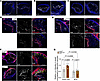
Figure 2Differential Lox expression patterns in mouse aortic vessels. (A–C) Differential expression patterns of Lox in aortic vessels, including the aortic root (A), BCA (B), and LC (C) from Apoe–/– mice after 16 weeks of HFD feeding using RNAscope ISH. Outlined dotted areas indicate the regions magnified in the next panels. (D–F) Comparison of Lox expression patterns between LoxWT and LoxR152Q mice after 16 weeks of HFD feeding using RNAscope ISH. Aortic roots (D and E) and BCAs (F) were used and costained for the VSMC marker SMα-actin. Outlined areas indicate the regions magnified in the next 2 panels. Gray arrows (shown in regions 1 and 2 in D and E) indicate basal expression of Lox by VSMCs in the medial layer of aortic roots compared with its expression in fibrous cap from both genotypes. Yellow arrows (shown in F) indicate higher expression of Lox by VSMCs in the medial layer of BCAs along with its expression in fibrous cap from both genotypes. (G) Quantification of relative Lox intensity in aortic vessels normalized to SMα-actin intensity. Scale bars: 200 μm (A–C) and 100 μm (D–F). Data were analyzed with 2-way ANOVA test, and are shown as the mean ± SEM.
We next asked whether the R158Q polymorphism was associated with regional changes in expression using Lox RNAscope ISH in the aortic root (Figure 2, D and E) and BCA plaque tissues (Figure 2F) of LoxWT and LoxR152Q mice. Most of the Lox expression in the fibrous caps of plaques colocalized with neointimal cells that costained with the contractile VSMC maker SMα-actin. Interestingly, we found that the expression levels of both Lox and SMα-actin were significantly lower in the medial VSMC layer beneath plaques from the aortic root tissues (Figure 2, D and E) as compared with the medial layer beneath plaques from BCAs (Figure 2F). However, when normalized to SMα-actin intensity, Lox expression was significantly increased in the aortic root compared with BCA from LoxR152Q mice (Figure 2G). These findings suggest that the aortic root would be the region most susceptible to the R158Q polymorphism and support our observation that atherosclerotic plaque formation was more affected in the aortic root than rest of the arteries.
Aortic root VSMCs from LoxR152Q mice have greater dedifferentiation, proliferation, and calcification. To dissect the transcriptional changes induced by the R158Q polymorphism during the development of atherosclerosis, we performed single-cell RNA sequencing on sorted cells from separately isolated aortic roots and ascending aortas (defined as aortic tissue from its origin through the BCA) from 16-week HFD-fed LoxR152Q and LoxWT mice (Figure 3A). Enzymatically dissociated atherosclerotic plaque cells from each tissue region were pooled (n = 5 mice/genotype) and labeled with Deep Red Anthraquinone 5 (DRAQ5) and 4′,6-diamidino-2-phenylindole (DAPI); DRAQ5+DAPI– live cells isolated by fluorescence-activated cell sorting (FACS) were subsequently used for analysis (Supplemental Figure 5A). Through clustering and differential gene expression analysis of all cells, single-cell RNA sequencing analysis identified 9 major vascular and immune cell types that were annotated using canonical gene expression markers (Figure 3, B and C). Across all cells, Lox was most highly expressed in VSMCs and fibroblasts (Figure 3D), similar to expression patterns in human coronaries (Supplemental Figure 3D). We found a large number of differentially regulated genes between stromal cells of the aortic root and ascending aorta (Figure 3E). To prioritize cell types that were transcriptionally most distinct between LoxWT and LoxR152Q mice, we performed differential expression analysis in each cell compartment split by aortic root and ascending aorta. We found a minimal number of differentially expressed genes by genotype in the ascending aorta. In contrast, there were a large number of differentially expressed genes by genotype in the aortic root, most prominently seen in VSMCs (Figure 3F). Accordingly, we examined differentially expressed genes per cell type in the aortic arch (Supplemental Table 1), aortic root (Supplemental Table 2), and comparison between tissues (Supplemental Table 3). Gene Ontology analysis of differentially expressed genes in the aortic root VSMCs between LoxR152Q and LoxWT mice demonstrated a strong enrichment of cyclase- and GTPase-related pathways in LoxR152Q mice, whereas ribosome- and ubiquitin-related pathways were enriched in the genes upregulated in the VSMCs of LoxWT mice (Figure 3G). To characterize regulatory changes between LoxR152Q and LoxWT, we used PROGENy to determine differential transcription factor (TF) enrichment and found a greater enrichment for hypoxia, inflammation, and cell growth TFs in LoxR152Q relative to LoxWT mice (Figure 3H). Additionally, differential expression analysis found that Stat3 (involved in cell growth and proliferation) (31–33) and Sox9 (involved in vascular calcification) (34–36) were highly upregulated in the aortic root of LoxR152Q compared with LoxWT mice (Figure 3, I and L). In contrast, there were no differences in the expression levels of these 2 transcripts in the ascending aorta between genotypes (Supplemental Figure 5B). To experimentally validate these findings, we performed RNAscope ISH for Stat3 (Figure 3J) and Sox9 (Figure 3M) in mouse aortic root plaques. We found that both the intensity of Stat3 (Figure 3K) and the number of Sox9-positive cells (Figure 3N) were significantly increased in the aortic root of LoxR152Q mice compared with LoxWT mice.
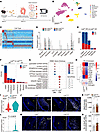
Figure 3Transcriptomic characterization of atherosclerotic plaques in aortic root and ascending aorta. (A) Study design for single-cell RNA sequencing using each aortic root and ascending aorta (from beginning of aorta up to the BCA) isolated from HFD-fed mice. (B) UMAP embedding plot with cell types present in the mouse aortic root and ascending aorta (n = 5/group). (C) Heatmap of differentially expressed genes between cell types. (D) Violin plot of Lox genes across genotypes and tissues. (E) Enrichment of overlapping genes across cell types in tissues of LoxWT. (F) Enrichment of overlapping genes across cell types in each tissue between genotypes. (G) VSMC Gene Ontology analysis in the aortic root based on differentially expressed marker genes identified unique pathway enrichment across states. (H) TF enrichment analysis for smooth muscle cell/fibroblast states in the aortic root between genotypes showing key regulators. (I–N) Comparison of Stat3 (I) and Sox9 (L) expression in the aortic root between genotypes. Validation of Stat3 (K) and Sox9 (N) expression in the aortic root using RNAscope ISH. (K) Quantification of Stat3 intensity calculated by ratio to plaque (n = 8–9/group), and (N) number of Sox9+ cells in plaque (n = 7/group). Scale bars: 200 μm. Data were analyzed with unpaired nonparametric Mann-Whitney U test (K and N) and are shown as the mean ± SEM. P, plaque; L, lumen.
Finally, we asked whether the LOX R158Q polymorphism affected the expression of other LOX family members, including LOXL1 through LOXL4, by examining their expression in the aortic root as well as ascending aorta from both groups of mice. In aortic root tissues, all Lox family members were highly upregulated in LoxR152Q compared with LoxWT mice, although expression levels of Lox13 and Loxl4 were relatively low in both groups (Supplemental Figure 5C). In contrast, the expression of these genes in the ascending aorta was not altered between genotypes with the exception of Loxl1 (Supplemental Figure 5D). As fibroblasts also showed high level of Lox expression comparable to VSMCs in our single-cell RNA sequencing data, we examined the expression of Lox family genes in fibroblasts from the aortic root and ascending aorta, observing similar expression patterns (Supplemental Figure 6, A and B). Differential expression and pathway analyses in the aortic root fibroblasts (Supplemental Table 2 and Supplemental Figure 6C) demonstrated more changes in ECM regulation in the aortic root fibroblasts from LoxR152Q mice compared with controls. Taken together, these data suggest that the aortic root provides environmental cues for the LOX R158Q polymorphism–mediated disease development through VSMC dedifferentiation, proliferation, and calcification in the setting of atherosclerosis.
Human genetics has repeatedly illuminated novel mechanisms underlying cardiovascular disease, guiding the development of transformative therapies such as PCSK9 inhibitors, which were supported by the discovery of loss-of-function variants that dramatically lower LDL cholesterol and cardiovascular risk (37, 38). Here, our identification of a functional variant in LOX-PP reveals what appears to be a previously unrecognized pathway involving impaired propeptide-mediated ECM remodeling, offering not only mechanistic insights into CAD pathogenesis but also a potential new therapeutic target. Translational research plays an indispensable role in bridging fundamental discoveries in cardiovascular biology with clinical advancements in the prevention, diagnosis, and management of CAD. Given the multifactorial nature of CAD, which involves complex interactions, a translational approach enables the integration of mechanistic insights with patient-centered interventions. This paradigm is especially valuable in accelerating the transition from bench to bedside, effectively addressing the application of basic research in clinical settings (39). Furthermore, the emergence of advanced omics technologies including single-cell RNA sequencing has enabled unprecedented granularity in phenotyping CAD, facilitating the development of precision medicine approaches.
Studies in both human and animal models have shown that expression of the ECM-modifying protein LOX is dysregulated during the development of cardiovascular diseases (8–11), highlighting VSMCs as the target cell type of LOX on mediating disease progression through ECM remodeling (27, 40), VSMC proliferation (41), and neointimal thickening (42). However, these prior studies examined changes in the expression of full-length LOX, which encompasses both the propeptide and catalytic/enzymatic domains, leaving the specific pathophysiological contribution of the propeptide domain largely unexplored. In contrast, the R158Q variant investigated here is uniquely located in the propeptide domain and does not alter the enzymatic domain or catalytic activity of LOX. By demonstrating that this propeptide-specific loss-of-function variant promotes greater atherosclerotic plaque development in mice — consistent with the increased atherosclerosis risk observed in human carriers of the R158Q variant (13) that impairs endogenous LOX-PP function (6) — our study provides what we believe to be the first direct evidence for an independent atheroprotective role of LOX-PP, distinct from the enzymatic functions of mature LOX.
It is noteworthy that rs1800449 lies within the overlapping region of LOX (propeptide domain, reverse strand) and SRFBP1 (3′ UTR, forward strand). Although the respective mouse homologs do not overlap, this genomic arrangement in humans raises the possibility of pleiotropic effects. A separate intronic variant in SRFBP1 (rs12657394) has been linked to visceral adiposity in certain populations (43), suggesting that rs1800449 could potentially influence SRFBP1 expression or function. However, existing functional data predominantly support a role for rs1800449 in impairing LOX-PP tumor suppressor activity and ECM cross-linking, consistent with the mechanisms observed in our study. Future studies examining SRFBP1-specific effects would help clarify any additional contributions.
Fate-mapping studies have established that VSMCs in the arterial wall originate from multiple progenitors from different developmental origins (29, 30). These distinct origins have functional consequences, as the progression of atherosclerotic lesions in response to systemic risk factors differs in 4 vascular regions, including the coronary arterial bed, the branches of the aorta, the abdominal visceral arteries, and the terminal abdominal aorta and its major branches (44), suggesting that distinct VSMC lineages based on embryonic origins might influence the development of atherosclerosis. Although LOX is expressed by VSMCs in the arterial wall, the effect of regional VSMC embryonic origins on LOX expression has not been reported. In this study, we found that the transcriptional level of Lox is regionally regulated in VSMCs according to their embryonic origins. We believe this regional expression pattern may explain why we observed increased atherosclerotic burden in the aortic root of animals harboring the partial loss-of-function R158Q mutation. Our single-cell RNA sequencing analysis supports this conclusion, finding that the aortic root provides environmental cues for LOX R158Q polymorphism–mediated disease development through enhanced VSMC dedifferentiation, proliferation, and calcification. Through integrated single-cell RNA sequencing analysis, we also found that VSMCs harbor the greatest transcriptional differences in LoxR152Q mice, suggesting that this population is dysregulated in disease progression. Although our data indicate that VSMCs are changed likely through cell-autonomous and non–cell-autonomous mechanisms, we cannot directly causally implicate VSMC-derived LOX as a catalyst for plaque progression in the absence of cell-type-specific models. Future studies are necessary to implicate the broader effects of cell-specific-derived LOX.
With development of atherosclerotic plaque, phenotypic modulation mostly has been focused on intimal VSMCs. However, atherosclerotic damage factors may cause DNA damage to medial VSMCs, leading to senescence (45, 46). A decrease in contractile markers (including SMα-actin and calponin) and an increase in synthetic markers (including osteopontin) were observed in mouse models of aging and hypertension (47), and these changes may be related to the observation that aging vasculature is prone to calcification. Our in vivo data strengthen this concept, as the calcification-related gene Sox9 was highly upregulated in the atherosclerosis-prone aortic root of LoxR152Q compared with LoxWT mice. Therefore, our results suggest that loss of function in LOX R158Q polymorphism promotes vascular calcification.
Based on the observation that LOX R158Q polymorphism was found to be associated with high risk of breast cancer (48, 49), ovarian cancer (50), oral cancer (51), as well as lung and colon cancer (52), the LOX R158Q polymorphism has been studied in several solid cancer models. Notably, the R158Q polymorphism attenuates the ability of LOX-PP to function as an endogenous tumor suppressor without reducing LOX enzymatic activity (6, 12). To examine the effect of the LOX-PP on cellular proliferation under the atherosclerotic conditions, we generated a mouse model of the LOX R158Q polymorphism and found that LoxR152Q mice had a profound increase in the proliferation of VSMCs as well as macrophages in plaque burden. These data suggest that LOX-PP phenocopies the tumor-suppressive phenotype and that loss of LOX-PP function through the R158Q polymorphism results in cellular proliferation, leading to exacerbated atherosclerotic plaque development. Future studies will be needed to determine whether there are means to leverage the proliferative inhibitory function of LOX-PP as a potential therapeutic pathway for the prevention or management of CAD.
Sex as a biological variable. Male mice were used for the in vivo atherosclerosis studies to reduce biological variability in atherosclerotic plaque development in the AAV8-D377Y-mPcsk9/HFD model and to provide sufficient power to detect genotype-dependent effects. Therefore, sex was not analyzed as an independent biological variable in the animal studies. Sex was also not considered as a biological variable in the in vitro studies using commercially available primary human VSMCs. Although the mechanisms related to LOX-PP function, VSMC proliferation, and ECM remodeling may be relevant to more than one sex, future studies including female mice will be required to determine whether the effects of the LOX R158Q polymorphism differ by sex.
Mice. CRISPR-Cas9 genome editing technology was used in collaboration with the Washington University School of Medicine Genome Engineering and Transgenic Micro-Injection Cores to generate heterozygous mice harboring an arginine to glutamine exchange at a position 152 (which corresponds to human residue 158 of the LOX-PP domain) on a C57BL/6 background. All mice were housed in separate cages in a pathogen-free environment at Washington University School of Medicine animal facility, and maintained on a 12-hour light/12-hour dark cycle with a room temperature of 22°C ± 1°C.
Diet and assessment of atherosclerosis. All experimental mice were fed an HFD containing 21% fat and 0.2% cholesterol (TD.88137, Envigo Teklad) at 8 weeks of age for designed times. To induce hypercholesterolemia in LoxR152Q and LoxWT mice, mice were intravenously injected with 5 × 10¹¹ vector genome copies of AAV8-D377Y-mPcsk9 (Vector Biolabs) at 8 weeks of age and immediately placed on an HFD. After HFD feeding, blood was collected from the retro-orbital plexus after 12-hour fasting, and mice were euthanized by carbon dioxide inhalation. Plasma samples were prepared from the collected blood by centrifugation at 15,000 g for 10 minutes at 4°C. Total cholesterol (catalog STA-384), triglycerides (catalog STA-397), and glucose (catalog STA-681) in mouse plasma were determined using each kit (all purchased from Cell Biolabs, Inc). Hearts and whole aortas (from the aortic arch to the iliac artery) were harvested after perfusion with phosphate-buffered saline (PBS). For en face analysis, isolated aortas were cleaned by removal of perivascular fat tissues, opened longitudinally, and pinned onto black wax plates. After fixation with 4% paraformaldehyde (PFA) overnight at 4°C, aortas were washed with PBS for 1 hour and stained with 0.5% Oil Red O in propylene glycol (O1516, Sigma-Aldrich) for 3 hours at room temperature. After staining, aortas were destained with 85% propylene glycol in distilled water for 5 minutes to reduce background staining, and washed with distilled water for 15 minutes. For analysis of plaques in aortic roots, hearts were fixed overnight with 4% PFA at 4°C, washed with PBS for 1 hour, and embedded into OCT compound (4583, Sakura Finetek). For serial cross sectioning of the aortic root, heart tissues were cut at 5 μm thickness. A total of 44 serial sections per heart were obtained and mounted on 13 microscopic slides. The first slide, containing 8 sections located at 5, 20, 35, 100, 115, 130, 195, and 210 μm from the aortic sinus, was stained overnight with 0.5% Oil Red O in propylene glycol, then destained with 85% propylene glycol in distilled water for 5 minutes and washed with distilled water for 15 minutes. Plaque areas were quantified at multiple levels throughout the aortic sinus, and the average plaque size was calculated. The atherosclerotic plaque area was digitized and calculated using AxioVison (Carl Zeiss).
Antibodies and reagents. For immunofluorescent staining, anti–Mac-3 (550292, BD Biosciences; 1:100), anti–SMα-actin-Cy3 (C6198, Sigma-Aldrich; 1:1000), anti-iNOS (PA3-030A, Thermo Fisher Scientific; 1:100), and anti-CD206 (PA5-46994, Thermo Fisher Scientific; 1:40) were used and then visualized with anti-rat (catalog A21208), anti-rabbit (catalog A21206) and anti-goat–Alexa Fluor 488 (catalog A11055) (all from Invitrogen; 1:400). Nuclei were visualized using Vectashield Vibrance antifade mounting media with DAPI (H-1800, Vector Laboratories). For immunocytochemistry, anti-mouse Myc (2276, Cell Signaling Technology; 1:8000) was used. For immunohistochemistry, hematoxylin solution (catalog HHS80), eosin solution (catalog HT110180), Masson’s trichrome staining kit (catalog HT15-1KT), Masson’s Goldner staining kit (catalog 1.00485.0001) (all purchased from Sigma-Aldrich), and Permount solution (SP15-500, Fisher Chemicals) were used.
Immunohistochemistry and immunofluorescent staining. For all immunohistochemistry and immunofluorescence experiments, 4% PFA–fixed frozen sections at 5 μm thickness were used. Briefly, slides were air dried for 1 hour at room temperature and then hydrated with PBS for 10 minutes. After permeabilization with 0.5% Triton X-100 for 10 minutes, sections were blocked with PBS containing 5% donkey serum (D9663, Sigma-Aldrich) with 0.5% Triton X-100 for 1 hour at room temperature. Slides were subsequently incubated with the indicated antibodies. For hematoxylin and eosin (H&E) staining, air-dried slides were hydrated in PBS for 10 minutes, placed in hematoxylin solution for 10 minutes, and then rinsed in running tap water. After destaining in 1% acetic acid for 5 minutes, slides were rinsed in tap water and placed in 90% ethanol for 5 minutes. Slides were stained with eosin solution for 8 minutes, gradually dehydrated in ethanol solution (from 80% to 100%), and then incubated with xylene for 10 minutes followed by mounting with Permount solution. To detect collagen, Masson’s trichrome and Goldner staining kits were used according to the manufacturer’s instructions.
ISH. To detect RNA transcripts for LOX in both human and mouse vascular tissues, a commercially available kit (323100, RNAscope Multiplex Fluorescent Reagent Kit v2, Advanced Cell Diagnostics) was used according to the manufacturer’s instructions. Briefly, 4% PFA–fixed human and mouse frozen sections (5 μm) were air dried for 1 hour at room temperature and treated with hydrogen peroxide for 10 minutes to block endogenous peroxidase activity. After antigen retrieval by boiling in target antigen retrieval solution for 5 minutes at 95°C–100°C, slides were treated with protease III for 30 minutes at 40°C. Target probes (415941, human LOX; 425311, mouse Lox) were hybridized for 2 hours at 40°C, followed by a series of signal amplification and washing steps. Hybridization signals were detected by TSA Plus Cyanine 5, and costained with anti–SMα-actin-Cy3. For validation of single-cell RNA sequencing data, probes for mouse Sox9 (catalog 401051) and Stat3 (catalog 425641) were used.
EdU labeling/staining. Experimental mice received single intraperitoneal injection of EdU (A10044, Invitrogen) at a dose of 50 mg/kg body weight 96 hours before euthanasia. Fixed (4% PFA), frozen sections (5 μm) were stained using a Click & Go Click Chemistry Reaction Buffer Kit (1001, Click Chemistry Tools) according to the manufacturer’s protocol. Briefly, after washing with PBS, the sections were incubated with 2 M HCl for 30 minutes to denature DNA. The sections were washed again with 0.5% Triton X-100 in PBS for 15 minutes and blocked with 2% bovine serum albumin (BSA) in PBS for 5 minutes. The sections were incubated with a Click & Go reaction cocktail containing reaction buffer, CuSO4, Alexa Fluor 488 Azide plus, and reaction buffer additive for 30 minutes. The sections were washed with 2% BSA in PBS for 5 minutes then costained with the indicated antibodies.
Analysis of MMP activity. To evaluate MMP activities in atherosclerotic plaques, we performed in situ zymography. Sections were incubated with a green fluorogenic gelatin substrate (DQ gelatin; D12054, Molecular Probes) according to the manufacturer’s protocol. Briefly, slides were air-dried for 1 hour at room temperature and then hydrated with PBS for 10 minutes. After incubation with 100 mg/mL DQ-gelatin diluted in reaction buffer, sections were washed with PBS for 15 minutes followed by mounting with Vectashield Vibrance antifade mounting media with DAPI.
Mouse cell isolation for single-cell RNA sequencing. The aortic root and ascending aorta (from beginning of aorta up to BCA) were isolated and transferred to an enzymatic dissociation cocktail with 4 U/mL Liberase (catalog 5401127001), 60 U/mL hyaluronidase (catalog H3506), and 120 U/mL DNase I (catalog D4527) (all purchased from Sigma-Aldrich) in Dulbecco’s modified Eagle medium (DMEM) and slightly minced. After incubation at 37°C for 70 minutes with agitation, the digestion reaction was quenched with 6 mL of HBB buffer (2% FBS and 0.2% BSA in Hank’s balanced salt solution [HBSS]), then filtered through 70 μm filters. Samples were pelleted by centrifugation at 4°C, 400 g for 5 minutes and the supernatant was discarded. Cells were resuspended in FACS buffer (2% FBS and 2 mM EDTA in calcium/magnesium–free PBS) and centrifugation was repeated in above conditions and supernatant aspirated. Cells were resuspended in FACS buffer with the addition of DRAQ5 (62251, Thermo Fisher Scientific; 1:100) and incubated on ice for 30 minutes. Cells were washed 3 times with FACS buffer following same centrifugation as above and then resuspended in FACS buffer with DAPI (564907, BD Biosciences) and filtered into filter-top FACS tubes. First singlets were gated and subsequent DRAQ5+DAPI– events were collected in 300 μL cell resuspension buffer (0.04% BSA in PBS) – collected cells were centrifuged as above and resuspended in collection buffer to a target concentration of 1,000 cells/μL. Cells were counted on a hemocytometer before proceeding with the 10X Genomics protocol.
Single-cell RNA sequencing. Cells were processed using the Chromium Single Cell 5′ Reagent V1.1 kit from 10X Genomics. CellRanger v7 was used to align sequencing fastq files to the mouse genome. All CellRanger output count files were analyzed using Seurat v4 (https://satijalab.org/seurat/). For quality control, cells with the following criteria were kept for analysis: nFeature_RNA > 200 and nFeature_RNA < 6000 and percent.mt < 10 and nCount_RNA < 25,000. After quality control 19,712 cells were used for subsequent analysis. Data were subsequently normalized with SCTransform (regressing out nCount and percent.mt), dimensional reduction was calculated using PCA, integration using harmony, followed by nearest neighbor analysis, UMAP embedding construction, and clustering. Differential expression analysis using the FindAllMarkers function was used, and cells were annotated using canonical marker genes. Differential expression analysis between LoxR152Q and LoxWT was performed using FindAllMarkers within each cell type and aortic root/ascending aorta data. PROGENy (https://bioconductor.org/packages/progeny/) and cluster profiler (https://bioconductor.org/packages/release/bioc/html/clusterProfiler.html) were used for the pathway analysis.
LOX enzyme activity. LOX enzymatic activity was determined using a LOX Activity Fluorometric Assay Kit (ab112139, Abcam) according to the manufacturer’s instructions. Briefly, aortic root tissues were carefully dissected from perfused hearts and rinsed in ice-cold PBS to remove residual blood. Tissues were homogenized in ice-cold RIPA lysis buffer (50 mM Tris-HCl pH 7.4, 150 mM NaCl, 1% NP-40, 0.5% sodium deoxycholate, 0.1% SDS) supplemented with protease and phosphatase inhibitor cocktails (4693116001, Roche) using a bead homogenizer. Tissue extracts from aortic roots from LoxR152Q and LoxWT mice fed HFD for 16 weeks were centrifuged at 13,000g for 5 minutes at 4°C. Then, 50 μL of supernatant was distributed in duplicate into a clear-bottom 96-well plate (50 μL of assay buffer was used as a blank) and 50 μL of LOX reaction mix was added to each well. The plate was incubated for 30 minutes at 37°C in the dark. Fluorescence was then monitored on a microplate reader at Ex/Em = 560/590 nm.
LOX cDNA site-directed mutagenesis and cDNA production. Human LOX cDNA (RG13323, Origene) was inserted into a pCMV6-Myc-His vector. Site-directed mutagenesis was then used to insert the mutation of interest at residue 158. To complete this mutagenesis, primers were constructed to change nucleotide 473 (position 1504 in plasmid) from guanine (G) to adenine (A). The primers used were 5′-ACGCGTACGCGGCCGCTCGAG-3′ and 5′-GCCCACCATGCCGTCCACGC-3′. This resulted in an amino acid change of arginine to glutamine at residue 158. LOX-PP was then subcloned in a separate construct with the native signal peptide. The tobacco etch virus protease site was inserted into the LOX-PP construct to remove the tags at a later time. Both plasmids were then treated with Dpn 1 to digest any non-mutated plasmids and incubated at 37°C. The LOX-PP constructs were transformed by electroporation in DH10B E. coli cells and incubated with shaking for 1 hour at 37°C. Cells were then plated on LB agar plates containing kanamycin (LB-kanamycin) and incubated overnight. Colonies were then picked from each plate and incubated in LB-kanamycin media overnight. A plasmid prep using the QIAprep Spin Miniprep Kit (27104, Qiagen) was then performed to obtain purified plasmids. The mutation of interest was then verified by sending the samples for sequencing at GeneWiz.
Expression of full-length LOX constructs. HEK-239T cells were cultured in 6-well plates in 90% DMEM, 10% FBS, and 0.1× Pen/Strep solution (Gibco; 10,000 U/mL penicillin, 10,000 μg/mL streptomycin) at 37°C and 5% CO2. The cells were transfected with 2 μg of either WT LOX, LOX R158Q, or vehicle alone using 6 μg Lipofectamine 2000 and Opti-MEM. At 48 hours after transfection, cell lysates and/or conditioned media were collected for analysis of LOX expression and secretion.
Production and use of LOX-PP proteins. WT and R158Q LOX-PP constructs were expressed in a 293F expression system. LOX-PP proteins were collected from conditioned media, purified using immobilized affinity chromatography (IMAC) as a capture step and size exclusion chromatography (SEC) as a polishing step, and then treated with TEV protease when removal of the epitope tag was required. Purified proteins were quantified by the bicinchoninic acid method (Thermo Fisher Scientific), and equal amounts of WT or R158Q LOX-PP were applied for the proliferation assay.
In vitro proliferation assay. A Click-iT EdU Cell Proliferation Kit for Imaging, Alexa Fluor 488 dye (10337, Invitrogen) was used. Human CASMCs and AoSMCs were seeded at 1 × 10⁵ cell/well in 6-well plates. After reaching 60% confluence, cells were treated with 10 μg/mL WT or R158Q LOX-PP for 24 hours or 48 hours in complete media (SmGM-2 Smooth Muscle Cell Growth Medium-2 BulletKit, CC-3182, Lonza). After treatment, cells were labeled with 10 μM EdU in complete media for 24 hours, and then fixed in 4% PFA for 2 hours at room temperature. Fixed cells were incubated overnight at 4°C in Alexa Fluor 488 donkey anti-rabbit antibody (1:400 in PBS containing 10% normal donkey serum and 0.1% Tween 20). The cells were washed 3 times with PBS with 0.1% Tween 20 for 5 minutes each and then washed 2 times for 5 minutes each with PBS. The coverslips with cells were then mounted on glass slides using 1 drop of Vectashield Vibrance antifade mounting media with DAPI. A Zeiss LSM 700 laser-scanning confocal microscope with ZEN software was used to take the images.
Conditioned media and cell lysate preparation. The conditioned media were collected from the plates of cells using a syringe and then filtered using 0.22 μm filter. The filtered conditioned media were concentrated 20-fold by centrifugation. The plates were then washed once with PBS. The cells were then collected using 10% 10× radioimmunoprecipitation assay (RIPA) buffer with 10% protease inhibitor and 0.1% nuclease inhibitor and scraping. The cells in lysis buffer were then centrifuged at 15,000 g for 10 minutes at 4°C. The supernatant solution was removed and placed in a new tube. The cell lysate was then used to measure total protein concentration using a bicinchoninic acid assay (Thermo Fisher Scientific).
Immunoblot assay. The cell lysate and conditioned media samples were mixed with NuPAGE LDS Sample Buffer plus 50 mM dithiothreitol (DTT) and heated at 75°C for 10 minutes. Samples were loaded and run on a Bio-Rad Mini-PROTEAN TGX gel (4%–15%) at 100–150 volts. The proteins in the gel were then transferred to a nitrocellulose membrane using the Trans-Blot Turbo Transfer System (Bio-Rad). The membrane was then rinsed with water and incubated with Odyssey Blocking Buffer in PBS (92770001, LICOR Bio) for 1 hour with gentle shaking. The membrane was then incubated with mouse anti-Myc antibody (2276, Cell Signaling Technology; 1:1000) for 1 hour with gentle shaking. The membrane was then rinsed and washed 3 times with PBS with 0.5% Tween 20 for 5 minutes with gentle shaking. The membrane was incubated with goat anti-mouse StarBright Blue 700 antibody (12004158, Bio-Rad; 1:5000) and anti-tubulin hFAB rhodamine antibody (12004166, Bio-Rad, 1:5000) for 1 hour with gentle shaking. The membrane was then rinsed and washed 3 times with PBS with 0.5% Tween 20 for 5 minutes. The membrane was then imaged using the Bio-Rad ChemiDoc MP imaging system.
Immunocytochemistry. HEK-239T cells were cultured in 90% DMEM with 10% FBS and 0.1× Pen/Strep solution at 37°C and 5% CO2. Cells were seeded in 6-well plates at 6 × 10⁵ cells/well with a glass coverslip the day before transfection. The cells were then transfected with 2 μg of WT LOX, LOX R158Q, or vehicle alone using 6 μg polyethyleneimine (PEI) and left overnight. The next day, the cells were rinsed twice with PBS. Cells were then fixed with ice-cold 4% PFA for 10 minutes and then washed 3 times for 5 minutes each with PBS. Cells were permeabilized with 0.25% Triton X-100 for 10 minutes and then washed 3 times for 5 minutes each with PBS. The wells were then blocked with 5% goat serum in 0.25% Triton X-100 for 30 minutes. The cells were then incubated with mouse anti-Myc antibody in blocking solution overnight at 4°C. The next day, cells were washed 3 times for 5 minutes each with PBS with 0.1% Tween 20. The cells were incubated in goat anti-mouse Alexa Fluor 594 antibody (Invitrogen; 1:400) in PBS with 0.1% Tween 20. The cells were washed 3 times with PBS with 0.1% Tween 20 for 5 minutes each and then washed 2 times for 5 minutes each with PBS. The coverslips with cells were then mounted on glass slides using 1 drop of Vectashield Vibrance antifade mounting media with DAPI. A Zeiss LSM 700 laser-scanning confocal microscope with ZEN software was used to take the images.
Statistics. Statistical tests applied and n sample numbers are detailed in the figure legends. Data were analyzed using GraphPad Prism software and shown as the mean ± SEM. For animal model data, the Mann-Whitney U test and 2-way ANOVA were used. A P value of less than 0.05 was considered statistically significant.
Study approval. All animal procedures were performed in accordance with the NIH Guide for the Care and Use of Laboratory Animals (National Academies Press, 2011) under protocols 2021-0345 and 2024-0328 approved by the Institutional Animal Care and Use Committee (IACUC) at Washington University School of Medicine in St. Louis, Missouri, USA.
Data availability. Raw and processed sequencing files are available in the NCBI Gene Expression Omnibus under accession GSE275723. Reviewers can access these data at https://www.ncbi.nlm.nih.gov/geo/query/acc.cgi?acc=GSE275723 using the private access token: szuhkmmqvjklrwb.
IHJ and NOS conceived the project. IHJ performed the main analysis and generated figures. JMA performed single-cell RNA sequencing analysis. IHJ and NOS designed experiments. IHJ, JMA, SEL, REW, AA, PCL, KHB, JO, HCPP, and CJK performed experiments. IHJ and NOS wrote and revised the manuscript. NOS acquired funds. All authors approved the final manuscript.
NOS has received consulting fees from Novo Nordisk unrelated to this study.
This work is the result of NIH funding, in whole or in part, and is subject to the NIH Public Access Policy. Through acceptance of this federal funding, the NIH has been given a right to make the work publicly available in PubMed Central.
SEL’s present address is: Institute for Stem Cell Biology and Regenerative Medicine, Stanford University School of Medicine, Stanford, California, USA.
Copyright: © 2026, Jung et al. This is an open access article published under the terms of the Creative Commons Attribution 4.0 International License.
Reference information: JCI Insight. 2026;11(14):e201535.https://doi.org/10.1172/jci.insight.201535.