
Research ArticleImmunologyInfectious diseaseInflammation
Open Access |  10.1172/jci.insight.199896
Caspase-11–mediated hyperinflammation impairs CD8+ T cell immunity and viral clearance in severe SARS-CoV-2 infection
Mostafa M. Eltobgy,1 Mohamed M. Shamseldin,1,2 Owen D. Whitham,1 Heba M. Amer,1 Jeffrey R. Atkinson,3,4 Asmaa Badr,1 Jesse M. Hall,1 Gauruv Gupta,1 Yara Y. Hassan,1 Rabab El-Mergawy,1 Richard Perez,1 Sarah E. Faber,1 Maciej Pietrzak,5 Amy Webb,5 Xiaoli Zhang,6 Adam D. Kenney,1 Destiny Bissell,1 Jihad I. Omran,1 Shady Estfanous,2 Kylene P. Daily,1 Amir Yousif,1 Marisa R. Joldrichsen,7 Andrew McNamara,1 Mahesh KC,8 Mark E. Peeples,8,9 Emily A. Hemann,1 Hazem E. Ghoneim,1 Shahid M. Nimjee,10 Estelle Cormet-Boyaka,7 Jianrong Li,7 Prosper N. Boyaka,7 Jacob S. Yount,1,9 Benjamin M. Segal,3,4 Purnima Dubey,1 and Amal O. Amer1,9
1Department of Microbial Infection and Immunity, College of Medicine, The Ohio State University, Columbus, Ohio, USA.
2Department of Microbiology and Immunology, Faculty of Pharmacy, Helwan University, Cairo, Egypt.
3Department of Neurology, College of Medicine and Wexner Medical Center,
4Neuroscience Research Institute, and
5Department of Biomedical Informatics, The Ohio State University, Columbus, Ohio, USA.
6College of Nursing, University of South Florida, Tampa, Florida, USA.
7Department of Veterinary Biosciences, College of Veterinary Medicine, The Ohio State University, Columbus, Ohio, USA.
8Abigail Wexner Research Institute at Nationwide Children’s Hospital, Columbus, Ohio, USA.
9Infectious Diseases Institute and
10Department of Neurological Surgery, College of Medicine and Wexner Medical Center, The Ohio State University, Columbus, Ohio, USA.
Address correspondence to: Amal O. Amer or Mostafa M. Eltobgy, Department of Microbial Infection and Immunity, College of Medicine, The Ohio State University, 706 Biomedical Research Tower (BRT)460 W 12th Ave, Columbus OH 43210, USA. Email: Amal.Amer@osumc.edu (AOA); Mostafa.eltobgy@osumc.edu (MME).
Authorship note: MME and MMS contributed equally to this work.
Find articles by
Eltobgy, M.
in:
PubMed
|
Google Scholar
|

1Department of Microbial Infection and Immunity, College of Medicine, The Ohio State University, Columbus, Ohio, USA.
2Department of Microbiology and Immunology, Faculty of Pharmacy, Helwan University, Cairo, Egypt.
3Department of Neurology, College of Medicine and Wexner Medical Center,
4Neuroscience Research Institute, and
5Department of Biomedical Informatics, The Ohio State University, Columbus, Ohio, USA.
6College of Nursing, University of South Florida, Tampa, Florida, USA.
7Department of Veterinary Biosciences, College of Veterinary Medicine, The Ohio State University, Columbus, Ohio, USA.
8Abigail Wexner Research Institute at Nationwide Children’s Hospital, Columbus, Ohio, USA.
9Infectious Diseases Institute and
10Department of Neurological Surgery, College of Medicine and Wexner Medical Center, The Ohio State University, Columbus, Ohio, USA.
Address correspondence to: Amal O. Amer or Mostafa M. Eltobgy, Department of Microbial Infection and Immunity, College of Medicine, The Ohio State University, 706 Biomedical Research Tower (BRT)460 W 12th Ave, Columbus OH 43210, USA. Email: Amal.Amer@osumc.edu (AOA); Mostafa.eltobgy@osumc.edu (MME).
Authorship note: MME and MMS contributed equally to this work.
Find articles by Shamseldin, M. in: PubMed | Google Scholar
1Department of Microbial Infection and Immunity, College of Medicine, The Ohio State University, Columbus, Ohio, USA.
2Department of Microbiology and Immunology, Faculty of Pharmacy, Helwan University, Cairo, Egypt.
3Department of Neurology, College of Medicine and Wexner Medical Center,
4Neuroscience Research Institute, and
5Department of Biomedical Informatics, The Ohio State University, Columbus, Ohio, USA.
6College of Nursing, University of South Florida, Tampa, Florida, USA.
7Department of Veterinary Biosciences, College of Veterinary Medicine, The Ohio State University, Columbus, Ohio, USA.
8Abigail Wexner Research Institute at Nationwide Children’s Hospital, Columbus, Ohio, USA.
9Infectious Diseases Institute and
10Department of Neurological Surgery, College of Medicine and Wexner Medical Center, The Ohio State University, Columbus, Ohio, USA.
Address correspondence to: Amal O. Amer or Mostafa M. Eltobgy, Department of Microbial Infection and Immunity, College of Medicine, The Ohio State University, 706 Biomedical Research Tower (BRT)460 W 12th Ave, Columbus OH 43210, USA. Email: Amal.Amer@osumc.edu (AOA); Mostafa.eltobgy@osumc.edu (MME).
Authorship note: MME and MMS contributed equally to this work.
Find articles by Whitham, O. in: PubMed | Google Scholar
1Department of Microbial Infection and Immunity, College of Medicine, The Ohio State University, Columbus, Ohio, USA.
2Department of Microbiology and Immunology, Faculty of Pharmacy, Helwan University, Cairo, Egypt.
3Department of Neurology, College of Medicine and Wexner Medical Center,
4Neuroscience Research Institute, and
5Department of Biomedical Informatics, The Ohio State University, Columbus, Ohio, USA.
6College of Nursing, University of South Florida, Tampa, Florida, USA.
7Department of Veterinary Biosciences, College of Veterinary Medicine, The Ohio State University, Columbus, Ohio, USA.
8Abigail Wexner Research Institute at Nationwide Children’s Hospital, Columbus, Ohio, USA.
9Infectious Diseases Institute and
10Department of Neurological Surgery, College of Medicine and Wexner Medical Center, The Ohio State University, Columbus, Ohio, USA.
Address correspondence to: Amal O. Amer or Mostafa M. Eltobgy, Department of Microbial Infection and Immunity, College of Medicine, The Ohio State University, 706 Biomedical Research Tower (BRT)460 W 12th Ave, Columbus OH 43210, USA. Email: Amal.Amer@osumc.edu (AOA); Mostafa.eltobgy@osumc.edu (MME).
Authorship note: MME and MMS contributed equally to this work.
Find articles by Amer, H. in: PubMed | Google Scholar
1Department of Microbial Infection and Immunity, College of Medicine, The Ohio State University, Columbus, Ohio, USA.
2Department of Microbiology and Immunology, Faculty of Pharmacy, Helwan University, Cairo, Egypt.
3Department of Neurology, College of Medicine and Wexner Medical Center,
4Neuroscience Research Institute, and
5Department of Biomedical Informatics, The Ohio State University, Columbus, Ohio, USA.
6College of Nursing, University of South Florida, Tampa, Florida, USA.
7Department of Veterinary Biosciences, College of Veterinary Medicine, The Ohio State University, Columbus, Ohio, USA.
8Abigail Wexner Research Institute at Nationwide Children’s Hospital, Columbus, Ohio, USA.
9Infectious Diseases Institute and
10Department of Neurological Surgery, College of Medicine and Wexner Medical Center, The Ohio State University, Columbus, Ohio, USA.
Address correspondence to: Amal O. Amer or Mostafa M. Eltobgy, Department of Microbial Infection and Immunity, College of Medicine, The Ohio State University, 706 Biomedical Research Tower (BRT)460 W 12th Ave, Columbus OH 43210, USA. Email: Amal.Amer@osumc.edu (AOA); Mostafa.eltobgy@osumc.edu (MME).
Authorship note: MME and MMS contributed equally to this work.
Find articles by
Atkinson, J.
in:
PubMed
|
Google Scholar
|
1Department of Microbial Infection and Immunity, College of Medicine, The Ohio State University, Columbus, Ohio, USA.
2Department of Microbiology and Immunology, Faculty of Pharmacy, Helwan University, Cairo, Egypt.
3Department of Neurology, College of Medicine and Wexner Medical Center,
4Neuroscience Research Institute, and
5Department of Biomedical Informatics, The Ohio State University, Columbus, Ohio, USA.
6College of Nursing, University of South Florida, Tampa, Florida, USA.
7Department of Veterinary Biosciences, College of Veterinary Medicine, The Ohio State University, Columbus, Ohio, USA.
8Abigail Wexner Research Institute at Nationwide Children’s Hospital, Columbus, Ohio, USA.
9Infectious Diseases Institute and
10Department of Neurological Surgery, College of Medicine and Wexner Medical Center, The Ohio State University, Columbus, Ohio, USA.
Address correspondence to: Amal O. Amer or Mostafa M. Eltobgy, Department of Microbial Infection and Immunity, College of Medicine, The Ohio State University, 706 Biomedical Research Tower (BRT)460 W 12th Ave, Columbus OH 43210, USA. Email: Amal.Amer@osumc.edu (AOA); Mostafa.eltobgy@osumc.edu (MME).
Authorship note: MME and MMS contributed equally to this work.
Find articles by Badr, A. in: PubMed | Google Scholar
1Department of Microbial Infection and Immunity, College of Medicine, The Ohio State University, Columbus, Ohio, USA.
2Department of Microbiology and Immunology, Faculty of Pharmacy, Helwan University, Cairo, Egypt.
3Department of Neurology, College of Medicine and Wexner Medical Center,
4Neuroscience Research Institute, and
5Department of Biomedical Informatics, The Ohio State University, Columbus, Ohio, USA.
6College of Nursing, University of South Florida, Tampa, Florida, USA.
7Department of Veterinary Biosciences, College of Veterinary Medicine, The Ohio State University, Columbus, Ohio, USA.
8Abigail Wexner Research Institute at Nationwide Children’s Hospital, Columbus, Ohio, USA.
9Infectious Diseases Institute and
10Department of Neurological Surgery, College of Medicine and Wexner Medical Center, The Ohio State University, Columbus, Ohio, USA.
Address correspondence to: Amal O. Amer or Mostafa M. Eltobgy, Department of Microbial Infection and Immunity, College of Medicine, The Ohio State University, 706 Biomedical Research Tower (BRT)460 W 12th Ave, Columbus OH 43210, USA. Email: Amal.Amer@osumc.edu (AOA); Mostafa.eltobgy@osumc.edu (MME).
Authorship note: MME and MMS contributed equally to this work.
Find articles by Hall, J. in: PubMed | Google Scholar
1Department of Microbial Infection and Immunity, College of Medicine, The Ohio State University, Columbus, Ohio, USA.
2Department of Microbiology and Immunology, Faculty of Pharmacy, Helwan University, Cairo, Egypt.
3Department of Neurology, College of Medicine and Wexner Medical Center,
4Neuroscience Research Institute, and
5Department of Biomedical Informatics, The Ohio State University, Columbus, Ohio, USA.
6College of Nursing, University of South Florida, Tampa, Florida, USA.
7Department of Veterinary Biosciences, College of Veterinary Medicine, The Ohio State University, Columbus, Ohio, USA.
8Abigail Wexner Research Institute at Nationwide Children’s Hospital, Columbus, Ohio, USA.
9Infectious Diseases Institute and
10Department of Neurological Surgery, College of Medicine and Wexner Medical Center, The Ohio State University, Columbus, Ohio, USA.
Address correspondence to: Amal O. Amer or Mostafa M. Eltobgy, Department of Microbial Infection and Immunity, College of Medicine, The Ohio State University, 706 Biomedical Research Tower (BRT)460 W 12th Ave, Columbus OH 43210, USA. Email: Amal.Amer@osumc.edu (AOA); Mostafa.eltobgy@osumc.edu (MME).
Authorship note: MME and MMS contributed equally to this work.
Find articles by Gupta, G. in: PubMed | Google Scholar
1Department of Microbial Infection and Immunity, College of Medicine, The Ohio State University, Columbus, Ohio, USA.
2Department of Microbiology and Immunology, Faculty of Pharmacy, Helwan University, Cairo, Egypt.
3Department of Neurology, College of Medicine and Wexner Medical Center,
4Neuroscience Research Institute, and
5Department of Biomedical Informatics, The Ohio State University, Columbus, Ohio, USA.
6College of Nursing, University of South Florida, Tampa, Florida, USA.
7Department of Veterinary Biosciences, College of Veterinary Medicine, The Ohio State University, Columbus, Ohio, USA.
8Abigail Wexner Research Institute at Nationwide Children’s Hospital, Columbus, Ohio, USA.
9Infectious Diseases Institute and
10Department of Neurological Surgery, College of Medicine and Wexner Medical Center, The Ohio State University, Columbus, Ohio, USA.
Address correspondence to: Amal O. Amer or Mostafa M. Eltobgy, Department of Microbial Infection and Immunity, College of Medicine, The Ohio State University, 706 Biomedical Research Tower (BRT)460 W 12th Ave, Columbus OH 43210, USA. Email: Amal.Amer@osumc.edu (AOA); Mostafa.eltobgy@osumc.edu (MME).
Authorship note: MME and MMS contributed equally to this work.
Find articles by Hassan, Y. in: PubMed | Google Scholar
1Department of Microbial Infection and Immunity, College of Medicine, The Ohio State University, Columbus, Ohio, USA.
2Department of Microbiology and Immunology, Faculty of Pharmacy, Helwan University, Cairo, Egypt.
3Department of Neurology, College of Medicine and Wexner Medical Center,
4Neuroscience Research Institute, and
5Department of Biomedical Informatics, The Ohio State University, Columbus, Ohio, USA.
6College of Nursing, University of South Florida, Tampa, Florida, USA.
7Department of Veterinary Biosciences, College of Veterinary Medicine, The Ohio State University, Columbus, Ohio, USA.
8Abigail Wexner Research Institute at Nationwide Children’s Hospital, Columbus, Ohio, USA.
9Infectious Diseases Institute and
10Department of Neurological Surgery, College of Medicine and Wexner Medical Center, The Ohio State University, Columbus, Ohio, USA.
Address correspondence to: Amal O. Amer or Mostafa M. Eltobgy, Department of Microbial Infection and Immunity, College of Medicine, The Ohio State University, 706 Biomedical Research Tower (BRT)460 W 12th Ave, Columbus OH 43210, USA. Email: Amal.Amer@osumc.edu (AOA); Mostafa.eltobgy@osumc.edu (MME).
Authorship note: MME and MMS contributed equally to this work.
Find articles by El-Mergawy, R. in: PubMed | Google Scholar
1Department of Microbial Infection and Immunity, College of Medicine, The Ohio State University, Columbus, Ohio, USA.
2Department of Microbiology and Immunology, Faculty of Pharmacy, Helwan University, Cairo, Egypt.
3Department of Neurology, College of Medicine and Wexner Medical Center,
4Neuroscience Research Institute, and
5Department of Biomedical Informatics, The Ohio State University, Columbus, Ohio, USA.
6College of Nursing, University of South Florida, Tampa, Florida, USA.
7Department of Veterinary Biosciences, College of Veterinary Medicine, The Ohio State University, Columbus, Ohio, USA.
8Abigail Wexner Research Institute at Nationwide Children’s Hospital, Columbus, Ohio, USA.
9Infectious Diseases Institute and
10Department of Neurological Surgery, College of Medicine and Wexner Medical Center, The Ohio State University, Columbus, Ohio, USA.
Address correspondence to: Amal O. Amer or Mostafa M. Eltobgy, Department of Microbial Infection and Immunity, College of Medicine, The Ohio State University, 706 Biomedical Research Tower (BRT)460 W 12th Ave, Columbus OH 43210, USA. Email: Amal.Amer@osumc.edu (AOA); Mostafa.eltobgy@osumc.edu (MME).
Authorship note: MME and MMS contributed equally to this work.
Find articles by Perez, R. in: PubMed | Google Scholar
1Department of Microbial Infection and Immunity, College of Medicine, The Ohio State University, Columbus, Ohio, USA.
2Department of Microbiology and Immunology, Faculty of Pharmacy, Helwan University, Cairo, Egypt.
3Department of Neurology, College of Medicine and Wexner Medical Center,
4Neuroscience Research Institute, and
5Department of Biomedical Informatics, The Ohio State University, Columbus, Ohio, USA.
6College of Nursing, University of South Florida, Tampa, Florida, USA.
7Department of Veterinary Biosciences, College of Veterinary Medicine, The Ohio State University, Columbus, Ohio, USA.
8Abigail Wexner Research Institute at Nationwide Children’s Hospital, Columbus, Ohio, USA.
9Infectious Diseases Institute and
10Department of Neurological Surgery, College of Medicine and Wexner Medical Center, The Ohio State University, Columbus, Ohio, USA.
Address correspondence to: Amal O. Amer or Mostafa M. Eltobgy, Department of Microbial Infection and Immunity, College of Medicine, The Ohio State University, 706 Biomedical Research Tower (BRT)460 W 12th Ave, Columbus OH 43210, USA. Email: Amal.Amer@osumc.edu (AOA); Mostafa.eltobgy@osumc.edu (MME).
Authorship note: MME and MMS contributed equally to this work.
Find articles by Faber, S. in: PubMed | Google Scholar
1Department of Microbial Infection and Immunity, College of Medicine, The Ohio State University, Columbus, Ohio, USA.
2Department of Microbiology and Immunology, Faculty of Pharmacy, Helwan University, Cairo, Egypt.
3Department of Neurology, College of Medicine and Wexner Medical Center,
4Neuroscience Research Institute, and
5Department of Biomedical Informatics, The Ohio State University, Columbus, Ohio, USA.
6College of Nursing, University of South Florida, Tampa, Florida, USA.
7Department of Veterinary Biosciences, College of Veterinary Medicine, The Ohio State University, Columbus, Ohio, USA.
8Abigail Wexner Research Institute at Nationwide Children’s Hospital, Columbus, Ohio, USA.
9Infectious Diseases Institute and
10Department of Neurological Surgery, College of Medicine and Wexner Medical Center, The Ohio State University, Columbus, Ohio, USA.
Address correspondence to: Amal O. Amer or Mostafa M. Eltobgy, Department of Microbial Infection and Immunity, College of Medicine, The Ohio State University, 706 Biomedical Research Tower (BRT)460 W 12th Ave, Columbus OH 43210, USA. Email: Amal.Amer@osumc.edu (AOA); Mostafa.eltobgy@osumc.edu (MME).
Authorship note: MME and MMS contributed equally to this work.
Find articles by
Pietrzak, M.
in:
PubMed
|
Google Scholar
|
1Department of Microbial Infection and Immunity, College of Medicine, The Ohio State University, Columbus, Ohio, USA.
2Department of Microbiology and Immunology, Faculty of Pharmacy, Helwan University, Cairo, Egypt.
3Department of Neurology, College of Medicine and Wexner Medical Center,
4Neuroscience Research Institute, and
5Department of Biomedical Informatics, The Ohio State University, Columbus, Ohio, USA.
6College of Nursing, University of South Florida, Tampa, Florida, USA.
7Department of Veterinary Biosciences, College of Veterinary Medicine, The Ohio State University, Columbus, Ohio, USA.
8Abigail Wexner Research Institute at Nationwide Children’s Hospital, Columbus, Ohio, USA.
9Infectious Diseases Institute and
10Department of Neurological Surgery, College of Medicine and Wexner Medical Center, The Ohio State University, Columbus, Ohio, USA.
Address correspondence to: Amal O. Amer or Mostafa M. Eltobgy, Department of Microbial Infection and Immunity, College of Medicine, The Ohio State University, 706 Biomedical Research Tower (BRT)460 W 12th Ave, Columbus OH 43210, USA. Email: Amal.Amer@osumc.edu (AOA); Mostafa.eltobgy@osumc.edu (MME).
Authorship note: MME and MMS contributed equally to this work.
Find articles by Webb, A. in: PubMed | Google Scholar
1Department of Microbial Infection and Immunity, College of Medicine, The Ohio State University, Columbus, Ohio, USA.
2Department of Microbiology and Immunology, Faculty of Pharmacy, Helwan University, Cairo, Egypt.
3Department of Neurology, College of Medicine and Wexner Medical Center,
4Neuroscience Research Institute, and
5Department of Biomedical Informatics, The Ohio State University, Columbus, Ohio, USA.
6College of Nursing, University of South Florida, Tampa, Florida, USA.
7Department of Veterinary Biosciences, College of Veterinary Medicine, The Ohio State University, Columbus, Ohio, USA.
8Abigail Wexner Research Institute at Nationwide Children’s Hospital, Columbus, Ohio, USA.
9Infectious Diseases Institute and
10Department of Neurological Surgery, College of Medicine and Wexner Medical Center, The Ohio State University, Columbus, Ohio, USA.
Address correspondence to: Amal O. Amer or Mostafa M. Eltobgy, Department of Microbial Infection and Immunity, College of Medicine, The Ohio State University, 706 Biomedical Research Tower (BRT)460 W 12th Ave, Columbus OH 43210, USA. Email: Amal.Amer@osumc.edu (AOA); Mostafa.eltobgy@osumc.edu (MME).
Authorship note: MME and MMS contributed equally to this work.
Find articles by Zhang, X. in: PubMed | Google Scholar
1Department of Microbial Infection and Immunity, College of Medicine, The Ohio State University, Columbus, Ohio, USA.
2Department of Microbiology and Immunology, Faculty of Pharmacy, Helwan University, Cairo, Egypt.
3Department of Neurology, College of Medicine and Wexner Medical Center,
4Neuroscience Research Institute, and
5Department of Biomedical Informatics, The Ohio State University, Columbus, Ohio, USA.
6College of Nursing, University of South Florida, Tampa, Florida, USA.
7Department of Veterinary Biosciences, College of Veterinary Medicine, The Ohio State University, Columbus, Ohio, USA.
8Abigail Wexner Research Institute at Nationwide Children’s Hospital, Columbus, Ohio, USA.
9Infectious Diseases Institute and
10Department of Neurological Surgery, College of Medicine and Wexner Medical Center, The Ohio State University, Columbus, Ohio, USA.
Address correspondence to: Amal O. Amer or Mostafa M. Eltobgy, Department of Microbial Infection and Immunity, College of Medicine, The Ohio State University, 706 Biomedical Research Tower (BRT)460 W 12th Ave, Columbus OH 43210, USA. Email: Amal.Amer@osumc.edu (AOA); Mostafa.eltobgy@osumc.edu (MME).
Authorship note: MME and MMS contributed equally to this work.
Find articles by Kenney, A. in: PubMed | Google Scholar
1Department of Microbial Infection and Immunity, College of Medicine, The Ohio State University, Columbus, Ohio, USA.
2Department of Microbiology and Immunology, Faculty of Pharmacy, Helwan University, Cairo, Egypt.
3Department of Neurology, College of Medicine and Wexner Medical Center,
4Neuroscience Research Institute, and
5Department of Biomedical Informatics, The Ohio State University, Columbus, Ohio, USA.
6College of Nursing, University of South Florida, Tampa, Florida, USA.
7Department of Veterinary Biosciences, College of Veterinary Medicine, The Ohio State University, Columbus, Ohio, USA.
8Abigail Wexner Research Institute at Nationwide Children’s Hospital, Columbus, Ohio, USA.
9Infectious Diseases Institute and
10Department of Neurological Surgery, College of Medicine and Wexner Medical Center, The Ohio State University, Columbus, Ohio, USA.
Address correspondence to: Amal O. Amer or Mostafa M. Eltobgy, Department of Microbial Infection and Immunity, College of Medicine, The Ohio State University, 706 Biomedical Research Tower (BRT)460 W 12th Ave, Columbus OH 43210, USA. Email: Amal.Amer@osumc.edu (AOA); Mostafa.eltobgy@osumc.edu (MME).
Authorship note: MME and MMS contributed equally to this work.
Find articles by Bissell, D. in: PubMed | Google Scholar
1Department of Microbial Infection and Immunity, College of Medicine, The Ohio State University, Columbus, Ohio, USA.
2Department of Microbiology and Immunology, Faculty of Pharmacy, Helwan University, Cairo, Egypt.
3Department of Neurology, College of Medicine and Wexner Medical Center,
4Neuroscience Research Institute, and
5Department of Biomedical Informatics, The Ohio State University, Columbus, Ohio, USA.
6College of Nursing, University of South Florida, Tampa, Florida, USA.
7Department of Veterinary Biosciences, College of Veterinary Medicine, The Ohio State University, Columbus, Ohio, USA.
8Abigail Wexner Research Institute at Nationwide Children’s Hospital, Columbus, Ohio, USA.
9Infectious Diseases Institute and
10Department of Neurological Surgery, College of Medicine and Wexner Medical Center, The Ohio State University, Columbus, Ohio, USA.
Address correspondence to: Amal O. Amer or Mostafa M. Eltobgy, Department of Microbial Infection and Immunity, College of Medicine, The Ohio State University, 706 Biomedical Research Tower (BRT)460 W 12th Ave, Columbus OH 43210, USA. Email: Amal.Amer@osumc.edu (AOA); Mostafa.eltobgy@osumc.edu (MME).
Authorship note: MME and MMS contributed equally to this work.
Find articles by Omran, J. in: PubMed | Google Scholar
1Department of Microbial Infection and Immunity, College of Medicine, The Ohio State University, Columbus, Ohio, USA.
2Department of Microbiology and Immunology, Faculty of Pharmacy, Helwan University, Cairo, Egypt.
3Department of Neurology, College of Medicine and Wexner Medical Center,
4Neuroscience Research Institute, and
5Department of Biomedical Informatics, The Ohio State University, Columbus, Ohio, USA.
6College of Nursing, University of South Florida, Tampa, Florida, USA.
7Department of Veterinary Biosciences, College of Veterinary Medicine, The Ohio State University, Columbus, Ohio, USA.
8Abigail Wexner Research Institute at Nationwide Children’s Hospital, Columbus, Ohio, USA.
9Infectious Diseases Institute and
10Department of Neurological Surgery, College of Medicine and Wexner Medical Center, The Ohio State University, Columbus, Ohio, USA.
Address correspondence to: Amal O. Amer or Mostafa M. Eltobgy, Department of Microbial Infection and Immunity, College of Medicine, The Ohio State University, 706 Biomedical Research Tower (BRT)460 W 12th Ave, Columbus OH 43210, USA. Email: Amal.Amer@osumc.edu (AOA); Mostafa.eltobgy@osumc.edu (MME).
Authorship note: MME and MMS contributed equally to this work.
Find articles by Estfanous, S. in: PubMed | Google Scholar
1Department of Microbial Infection and Immunity, College of Medicine, The Ohio State University, Columbus, Ohio, USA.
2Department of Microbiology and Immunology, Faculty of Pharmacy, Helwan University, Cairo, Egypt.
3Department of Neurology, College of Medicine and Wexner Medical Center,
4Neuroscience Research Institute, and
5Department of Biomedical Informatics, The Ohio State University, Columbus, Ohio, USA.
6College of Nursing, University of South Florida, Tampa, Florida, USA.
7Department of Veterinary Biosciences, College of Veterinary Medicine, The Ohio State University, Columbus, Ohio, USA.
8Abigail Wexner Research Institute at Nationwide Children’s Hospital, Columbus, Ohio, USA.
9Infectious Diseases Institute and
10Department of Neurological Surgery, College of Medicine and Wexner Medical Center, The Ohio State University, Columbus, Ohio, USA.
Address correspondence to: Amal O. Amer or Mostafa M. Eltobgy, Department of Microbial Infection and Immunity, College of Medicine, The Ohio State University, 706 Biomedical Research Tower (BRT)460 W 12th Ave, Columbus OH 43210, USA. Email: Amal.Amer@osumc.edu (AOA); Mostafa.eltobgy@osumc.edu (MME).
Authorship note: MME and MMS contributed equally to this work.
Find articles by Daily, K. in: PubMed | Google Scholar
1Department of Microbial Infection and Immunity, College of Medicine, The Ohio State University, Columbus, Ohio, USA.
2Department of Microbiology and Immunology, Faculty of Pharmacy, Helwan University, Cairo, Egypt.
3Department of Neurology, College of Medicine and Wexner Medical Center,
4Neuroscience Research Institute, and
5Department of Biomedical Informatics, The Ohio State University, Columbus, Ohio, USA.
6College of Nursing, University of South Florida, Tampa, Florida, USA.
7Department of Veterinary Biosciences, College of Veterinary Medicine, The Ohio State University, Columbus, Ohio, USA.
8Abigail Wexner Research Institute at Nationwide Children’s Hospital, Columbus, Ohio, USA.
9Infectious Diseases Institute and
10Department of Neurological Surgery, College of Medicine and Wexner Medical Center, The Ohio State University, Columbus, Ohio, USA.
Address correspondence to: Amal O. Amer or Mostafa M. Eltobgy, Department of Microbial Infection and Immunity, College of Medicine, The Ohio State University, 706 Biomedical Research Tower (BRT)460 W 12th Ave, Columbus OH 43210, USA. Email: Amal.Amer@osumc.edu (AOA); Mostafa.eltobgy@osumc.edu (MME).
Authorship note: MME and MMS contributed equally to this work.
Find articles by Yousif, A. in: PubMed | Google Scholar
1Department of Microbial Infection and Immunity, College of Medicine, The Ohio State University, Columbus, Ohio, USA.
2Department of Microbiology and Immunology, Faculty of Pharmacy, Helwan University, Cairo, Egypt.
3Department of Neurology, College of Medicine and Wexner Medical Center,
4Neuroscience Research Institute, and
5Department of Biomedical Informatics, The Ohio State University, Columbus, Ohio, USA.
6College of Nursing, University of South Florida, Tampa, Florida, USA.
7Department of Veterinary Biosciences, College of Veterinary Medicine, The Ohio State University, Columbus, Ohio, USA.
8Abigail Wexner Research Institute at Nationwide Children’s Hospital, Columbus, Ohio, USA.
9Infectious Diseases Institute and
10Department of Neurological Surgery, College of Medicine and Wexner Medical Center, The Ohio State University, Columbus, Ohio, USA.
Address correspondence to: Amal O. Amer or Mostafa M. Eltobgy, Department of Microbial Infection and Immunity, College of Medicine, The Ohio State University, 706 Biomedical Research Tower (BRT)460 W 12th Ave, Columbus OH 43210, USA. Email: Amal.Amer@osumc.edu (AOA); Mostafa.eltobgy@osumc.edu (MME).
Authorship note: MME and MMS contributed equally to this work.
Find articles by Joldrichsen, M. in: PubMed | Google Scholar
1Department of Microbial Infection and Immunity, College of Medicine, The Ohio State University, Columbus, Ohio, USA.
2Department of Microbiology and Immunology, Faculty of Pharmacy, Helwan University, Cairo, Egypt.
3Department of Neurology, College of Medicine and Wexner Medical Center,
4Neuroscience Research Institute, and
5Department of Biomedical Informatics, The Ohio State University, Columbus, Ohio, USA.
6College of Nursing, University of South Florida, Tampa, Florida, USA.
7Department of Veterinary Biosciences, College of Veterinary Medicine, The Ohio State University, Columbus, Ohio, USA.
8Abigail Wexner Research Institute at Nationwide Children’s Hospital, Columbus, Ohio, USA.
9Infectious Diseases Institute and
10Department of Neurological Surgery, College of Medicine and Wexner Medical Center, The Ohio State University, Columbus, Ohio, USA.
Address correspondence to: Amal O. Amer or Mostafa M. Eltobgy, Department of Microbial Infection and Immunity, College of Medicine, The Ohio State University, 706 Biomedical Research Tower (BRT)460 W 12th Ave, Columbus OH 43210, USA. Email: Amal.Amer@osumc.edu (AOA); Mostafa.eltobgy@osumc.edu (MME).
Authorship note: MME and MMS contributed equally to this work.
Find articles by McNamara, A. in: PubMed | Google Scholar
1Department of Microbial Infection and Immunity, College of Medicine, The Ohio State University, Columbus, Ohio, USA.
2Department of Microbiology and Immunology, Faculty of Pharmacy, Helwan University, Cairo, Egypt.
3Department of Neurology, College of Medicine and Wexner Medical Center,
4Neuroscience Research Institute, and
5Department of Biomedical Informatics, The Ohio State University, Columbus, Ohio, USA.
6College of Nursing, University of South Florida, Tampa, Florida, USA.
7Department of Veterinary Biosciences, College of Veterinary Medicine, The Ohio State University, Columbus, Ohio, USA.
8Abigail Wexner Research Institute at Nationwide Children’s Hospital, Columbus, Ohio, USA.
9Infectious Diseases Institute and
10Department of Neurological Surgery, College of Medicine and Wexner Medical Center, The Ohio State University, Columbus, Ohio, USA.
Address correspondence to: Amal O. Amer or Mostafa M. Eltobgy, Department of Microbial Infection and Immunity, College of Medicine, The Ohio State University, 706 Biomedical Research Tower (BRT)460 W 12th Ave, Columbus OH 43210, USA. Email: Amal.Amer@osumc.edu (AOA); Mostafa.eltobgy@osumc.edu (MME).
Authorship note: MME and MMS contributed equally to this work.
Find articles by KC, M. in: PubMed | Google Scholar
1Department of Microbial Infection and Immunity, College of Medicine, The Ohio State University, Columbus, Ohio, USA.
2Department of Microbiology and Immunology, Faculty of Pharmacy, Helwan University, Cairo, Egypt.
3Department of Neurology, College of Medicine and Wexner Medical Center,
4Neuroscience Research Institute, and
5Department of Biomedical Informatics, The Ohio State University, Columbus, Ohio, USA.
6College of Nursing, University of South Florida, Tampa, Florida, USA.
7Department of Veterinary Biosciences, College of Veterinary Medicine, The Ohio State University, Columbus, Ohio, USA.
8Abigail Wexner Research Institute at Nationwide Children’s Hospital, Columbus, Ohio, USA.
9Infectious Diseases Institute and
10Department of Neurological Surgery, College of Medicine and Wexner Medical Center, The Ohio State University, Columbus, Ohio, USA.
Address correspondence to: Amal O. Amer or Mostafa M. Eltobgy, Department of Microbial Infection and Immunity, College of Medicine, The Ohio State University, 706 Biomedical Research Tower (BRT)460 W 12th Ave, Columbus OH 43210, USA. Email: Amal.Amer@osumc.edu (AOA); Mostafa.eltobgy@osumc.edu (MME).
Authorship note: MME and MMS contributed equally to this work.
Find articles by Peeples, M. in: PubMed | Google Scholar
1Department of Microbial Infection and Immunity, College of Medicine, The Ohio State University, Columbus, Ohio, USA.
2Department of Microbiology and Immunology, Faculty of Pharmacy, Helwan University, Cairo, Egypt.
3Department of Neurology, College of Medicine and Wexner Medical Center,
4Neuroscience Research Institute, and
5Department of Biomedical Informatics, The Ohio State University, Columbus, Ohio, USA.
6College of Nursing, University of South Florida, Tampa, Florida, USA.
7Department of Veterinary Biosciences, College of Veterinary Medicine, The Ohio State University, Columbus, Ohio, USA.
8Abigail Wexner Research Institute at Nationwide Children’s Hospital, Columbus, Ohio, USA.
9Infectious Diseases Institute and
10Department of Neurological Surgery, College of Medicine and Wexner Medical Center, The Ohio State University, Columbus, Ohio, USA.
Address correspondence to: Amal O. Amer or Mostafa M. Eltobgy, Department of Microbial Infection and Immunity, College of Medicine, The Ohio State University, 706 Biomedical Research Tower (BRT)460 W 12th Ave, Columbus OH 43210, USA. Email: Amal.Amer@osumc.edu (AOA); Mostafa.eltobgy@osumc.edu (MME).
Authorship note: MME and MMS contributed equally to this work.
Find articles by Hemann, E. in: PubMed | Google Scholar
1Department of Microbial Infection and Immunity, College of Medicine, The Ohio State University, Columbus, Ohio, USA.
2Department of Microbiology and Immunology, Faculty of Pharmacy, Helwan University, Cairo, Egypt.
3Department of Neurology, College of Medicine and Wexner Medical Center,
4Neuroscience Research Institute, and
5Department of Biomedical Informatics, The Ohio State University, Columbus, Ohio, USA.
6College of Nursing, University of South Florida, Tampa, Florida, USA.
7Department of Veterinary Biosciences, College of Veterinary Medicine, The Ohio State University, Columbus, Ohio, USA.
8Abigail Wexner Research Institute at Nationwide Children’s Hospital, Columbus, Ohio, USA.
9Infectious Diseases Institute and
10Department of Neurological Surgery, College of Medicine and Wexner Medical Center, The Ohio State University, Columbus, Ohio, USA.
Address correspondence to: Amal O. Amer or Mostafa M. Eltobgy, Department of Microbial Infection and Immunity, College of Medicine, The Ohio State University, 706 Biomedical Research Tower (BRT)460 W 12th Ave, Columbus OH 43210, USA. Email: Amal.Amer@osumc.edu (AOA); Mostafa.eltobgy@osumc.edu (MME).
Authorship note: MME and MMS contributed equally to this work.
Find articles by Ghoneim, H. in: PubMed | Google Scholar
1Department of Microbial Infection and Immunity, College of Medicine, The Ohio State University, Columbus, Ohio, USA.
2Department of Microbiology and Immunology, Faculty of Pharmacy, Helwan University, Cairo, Egypt.
3Department of Neurology, College of Medicine and Wexner Medical Center,
4Neuroscience Research Institute, and
5Department of Biomedical Informatics, The Ohio State University, Columbus, Ohio, USA.
6College of Nursing, University of South Florida, Tampa, Florida, USA.
7Department of Veterinary Biosciences, College of Veterinary Medicine, The Ohio State University, Columbus, Ohio, USA.
8Abigail Wexner Research Institute at Nationwide Children’s Hospital, Columbus, Ohio, USA.
9Infectious Diseases Institute and
10Department of Neurological Surgery, College of Medicine and Wexner Medical Center, The Ohio State University, Columbus, Ohio, USA.
Address correspondence to: Amal O. Amer or Mostafa M. Eltobgy, Department of Microbial Infection and Immunity, College of Medicine, The Ohio State University, 706 Biomedical Research Tower (BRT)460 W 12th Ave, Columbus OH 43210, USA. Email: Amal.Amer@osumc.edu (AOA); Mostafa.eltobgy@osumc.edu (MME).
Authorship note: MME and MMS contributed equally to this work.
Find articles by Nimjee, S. in: PubMed | Google Scholar
1Department of Microbial Infection and Immunity, College of Medicine, The Ohio State University, Columbus, Ohio, USA.
2Department of Microbiology and Immunology, Faculty of Pharmacy, Helwan University, Cairo, Egypt.
3Department of Neurology, College of Medicine and Wexner Medical Center,
4Neuroscience Research Institute, and
5Department of Biomedical Informatics, The Ohio State University, Columbus, Ohio, USA.
6College of Nursing, University of South Florida, Tampa, Florida, USA.
7Department of Veterinary Biosciences, College of Veterinary Medicine, The Ohio State University, Columbus, Ohio, USA.
8Abigail Wexner Research Institute at Nationwide Children’s Hospital, Columbus, Ohio, USA.
9Infectious Diseases Institute and
10Department of Neurological Surgery, College of Medicine and Wexner Medical Center, The Ohio State University, Columbus, Ohio, USA.
Address correspondence to: Amal O. Amer or Mostafa M. Eltobgy, Department of Microbial Infection and Immunity, College of Medicine, The Ohio State University, 706 Biomedical Research Tower (BRT)460 W 12th Ave, Columbus OH 43210, USA. Email: Amal.Amer@osumc.edu (AOA); Mostafa.eltobgy@osumc.edu (MME).
Authorship note: MME and MMS contributed equally to this work.
Find articles by
Cormet-Boyaka, E.
in:
PubMed
|
Google Scholar
|
1Department of Microbial Infection and Immunity, College of Medicine, The Ohio State University, Columbus, Ohio, USA.
2Department of Microbiology and Immunology, Faculty of Pharmacy, Helwan University, Cairo, Egypt.
3Department of Neurology, College of Medicine and Wexner Medical Center,
4Neuroscience Research Institute, and
5Department of Biomedical Informatics, The Ohio State University, Columbus, Ohio, USA.
6College of Nursing, University of South Florida, Tampa, Florida, USA.
7Department of Veterinary Biosciences, College of Veterinary Medicine, The Ohio State University, Columbus, Ohio, USA.
8Abigail Wexner Research Institute at Nationwide Children’s Hospital, Columbus, Ohio, USA.
9Infectious Diseases Institute and
10Department of Neurological Surgery, College of Medicine and Wexner Medical Center, The Ohio State University, Columbus, Ohio, USA.
Address correspondence to: Amal O. Amer or Mostafa M. Eltobgy, Department of Microbial Infection and Immunity, College of Medicine, The Ohio State University, 706 Biomedical Research Tower (BRT)460 W 12th Ave, Columbus OH 43210, USA. Email: Amal.Amer@osumc.edu (AOA); Mostafa.eltobgy@osumc.edu (MME).
Authorship note: MME and MMS contributed equally to this work.
Find articles by Li, J. in: PubMed | Google Scholar
1Department of Microbial Infection and Immunity, College of Medicine, The Ohio State University, Columbus, Ohio, USA.
2Department of Microbiology and Immunology, Faculty of Pharmacy, Helwan University, Cairo, Egypt.
3Department of Neurology, College of Medicine and Wexner Medical Center,
4Neuroscience Research Institute, and
5Department of Biomedical Informatics, The Ohio State University, Columbus, Ohio, USA.
6College of Nursing, University of South Florida, Tampa, Florida, USA.
7Department of Veterinary Biosciences, College of Veterinary Medicine, The Ohio State University, Columbus, Ohio, USA.
8Abigail Wexner Research Institute at Nationwide Children’s Hospital, Columbus, Ohio, USA.
9Infectious Diseases Institute and
10Department of Neurological Surgery, College of Medicine and Wexner Medical Center, The Ohio State University, Columbus, Ohio, USA.
Address correspondence to: Amal O. Amer or Mostafa M. Eltobgy, Department of Microbial Infection and Immunity, College of Medicine, The Ohio State University, 706 Biomedical Research Tower (BRT)460 W 12th Ave, Columbus OH 43210, USA. Email: Amal.Amer@osumc.edu (AOA); Mostafa.eltobgy@osumc.edu (MME).
Authorship note: MME and MMS contributed equally to this work.
Find articles by Boyaka, P. in: PubMed | Google Scholar
1Department of Microbial Infection and Immunity, College of Medicine, The Ohio State University, Columbus, Ohio, USA.
2Department of Microbiology and Immunology, Faculty of Pharmacy, Helwan University, Cairo, Egypt.
3Department of Neurology, College of Medicine and Wexner Medical Center,
4Neuroscience Research Institute, and
5Department of Biomedical Informatics, The Ohio State University, Columbus, Ohio, USA.
6College of Nursing, University of South Florida, Tampa, Florida, USA.
7Department of Veterinary Biosciences, College of Veterinary Medicine, The Ohio State University, Columbus, Ohio, USA.
8Abigail Wexner Research Institute at Nationwide Children’s Hospital, Columbus, Ohio, USA.
9Infectious Diseases Institute and
10Department of Neurological Surgery, College of Medicine and Wexner Medical Center, The Ohio State University, Columbus, Ohio, USA.
Address correspondence to: Amal O. Amer or Mostafa M. Eltobgy, Department of Microbial Infection and Immunity, College of Medicine, The Ohio State University, 706 Biomedical Research Tower (BRT)460 W 12th Ave, Columbus OH 43210, USA. Email: Amal.Amer@osumc.edu (AOA); Mostafa.eltobgy@osumc.edu (MME).
Authorship note: MME and MMS contributed equally to this work.
Find articles by Yount, J. in: PubMed | Google Scholar
1Department of Microbial Infection and Immunity, College of Medicine, The Ohio State University, Columbus, Ohio, USA.
2Department of Microbiology and Immunology, Faculty of Pharmacy, Helwan University, Cairo, Egypt.
3Department of Neurology, College of Medicine and Wexner Medical Center,
4Neuroscience Research Institute, and
5Department of Biomedical Informatics, The Ohio State University, Columbus, Ohio, USA.
6College of Nursing, University of South Florida, Tampa, Florida, USA.
7Department of Veterinary Biosciences, College of Veterinary Medicine, The Ohio State University, Columbus, Ohio, USA.
8Abigail Wexner Research Institute at Nationwide Children’s Hospital, Columbus, Ohio, USA.
9Infectious Diseases Institute and
10Department of Neurological Surgery, College of Medicine and Wexner Medical Center, The Ohio State University, Columbus, Ohio, USA.
Address correspondence to: Amal O. Amer or Mostafa M. Eltobgy, Department of Microbial Infection and Immunity, College of Medicine, The Ohio State University, 706 Biomedical Research Tower (BRT)460 W 12th Ave, Columbus OH 43210, USA. Email: Amal.Amer@osumc.edu (AOA); Mostafa.eltobgy@osumc.edu (MME).
Authorship note: MME and MMS contributed equally to this work.
Find articles by
Segal, B.
in:
PubMed
|
Google Scholar
|
1Department of Microbial Infection and Immunity, College of Medicine, The Ohio State University, Columbus, Ohio, USA.
2Department of Microbiology and Immunology, Faculty of Pharmacy, Helwan University, Cairo, Egypt.
3Department of Neurology, College of Medicine and Wexner Medical Center,
4Neuroscience Research Institute, and
5Department of Biomedical Informatics, The Ohio State University, Columbus, Ohio, USA.
6College of Nursing, University of South Florida, Tampa, Florida, USA.
7Department of Veterinary Biosciences, College of Veterinary Medicine, The Ohio State University, Columbus, Ohio, USA.
8Abigail Wexner Research Institute at Nationwide Children’s Hospital, Columbus, Ohio, USA.
9Infectious Diseases Institute and
10Department of Neurological Surgery, College of Medicine and Wexner Medical Center, The Ohio State University, Columbus, Ohio, USA.
Address correspondence to: Amal O. Amer or Mostafa M. Eltobgy, Department of Microbial Infection and Immunity, College of Medicine, The Ohio State University, 706 Biomedical Research Tower (BRT)460 W 12th Ave, Columbus OH 43210, USA. Email: Amal.Amer@osumc.edu (AOA); Mostafa.eltobgy@osumc.edu (MME).
Authorship note: MME and MMS contributed equally to this work.
Find articles by Dubey, P. in: PubMed | Google Scholar
1Department of Microbial Infection and Immunity, College of Medicine, The Ohio State University, Columbus, Ohio, USA.
2Department of Microbiology and Immunology, Faculty of Pharmacy, Helwan University, Cairo, Egypt.
3Department of Neurology, College of Medicine and Wexner Medical Center,
4Neuroscience Research Institute, and
5Department of Biomedical Informatics, The Ohio State University, Columbus, Ohio, USA.
6College of Nursing, University of South Florida, Tampa, Florida, USA.
7Department of Veterinary Biosciences, College of Veterinary Medicine, The Ohio State University, Columbus, Ohio, USA.
8Abigail Wexner Research Institute at Nationwide Children’s Hospital, Columbus, Ohio, USA.
9Infectious Diseases Institute and
10Department of Neurological Surgery, College of Medicine and Wexner Medical Center, The Ohio State University, Columbus, Ohio, USA.
Address correspondence to: Amal O. Amer or Mostafa M. Eltobgy, Department of Microbial Infection and Immunity, College of Medicine, The Ohio State University, 706 Biomedical Research Tower (BRT)460 W 12th Ave, Columbus OH 43210, USA. Email: Amal.Amer@osumc.edu (AOA); Mostafa.eltobgy@osumc.edu (MME).
Authorship note: MME and MMS contributed equally to this work.
Find articles by Amer, A. in: PubMed | Google Scholar
Published September 22, 2026 - More info
JCI Insight. 2026;11(18):e199896. https://doi.org/10.1172/jci.insight.199896.
© 2026 Eltobgy et al. This work is licensed under the Creative Commons Attribution 4.0 International License. To view a copy of this license, visit http://creativecommons.org/licenses/by/4.0/.
Received: September 5, 2025; Accepted: August 7, 2026

-
Results
CASP11 deficiency protects against severe SARS-CoV-2 infection, limits early hyperinflammation, and enhances IFN-γ response. We infected 12–16-week-old WT and CASP11 KO (Casp11–/–) mice with 5 × 10⁵ TCID50 of the MA10 mouse-adapted SARS-CoV-2 (35). WT mice developed severe disease, losing approximately 30% of their body weight by day 7 after infection (Figure 1A). In contrast, Casp11–/– mice showed significantly milder disease, with recovery and notable weight regain starting at day 4 and continuing through day 14 after infection (Figure 1A). Daily body temperature monitoring revealed that Casp11–/– mice exhibited a transient but significant decrease in body temperature at day 2 after infection compared with WT controls (Supplemental Figure 1A; supplemental material available online with this article; https://doi.org/10.1172/jci.insight.199896DS1), although no significant differences were observed between groups at later time points. We next evaluated pulmonary function using whole-body plethysmography, a noninvasive method that measures enhanced pause (PenH) as a surrogate indicator of airway resistance in conscious, unrestrained mice. Casp11–/– mice exhibited significantly lower PenH compared with WT mice, indicating less respiratory distress and better lung function during infection (Figure 1B). This shows that in the absence of CASP11, mice are protected against clinically severe SARS-CoV-2 infection.
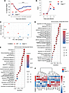 Figure 1
CASP11 deficiency protects against severe SARS-CoV-2 infection, limits early hyperinflammation, and enhances IFN-γ responses. (A) Body weight change in WT and Casp11–/– mice after i.n. infection with 5 × 10⁵ TCID50 of MA10 SARS-CoV-2. WT mice exhibited more severe weight loss and slower recovery through day 14 compared with Casp11–/– mice (n = 26/group). Statistical analysis: 1-way ANOVA with Šídák’s multiple-comparison test. (B) PenH, as a surrogate of airway resistance, measured by whole-body plethysmography at multiple time points after infection, showing significantly lower resistance in Casp11–/– mice, indicative of improved lung function (n = 10 WT, n = 12 Casp11–/–). Statistical analysis: 2-way ANOVA with Šídák’s multiple-comparison test. (C) Principal component analysis (PCA) of variance-stabilized RNA-Seq data revealed distinct transcriptional profiles between WT and Casp11–/– lungs at day 4 after infection (5 × 10⁵ TCID50 of MA10). PC1 accounted for 55% and PC2 for 17% of total variance. PERMANOVA analysis confirmed statistically significant separation between groups (R2 = 0.343, F = 3.135, P = 0.05, 999 permutations), indicating that genotype accounts for 34.3% of total transcriptional variance. Homogeneity of group dispersions was confirmed by betadisper analysis (F = 0.205, P = 0.554), validating the PERMANOVA result (n = 4/group). (D) GO enrichment analysis of genes with higher gene expression in Casp11–/– lungs compared with WT reveals enrichment in IFN signaling pathways and antiviral immune pathways (n = 4/group). (E) GO enrichment analysis of genes with lower gene expression in Casp11–/– lungs compared with WT highlights suppression of innate immune pathways, including myeloid cell chemotaxis and migration (n = 4/group). (F) Heatmap of differentially expressed cytokine and chemokine genes shows a shift in Casp11–/– lungs from pro-myeloid inflammation (e.g., Cxcl1, Il6, Ccl22) to lymphocyte-promoting responses (e.g., Ccl3, Ccl5, Ifng) (n = 4/group). ****P < 0.0001.
To further investigate the molecular mechanisms underlying the enhanced protection and recovery observed in Casp11–/– mice, we performed bulk RNA-Seq of lung tissue harvested at day 4 after infection. Principal component analysis (PCA) revealed distinct transcriptional profiles between infected WT and Casp11–/– lungs, highlighting a divergent host response (Figure 1C). PERMANOVA analysis confirmed statistically significant separation between groups (R2 = 0.343, F = 3.135, P = 0.05, 999 permutations), indicating that genotype accounts for 34.3% of total transcriptional variance. Homogeneity of group dispersions was confirmed by betadisper analysis (F = 0.205, P = 0.554), validating that the observed separation reflects true differences in group centroids rather than differences in within-group variance.
Gene Ontology (GO) enrichment analysis of genes with higher expression in the Casp11–/– group showed a marked enrichment of antiviral immune pathways. Notably, both type I and type II IFN response pathways were significantly upregulated at the transcription level, suggesting a robust antiviral state in the absence of CASP11 (Figure 1D). In contrast, genes with lower expression in the Casp11–/– lungs were predominantly related to myeloid cell chemotaxis and migration, suggesting a reduced innate inflammatory response in the absence of CASP11 (Figure 1E).
Further gene set enrichment analysis (GSEA) revealed significant enrichment of antiviral pathways in Casp11–/– lungs, including the IFN-γ response and lymphocyte-mediated immunity (Supplemental Figure 1B). We then examined the expression of cytokines and chemokines in infected lungs. In Casp11–/– mice, there was upregulation of IFN-γ and several chemokines known to promote effector T cell recruitment, including CCL3, CCL4, CCL5, and CXCL10 (36, 37) (Figure 1F and Supplemental Figure 2). In contrast, inflammatory mediators typically associated with neutrophil and monocyte recruitment such as CXCL1, CXCL5, CCL22, IL-6, IL-11, and IL1R1 were downregulated in Casp11–/– lungs relative to WT (38, 39) (Figure 1F), suggesting a shift away from proinflammatory innate immune signaling toward a more lymphocyte-driven antiviral response (Figure 1F and Supplemental Figure 2).
We then examined cytokine protein levels in lung homogenates using ELISA at day 4 after infection, the same time point used for the RNA-Seq analysis. Compared with WT, Casp11–/– lungs displayed markedly lower levels of IL-1β, CXCL1, and IL-6, and IFN-γ levels were significantly elevated (Figure 2A). These findings confirm the transcriptional signature observed by RNA-Seq and support a shift toward an enhanced IFN-γ response. We measured levels of type I IFNs (IFN-α and IFN-β) in lung homogenates at day 4 after infection using ELISA. We did not observe a significant difference in IFN-I protein levels between the WT and Casp11–/– infected groups (Supplemental Figure 3A).
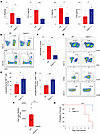 Figure 2
CASP11 deficiency limits early inflammation and neutrophil infiltration, enhances effector CD8+ T cell responses, promotes viral clearance, and protects against lethal SARS-CoV-2 infection. (A) Cytokine protein levels in lung homogenates at day 4 after infection (5 × 10⁵ TCID50 MA10), measured by ELISA and normalized to lung tissue weight (pg/mg tissue). Casp11–/– lungs exhibited reduced levels of IL-1β, CXCL1, and IL-6, and elevated IFN-γ compared with WT (n = 5/group). Statistical significance was determined by unpaired Student’s t test. (B and C) Flow cytometry of lung tissue at day 4 after infection (5 × 10⁵ TCID50 MA10) shows significantly fewer neutrophils (CD45hiCD11bhiLy6Ghi) in Casp11–/– mice compared with WT. Gating strategy (B) and quantification (C) (n = 5/group). Statistical significance was determined by unpaired Student’s t test. (D and E) At day 7 after infection, flow cytometry after intravascular anti-CD45 labeling revealed higher frequencies of lung parenchymal (CD45lo) CD8+ T cells and activated effector CD8+ T cells (CD62LloCD44hiCD69hi) in Casp11–/– lungs. Gating strategy (D) and quantification (E) (n = 6/group). Statistical significance was determined by unpaired Student’s t test. (F) Viral titers in lung homogenates assessed by plaque assay at day 7 after infection (5 × 10⁵ TCID50 MA10), expressed as PFU/g tissue. Casp11–/– mice completely cleared the virus by day 7 in contrast to WT (n = 6/group). Statistical significance was determined by unpaired Student’s t test. (G) Kaplan-Meier survival analysis after high-dose SARS-CoV-2 challenge (1 × 10⁶ TCID50 MA10). All WT mice died by day 7; 72.7% of Casp11–/– mice survived (n = 9 WT, n = 11 Casp11). Statistical significance was determined by log-rank (Mantel-Cox) test. *P < 0.05; **P < 0.01; ***P < 0.001.
CASP11 deficiency limits neutrophil infiltration, enhances effector CD8+ T cell responses, promotes viral clearance, and protects against lethal SARS-CoV-2 infection. Given the distinct differences in immune responses between WT and Casp11–/– mice, we next investigated the immune cell composition in the lungs after infection. Lungs were harvested on day 4 after infection and analyzed by flow cytometry. Consistent with the observed downregulation of neutrophil-attracting chemokines, Casp11–/– lungs exhibited significantly reduced neutrophil infiltration compared with WT-infected lungs (Figure 2C, gating strategy shown in Figure 2B), supporting a reduced innate inflammatory response in the absence of CASP11.
Given the significantly elevated levels of IFN-γ protein in Casp11–/– lungs at day 4 after infection, we sought to identify the primary cellular source of this cytokine during the early acute phase. Flow cytometric analysis with intracellular cytokine staining revealed that NK cells, rather than early infiltrating T cells, were the predominant producers of IFN-γ at this time point (Supplemental Figure 3, B and C).
Although both myeloid cells and NK cells were identified as baseline producers of IFN-γ at day 4 after infection, quantification revealed a compartment-specific enhancement in the Casp11–/– mice. As shown in Supplemental Figure 3C, the frequency of IFN-γ+ NK cells was significantly increased in Casp11–/– mice compared with WT controls. In contrast, IFN-γ production by myeloid cells and T cells remained unchanged between the genotypes. These data demonstrate that the CASP11-regulated elevation of early antiviral IFN-γ in Casp11–/– mice is specifically attributable to NK cells.
We then performed flow cytometry on lung tissue harvested at day 7 after infection, a time point selected to allow for the development of an effector T cell response. To distinguish lung-resident (parenchymal) T cells from circulating peripheral T cells, mice were retro-orbitally injected with fluorescent anti-CD45 antibody 10 minutes before euthanization, labeling CD45+ cells in the intravascular space while leaving tissue-resident cells (lung parenchymal cells) unlabeled. Peripheral blood cells were more than 90% CD45+, demonstrating efficient labeling of circulating cells (data not shown). Flow cytometric analysis revealed that Casp11–/– lungs contained significantly more lung parenchymal CD8+ T cells compared with WT controls (gating strategy in Figure 2, D and E). Further analysis of T cell activation status, based on surface marker expression, demonstrated an increased proportion of activated effector CD8+ T cells (CD62L–CD44+CD69+) in Casp11–/– lungs relative to WT lungs (gating strategy in Figure 2, D and E). No significant differences were observed in CD4+ T cell populations between the 2 groups (Supplemental Figure 4A).
To test whether this enhanced T cell response translates into improved viral clearance, we performed plaque assays on lungs collected from WT and Casp11–/– mice at days 4 and 7 after infection. Although viral loads were similar between groups at day 4 (Supplemental Figure 4B), by day 7, Casp11–/– mice had completely cleared the virus, in contrast to WT mice, which still had detectable viral titers (Figure 2F). These results suggest that the robust activated effector CD8+ T cell response in Casp11–/– mice is associated with enhanced viral clearance.
To gain a comprehensive understanding of the global adaptive immune response, we also evaluated the circulating intravascular CD8+ cell compartment, distinguished by in vivo intravascular CD45 labeling. Interestingly, in stark contrast to the enhanced effector response observed within the lung parenchyma, there were no significant differences between WT and Casp11–/– mice in the frequencies of total circulating CD45+CD8+ T cells, CD69+ activated circulating CD8+ T cells, or IFN-γ+ circulating CD8+ T cells (Supplemental Figure 4, C–E).
We next asked whether CASP11 deficiency protects against lethal SARS-CoV-2 infection. WT and Casp11–/– mice were infected with a higher dose of MA10 (1 × 10⁶ TCID50). All WT mice (n = 9) died from the infection by day 7, whereas 72.7% of the Casp11–/– mice (8 of 11 mice) survived (Figure 2G). These data demonstrate that CASP11 deficiency confers protection from lethal SARS-CoV-2 challenge.
CASP11 deficiency promotes functional, antigen-specific CD8+ T cell response independent of SARS-CoV-2 disease severity. Next, we investigated whether the absence of CASP11 similarly promotes a robust effector CD8+ T cell response during moderate SARS-CoV-2 infection. To address this, we used a low-dose infection model in which WT and Casp11–/– mice were infected with 1 × 10⁵ TCID50 of the mouse-adapted SARS-CoV-2 strain MA10. As previously reported, both WT and Casp11–/– mice began regaining weight by day 4 and continued to recover through day 7, although WT mice exhibited a slower recovery trajectory (Supplemental Figure 5A) (32). Casp11–/– mice also had reduced early inflammatory cytokines (IL-1β, CXCL1, and IL-6) and decreased neutrophil infiltration at days 2 and 4 after infection (32). Importantly, viral loads were comparable between strains, becoming nearly undetectable by day 4 and fully cleared by day 7 (32).
To further evaluate the adaptive immune response in this less-severe SARS-CoV-2 infection model, we performed flow cytometry on lung tissue at day 7 after infection, a time point when both groups had cleared the virus. As in previous experiments, mice received a retro-orbital injection of fluorescent anti-CD45 antibody prior to euthanasia to label intravascular immune cells, enabling us to distinguish infiltrating lung parenchymal from intravascular T cells.
We found that Casp11–/– mice harbored a higher number of lung parenchymal CD8+ T cells compared with WT cells (Figure 3B, gating strategy shown in Figure 3A), including a significantly increased population of activated effector CD8+ T cells (CD62LloCD44hiCD69hi) (Figure 3C, gating strategy shown in Figure 3A). These findings confirm that the enhanced CD8+ T cell response observed in Casp11–/– mice is not merely a consequence of higher viral burden in WT mice. Instead, it is intrinsically driven by the absence of CASP11.
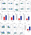 Figure 3
CASP11 deficiency promotes functional antigen-specific CD8+ T cell responses independent of SARS-CoV-2 disease severity. (A–C) Representative flow cytometry plots showing lung parenchymal CD8+ T cells (CD45loCD8+) and activated effector CD8+ T cells (CD62LloCD44hiCD69hi) at day 7 after infection in WT and Casp11–/– mice following low-dose (1 × 10⁵ TCID50) MA10 SARS-CoV-2 infection. (A) Gating strategy. Quantification of lung parenchymal CD8+ T cells (B) and activated effector CD8+ T cells (C) as a percentage of live cells showing increased frequency of functional effector CD8+ T cells in Casp11–/– lungs (n = 6/group). Statistical significance was determined by unpaired Student’s t test. (D and E) Flow cytometry analysis of intracellular IFN-γ production by CD8+ T cells after ex vivo stimulation with PMA/ionomycin (D) or SARS-CoV-2 spike peptide (E), showing increased frequency of functional IFN-γ–producing effector CD8+ T cells in Casp11–/– lungs. Gating strategy shown in A (n = 6/group). Statistical significance was determined by unpaired Student’s t test. (F and G) Representative flow cytometry analysis of tetramer staining of lung CD8+ T cells using SARS-CoV-2–specific tetramer (H-2Db N219-227). (F) Gating strategy. (G) Quantification of tetramer+ CD8+ T cells among lung parenchymal T cells. Casp11–/– mice exhibit a significantly greater population of antigen-specific CD8+ T cells (n = 6 WT, n = 8 Casp11–/–). Statistical significance was determined by unpaired Student’s t test. **P < 0.01; ****P < 0.0001.
Because CASP11 is a central effector in the inflammasome pathway, we also investigated whether its absence affects the T cell phenotype in response to SARS-CoV-2 infection (40, 41). To explore this question, we isolated lungs from WT and Casp11–/– mice at day 7 after infection and stimulated single-cell suspensions ex vivo with PMA/ionomycin or an overlapping peptide pool derived from SARS-CoV-2 spike protein. After stimulation, cells were stained for T cell activation markers and intracellular IFN-γ, since IFN-γ–producing CD8+ T cells are generally critical for respiratory virus clearance and infection control. Casp11–/– mice showed increased numbers of IFN-γ–producing effector CD8+ T cells in response to both stimuli (Figure 3, D and E; gating strategy shown in Figure 3A), suggesting that Casp11 deficiency promotes expansion or maintenance of an antigen-specific CD8+ T cell population with heightened effector function.
Furthermore, to evaluate the antigen specificity of the CD8+ T cell response, we performed SARS-CoV-2 tetramer staining, which allows direct detection of CD8+ T cells specific for defined viral epitopes presented by MHC class I (42). This approach is especially useful for distinguishing true antigen-driven responses from bystander T cell activation, an important distinction in the context of viral infections like SARS-CoV-2. Casp11–/– mice exhibited an increased number of lung parenchymal CD8+ T cells positive for tetramer (H-2Db N219-227) (Figure 3G, gating strategy shown in Figure 3F), indicating that the enhanced CD8+ T cell response detected in the absence of CASP11 is composed of virus antigen-specific effector cells. Overall, these findings demonstrate that CASP11 deficiency enhances the quantity and functional quality of virus-specific effector CD8+ T cells in the lungs after SARS-CoV-2 infection.
CASP11 mediates persistent inflammatory and T cell responses after SARS-CoV-2 infection, indicating ongoing inflammation and immune activation despite viral clearance. Persistent inflammation and sustained T cell activation have been implicated in postviral sequelae, including tissue remodeling; immune dysregulation; and long-term complications such as fibrosis, impaired lung function, and long COVID (43–48). Therefore, we investigated whether CASP11 influences the persistence of inflammatory responses and immune cell activation after viral clearance and whether it contributes to the prolonged immune activity often observed beyond the virus replication phase in SARS-CoV-2 infection. To address this question, we infected WT and Casp11–/– mice with MA10 (5 × 10⁵ TCID50). Mice were then euthanized at day 14, and lung tissue was collected for cytokine profiling and flow cytometric analysis. On day 14 after infection, WT lungs exhibited persistently elevated levels of inflammatory cytokines (Supplemental Figure 5B) and a higher number of CD8+ and CD4+ T cells than the lungs of Casp11–/– mice (Supplemental Figure 5, C and D). In contrast, Casp11–/– lungs demonstrated a more resolved immune state, characterized by lower cytokine levels and reduced T cell numbers.
To determine whether this sustained immunopathology correlates with chronic inflammasome activation, we analyzed Casp11 mRNA expression at a late time point. Strikingly, qPCR analysis revealed a sustained upregulation of Casp11 mRNA in the lung tissues of infected WT mice at 30 days after infection (Supplemental Figure 5E). Together, these findings indicate that Casp11 expression persists long after viral clearance, suggesting a potential ongoing role in chronic immune dysregulation.
CASP11 deficiency in hematopoietic immune cells enhances survival, reduces inflammation, and promotes T cell response after SARS-CoV-2 infection. To further elucidate the mechanism by which CASP11 deficiency enhances the adaptive immune response and protects against severe disease, we generated hematopoietic BM chimeric mice (Supplemental Figure 6A). In this model, lethally irradiated WT recipient mice were reconstituted with BM from either WT or Casp11–/– donors, generating 2 groups: WT → WT chimeras (WT chimera) and Casp11–/– → WT chimeras (hereafter referred to as Casp11–/– chimeras). This strategy enabled us to isolate the role of CASP11 within radiosensitive hematopoietic cells, such as monocytes, DCs, and lymphocytes, while preserving CASP11 expression in radioresistant nonhematopoietic compartments, including lung epithelial and endothelial cells. We assessed the contribution of hematopoietic CASP11 to SARS-CoV-2–induced immunopathology, T cell responses, and viral clearance. Importantly, interstitial macrophages and monocyte-derived macrophages in the lung are donor-derived, whereas alveolar macrophages are largely host-derived and remain WT due to their radioresistant, self-renewing nature (49). This setup enables a compartment-specific interpretation of CASP11 function in the lung immune microenvironment.
After a full reconstitution period, chimeric mice were infected with 1 × 10⁵ TCID50 of MA10. This dose, in the context of relatively aged chimeras (approximately 6 months old at infection), consistently induced severe disease. Only 41.7% (10 of 24) of WT chimeras survived by 7 days after infection, compared with 76.7% (23 of 30) of the Casp11–/– chimeras (Figure 4A). Respiratory functions in infected mice were assessed by plethysmography, which revealed that Casp11–/– infected chimera mice had decreased PenH values indicative of decreased airway resistance compared with infected WT chimeras (Figure 4B). This indicates that the deficiency of CASP11 in hematopoietic cells significantly alters lung functions and clinical severity in SARS-CoV-2 infection. Consistent with these findings, H&E staining of lung sections demonstrated that WT chimeras had significantly more lung consolidation, with increased edema, inflammation, and cellular infiltrates at day 4 after infection compared with Casp11–/– chimeras (Figure 4. C and D). Flow cytometric profiling of lung myeloid cells at day 4 after infection showed fewer infiltrating neutrophils in Casp11–/– chimeras compared with WT (Figure 4E).
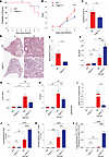 Figure 4
CASP11 deficiency in hematopoietic immune cells enhances survival, reduces inflammation, and promotes T cell response after SARS-CoV-2 infection. (A) Kaplan-Meier survival analysis of WT chimeras (WT → WT) and Casp11–/– chimeras (Casp11–/– → WT) after i.n. infection with 1 × 10⁵ TCID50 of MA10 SARS-CoV-2. Casp11–/– chimeras (Casp11–/– → WT) after i.n. infection with 1 × 10⁵ TCID50 of MA10 SARS-CoV-2(n = 24 WT chimeras, n = 30 Casp11–/– chimeras). (B) PenH, a surrogate of airway resistance, measured by whole-body plethysmography at day 3 after infection (n = 18/group). (C and D) Cellularity scores (C) and representative H&E-stained lung sections (original magnification, ×2 and ×10) (D) at day 4 after infection (n = 4/group). (E) Flow cytometric quantification of neutrophils (LY6G+) in lung tissue at day 4 after infection (n = 4/group). (F–H) RT-qPCR analysis of inflammatory mediators in lung tissue at day 4 after infection Ifng (F) Cxcl1 (G) Il6 (H) in Casp11–/– chimeras compared with WT chimeras (n = 5 infected WT chimeras, n = 6 infected Casp11–/– chimeras, n = 3 noninfected WT chimeras, n = 3 noninfected Casp11–/– chimeras). (I) SARS-CoV-2 N gene RNA copy number (RCN) in lung homogenates at day 7 after infection, quantified by RT-qPCR (n = 5 infected WT chimeras, n = 6 infected Casp11–/– chimeras, n = 4 noninfected WT chimeras, n = 4 noninfected Casp11–/– chimeras). (J and K) Flow cytometric quantification with intravascular CD45 labeling of lung parenchymal CD8+ T cells (J) and activated effector CD8+ T cells (CD62LloCD44hiCD69hi) (K) at day 7 after infection (n = 4 infected WT chimeras, n = 6 infected Casp11–/– chimeras, n = 4 noninfected WT chimeras, n = 4 noninfected Casp11–/– chimeras). (L) Frequency of IFN-γ+ CD8+ T cells among lung parenchymal CD8+ T cells after ex vivo stimulation with SARS-CoV-2 SN peptide mixture at day 7 after infection. (n = 4 infected WT chimeras, n = 6 infected Casp11–/– chimeras, n = 4 noninfected WT chimeras, n = 4 noninfected Casp11–/– chimeras). Statistical significance determined by (A) log-rank (Mantel-Cox) test, (B) 2-way ANOVA with Šídák’s multiple-comparison test, (C and E) unpaired Student’s t test, and (F–H, J, K, and L) 1-way ANOVA with Tukey’s multiple-comparison test. Noninfected controls were age-matched, naive chimeric mice that underwent identical irradiation and BM reconstitution. Data shown as mean ± SEM. *P < 0.05; **P < 0.01; ***P < 0.001; ****P < 0.0001.
Cytokine mRNA analysis by qPCR at day 4 after infection revealed Ifng was significantly upregulated while Il6 and Cxcl1 were downregulated in Casp11–/– chimera mice (Figure 4, F–H), consistent with earlier observations in global KO. To investigate broader transcriptional changes, we performed RNA-Seq on lung tissue at day 4 after infection. GSEA revealed enrichment of gene sets related to T cell activation and antigen presentation pathways in Casp11–/– chimeras (Supplemental Figure 6B).
By day 7 after infection, Casp11–/– chimeras exhibited significantly lower SARS-CoV-2 N gene RNA copy numbers in the lungs compared with WT chimeras, indicating enhanced viral clearance (Figure 4I). To assess T cell response, we performed in vivo labeling of intravascular immune cells via retro-orbital injection of fluorescent anti-CD45 antibody, as previously described. This allowed us to selectively quantify lung parenchymal CD8+ T cells. Flow cytometric analysis revealed a higher number of parenchymal CD8+ T cells in Casp11–/– chimeras relative to WT chimera controls (Figure 4J), along with a greater frequency of activated effector CD8+ T cells characterized by a CD62LloCD44hiCD69hi phenotype (Figure 4K).
To further evaluate CD8+ T cell effector function, we stimulated lung-derived CD8+ T cells ex vivo with a SARS-CoV-2 spike and nucleocapsid (SN) peptide mixture. Casp11–/– chimeras demonstrated a higher frequency of IFN-γ–producing CD8+ T cells in response to ex vivo SN peptide stimulation, indicating preservation of effector function and enhanced antiviral potential (gating strategy shown in Supplemental Figure 7 and Figure 4L).
Mononuclear phagocyte system cell–specific CASP11 deletion enhances antiviral CD8+ T cell responses, accelerates recovery, and improves viral clearance during SARS-CoV-2 infection. The enhanced adaptive immune response observed in Casp11–/– chimeras, along with reduced disease severity and improved viral clearance, suggests that CASP11 exerts its effects through hematopoietic immune cells. However, it remains unclear whether this regulation is primarily mediated by myeloid cells via modulation of the innate immune response, or whether CASP11 also plays a direct, intrinsic role in T cells.
To address this question, we generated mice with CASP11 deficiency specifically in Cx3cr1-expressing mononuclear phagocyte system (MPS) cells. We utilized Cx3cr1-CreERT2 mice, in which Cre-ERT2 and EYFP are knocked into the Cx3cr1 locus, to generate CASP11-deficient mice in Cx3cr1-expressing lineages. The main cell types targeted by this system include patrolling Ly6C-LOW monocytes, interstitial macrophages, monocyte-derived macrophages, and a subset of DCs, all of which express CX3CR1 at moderate-to-high levels (50). Classical Ly6Chi inflammatory monocytes, which express lower levels of CX3CR1, are less efficiently targeted by this system. Alveolar macrophages are minimally affected due to their low CX3CR1 expression (50, 51). Neutrophils and lymphocytes do not express CX3CR1 at high levels and are therefore not targeted (50, 52).
Importantly, both control and experimental mice are homozygous for the Cx3cr1-CreERT2 knockin allele, rendering both groups CX3CR1-deficient (Supplemental Figure 8A). This shared genetic background ensures that any observed differences between groups are attributable specifically to CASP11 deletion rather than CX3CR1 deficiency. For simplicity, we refer to Cx3cr1-CreERT2 Casp11WT/WT mice as Cx3cr1-Cre control and Cx3cr1-CreERT2 Casp11fl/fl mice as Cx3cr1-MPS-Casp11–/–.
To validate the specificity and efficiency of our inducible KO model, we utilized the YFP fluorescent reporter to track Cre recombinase activity in Cx3cr1-expressing cells. Flow cytometric analysis of noninfected lungs from Cx3cr1-Cre control and Cx3cr1-MPS-Casp11–/– mice was performed using a gating strategy that sequentially excluded dead cells and LY6G+ neutrophils, followed by identification of CD11B+F480+ macrophages and CD4+ and CD8+ T cells (Supplemental Figure 8B).
This analysis confirmed robust and selective YFP reporter expression specifically within the CD11B+F480+ lung macrophage compartment, which includes interstitial macrophages and monocyte-derived macrophages (95% YFP+). Conversely, LY6G+ neutrophils (4.5% YFP+), CD4+ T cells (0.5% YFP+), and CD8+ T cells (3.3% YFP+) remained largely YFP–, confirming precise cellular targeting within the mononuclear phagocyte compartment and a lack of appreciable off-target recombination in innate or adaptive immune cells (Supplemental Figure 8C).
To functionally verify targeted CASP11 deletion during acute infection, YFP+ cells were FACS-sorted from lung tissue at day 3 after SARS-CoV-2 infection. qPCR confirmed an approximately 75% reduction in Casp11 mRNA expression specifically within the sorted YFP+ macrophage compartment of Cx3cr1-MPS-Casp11–/– mice relative to Cx3cr1-Cre matched controls (Supplemental Figure 8D). Together, these data demonstrate specific and efficient CASP11 deletion within the Cx3cr1-expressing lung macrophage compartment, with no substantial targeting of neutrophils or lymphocytes.
Cx3cr1-MPS-Casp11–/– and Cx3cr1-Cre control mice were infected with 5 × 10⁵ TCID50 of MA10 virus to establish a severe SARS-CoV-2 infection model. Cx3cr1-MPS-Casp11–/– mice exhibited enhanced recovery, indicating milder disease severity (Figure 5A). Histological analysis (H&E staining) and cellularity scoring of lung sections at day 4 after infection revealed reduced inflammatory infiltrates and edema in Cx3cr1-MPS-Casp11–/– mice compared with Cx3cr1-Cre controls (Figure 5, B and C).
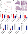 Figure 5
Cx3cr1-expressing MPS cell-specific CASP11 deletion enhances antiviral CD8+ T cell responses, accelerates recovery, and improves viral clearance during SARS-CoV-2 infection. (A) Body weight kinetics in Cx3cr1-Cre control and Cx3cr1-MPS-Casp11–/– mice after i.n. infection with 5 × 10⁵ TCID50 of MA10 SARS-CoV-2. Cx3cr1-MPS-Casp11–/– mice showed significantly improved recovery compared with Cx3cr1-Cre controls (n = 6/ Cx3cr1-Cre controls, n = 11/ Cx3cr1-MPS-Casp11–/–). (B) Representative H&E-stained lung sections at day 4 after infection showing reduced inflammatory infiltrates and edema in Cx3cr1-MPS-Casp11–/– mice versus Cx3cr1-Cre controls. Original magnification, ×2 and ×10. (C) Histopathological cellularity scoring of H&E-stained lung sections confirms significantly reduced pulmonary inflammation in Cx3cr1-MPS-Casp11–/– mice compared with Cx3cr1-Cre controls at day 4 after infection (n = 6/group). (D–F) Cytokine protein levels measured by ELISA in lung homogenates at day 4 after infection. Cx3cr1-MPS-Casp11–/– mice demonstrate significantly reduced CXCL1 (D, pg/mg tissue) and IL-1β (E, pg/mg tissue), and elevated IFN-γ (F, pg/mg tissue) compared with Cx3cr1-Cre controls (n = 6). (G) Viral titers in lung homogenates at day 7 after infection quantified by plaque assay on Vero E6-TMPRSS2 cells reveal significantly lower viral loads in Cx3cr1-MPS-Casp11–/– mice compared with Cx3cr1-Cre controls (PFU/g) (n = 4). (H) Quantification of lung parenchymal CD8+ T cells (CD45–CD8+) by flow cytometry with intravascular CD45 labeling at day 7 after infection. Cx3cr1-MPS-Casp11–/– mice harbor significantly greater numbers of lung parenchymal CD8+ T cells compared with Cx3cr1-Cre controls (n = 5/Cx3cr1-Cre controls, n = 4/Cx3cr1-MPS-Casp11–/–). (I) Frequency of activated effector CD8+ T cells (CD62Llo CD44hi CD69hi) among lung parenchymal CD8+ T cells at day 7 after infection. Cx3cr1-MPS-Casp11–/– mice show a significantly higher proportion of activated effector CD8+ T cells than Cx3cr1-Cre controls (n = 5/Cx3cr1-Cre controls, n = 4/Cx3cr1-MPS-Casp11–/–). (J and K) Frequency of IFN-γ+ CD8+ T cells among lung parenchymal CD8+ T cells after ex vivo stimulation with PMA/ionomycin (J) or SARS-CoV-2 SN peptide mixture (K) at day 7 after infection. Cx3cr1-MPS-Casp11–/– mice demonstrate a significantly higher frequency of IFN-γ–producing CD8+ T cells than Cx3cr1-Cre controls under both stimulation conditions (n = 5/Cx3cr1-Cre controls, n = 4/Cx3cr1-MPS-Casp11–/–). Statistical significance determined by (A) 2-way ANOVA with Šídák’s multiple-comparison test and (C–K) unpaired Student’s t test. Data shown as mean ± SEM. *P < 0.05; **P < 0.01; ***P < 0.001; ****P < 0.0001.
We next investigated the cytokine release profile in lung tissue harvested at day 4 after infection. Cx3cr1-MPS-Casp11–/– mice showed reduced release of inflammatory cytokines, including CXCL1 and IL-1β (Figure 5, D and E), while exhibiting elevated levels of IFN-γ (Figure 5F), consistent with observations in global Casp11–/– KO and chimera mice. These findings confirm that modulation of the early innate immune response and the resulting lung protection are mainly mediated by CASP11 within MPS cells.
We then used a plaque assay to quantify the viral load in the lungs of infected mice at day 7 after infection. We found that Cx3cr1-MPS-Casp11–/– mice had significantly lower viral loads compared with Cx3cr1-Cre control mice, indicating that the enhanced viral clearance observed in global Casp11–/– KO mice and Casp11–/– chimeras is due to the immunomodulatory effect of CASP11 in monocyte-derived cells (Figure 5G).
Next, we investigated changes in the CD8+ T cell response mediated by Casp11 deficiency in mononuclear phagocyte cells. Cx3cr1-MPS-Casp11–/– and Cx3cr1-Cre control mice were infected with the same dose of MA10 (5 × 10⁵ TCID50) and euthanized at day 7 after infection, after retro-orbital injection of fluorescent anti-CD45 antibody, as described before. Single-cell suspensions were prepared from the lungs, stained for surface markers, and subsequently stimulated ex vivo with PMA/ionomycin and a mixture of SN peptides, followed by permeabilization and intracellular IFN-γ staining. Flow cytometric analysis revealed that Cx3cr1-MPS-Casp11–/– mice had a higher number of lung parenchymal CD8+ T cells compared with Cx3cr1-Cre control mice (Figure 5H) (gating strategy shown in Supplemental Figure 9). Additionally, analysis of CD69+ activated effector CD8+ T cells showed significantly increased numbers in Cx3cr1-MPS-Casp11–/– mice (Figure 5I).
Intracellular cytokine staining demonstrated a higher frequency of IFN-γ–producing CD8+ T cells in the lungs of Cx3cr1-MPS-Casp11–/– mice compared with Cx3cr1-Cre control mice after stimulation with both PMA/ionomycin and SN peptide mixtures (Figure 5, J and K) (gating strategy shown in Supplemental Figure 9). Moreover, bulk RNA-Seq of lung tissue at day 4 after infection, coupled with GSEA, revealed enhanced antigen presentation and upregulation of lymphocyte activation and T cell activation pathways (Supplemental Figure 10). Collectively, these findings demonstrate that the upregulated IFN-γ response and the more robust adaptive CD8+ T cell response, leading to accelerated recovery and viral clearance, are mediated by the absence of CASP11 in myeloid cells, primarily lung interstitial macrophages and monocyte-derived macrophages.


