Research ArticleEndocrinologyGeneticsOncology
Open Access | ![]() 10.1172/jci.insight.198338
10.1172/jci.insight.198338
Tracing the molecular route to progression in miRNA-biogenesis-defective thyroid lesions
Anne-Sophie Chong,1,2 Carla Roca,1,3 Paula Morales-Sánchez,3 Eduard Dorca,4 Verónica Barea,5 Ignacio Ruz-Caracuel,6,7 Pablo Valderrabano,8 Carlota Rovira,9 Cristina Jou,9 Dorothée Bouron-Dal Soglio,10 Rebecca D. Chernock,11,12 Giovana T. Torrezan,13 Marc Pusztaszeri,14 José M. Cameselle-Teijeiro,15 Xavier Matias-Guiu,16 Clara V. Alvarez,17 Héctor Salvador,18 Jonathan D. Wasserman,19 Luis Javier Leandro-García,20 William D. Foulkes,21,22 Eduardo Andrés-León,23 Paula Casano-Sancho,24,25 and Barbara Rivera1,22
1Program in Molecular Mechanisms and Experimental Therapy in Oncology (Oncobell), Bellvitge Biomedical Research Institute (IDIBELL), L’Hospitalet de Llobregat, Barcelona, Spain.
2Genetics Program, Faculty of Biology, and
3Department of Biomedical Sciences, Faculty of Medicine and Health Sciences, University of Barcelona, Barcelona, Spain.
4Pathology Department, Bellvitge University Hospital, L’Hospitalet de Llobregat, Barcelona, Spain.
5Genetics and Genomics, Faculty of Biology, University of Barcelona, Barcelona, Spain.
6Ramón y Cajal Health Research Institute (IRYCIS), Ramón y Cajal University Hospital, CIBERONC, Madrid, Spain.
7Department of Pathology, Ramón y Cajal University Hospital, Madrid, Spain.
8Department of Endocrinology and Nutrition, Hospital Universitario Ramón y Cajal, IRYCIS, Madrid, Spain.
9Department of Pathology, Hospital Sant Joan de Déu, University of Barcelona, Barcelona, Spain.
10Centre Hospitalier Universitaire Sainte-Justine Research Center, Université de Montréal, Montréal, Quebec, Canada.
11Department of Pathology and Immunology, and
12Department of Otolaryngology Head and Neck Surgery, Washington University School of Medicine, St. Louis, Missouri, USA.
13Clinical and Functional Genomics Group, International Research Center/CIPE, A.C. Camargo Cancer Center, São Paulo, Brazil.
14Department of Pathology, Jewish General Hospital, McGill University, Montreal, Quebec, Canada.
15Department of Pathology, Clinical University Hospital of Santiago de Compostela, Health Research Institute of Santiago de Compostela (IDIS), University of Santiago de Compostela, Santiago de Compostela, Spain.
16Department of Pathology, Hospital Universitari Arnau de Vilanova, Universitat de Lleida, IRBLLEIDA, Lleida, Spain.
17Neoplasia & Endocrine Differentiation P0L5, Centre for Research in Molecular Medicine and Chronic Disease (CIMUS), Santiago de Compostela, Spain.
18Department of Oncology, Hospital Sant Joan de Déu, University of Barcelona, Barcelona, Spain.
19Division of Endocrinology, Department of Paediatrics, The Hospital for Sick Children, University of Toronto, Toronto, Ontario, Canada.
20Hereditary Endocrine Cancer Group, Human Cancer Genetics Program, Spanish National Cancer Research Centre (CNIO), Madrid, Spain.
21Department of Human Genetics, and
22Gerald Bronfman Department of Oncology, McGill University, Montreal, Quebec, Canada.
23Bioinformatics Unit, Institute of Parasitology and Biomedicine López-Neyra (IPBLN), CSIC, Granada, Spain.
24Pediatric Endocrinology Department, Institut de Recerca Sant Joan de Déu, University of Barcelona, Barcelona, Spain.
25Centro de Investigación Biomédica en Red de Diabetes y Enfermedades Metabólicas Asociadas (CIBERDEM), Instituto de Salud Carlos III, Madrid, Spain.
Address correspondence to: Bárbara Rivera, Bellvitge Biomedical Research Institute, Avinguda de la Granvia de L’Hospitalet, 199, L’Hospitalet de Llobregat, Barcelona, Spain. Email: brivera@idibell.cat.
Find articles by Chong, A. in: PubMed | Google Scholar
1Program in Molecular Mechanisms and Experimental Therapy in Oncology (Oncobell), Bellvitge Biomedical Research Institute (IDIBELL), L’Hospitalet de Llobregat, Barcelona, Spain.
2Genetics Program, Faculty of Biology, and
3Department of Biomedical Sciences, Faculty of Medicine and Health Sciences, University of Barcelona, Barcelona, Spain.
4Pathology Department, Bellvitge University Hospital, L’Hospitalet de Llobregat, Barcelona, Spain.
5Genetics and Genomics, Faculty of Biology, University of Barcelona, Barcelona, Spain.
6Ramón y Cajal Health Research Institute (IRYCIS), Ramón y Cajal University Hospital, CIBERONC, Madrid, Spain.
7Department of Pathology, Ramón y Cajal University Hospital, Madrid, Spain.
8Department of Endocrinology and Nutrition, Hospital Universitario Ramón y Cajal, IRYCIS, Madrid, Spain.
9Department of Pathology, Hospital Sant Joan de Déu, University of Barcelona, Barcelona, Spain.
10Centre Hospitalier Universitaire Sainte-Justine Research Center, Université de Montréal, Montréal, Quebec, Canada.
11Department of Pathology and Immunology, and
12Department of Otolaryngology Head and Neck Surgery, Washington University School of Medicine, St. Louis, Missouri, USA.
13Clinical and Functional Genomics Group, International Research Center/CIPE, A.C. Camargo Cancer Center, São Paulo, Brazil.
14Department of Pathology, Jewish General Hospital, McGill University, Montreal, Quebec, Canada.
15Department of Pathology, Clinical University Hospital of Santiago de Compostela, Health Research Institute of Santiago de Compostela (IDIS), University of Santiago de Compostela, Santiago de Compostela, Spain.
16Department of Pathology, Hospital Universitari Arnau de Vilanova, Universitat de Lleida, IRBLLEIDA, Lleida, Spain.
17Neoplasia & Endocrine Differentiation P0L5, Centre for Research in Molecular Medicine and Chronic Disease (CIMUS), Santiago de Compostela, Spain.
18Department of Oncology, Hospital Sant Joan de Déu, University of Barcelona, Barcelona, Spain.
19Division of Endocrinology, Department of Paediatrics, The Hospital for Sick Children, University of Toronto, Toronto, Ontario, Canada.
20Hereditary Endocrine Cancer Group, Human Cancer Genetics Program, Spanish National Cancer Research Centre (CNIO), Madrid, Spain.
21Department of Human Genetics, and
22Gerald Bronfman Department of Oncology, McGill University, Montreal, Quebec, Canada.
23Bioinformatics Unit, Institute of Parasitology and Biomedicine López-Neyra (IPBLN), CSIC, Granada, Spain.
24Pediatric Endocrinology Department, Institut de Recerca Sant Joan de Déu, University of Barcelona, Barcelona, Spain.
25Centro de Investigación Biomédica en Red de Diabetes y Enfermedades Metabólicas Asociadas (CIBERDEM), Instituto de Salud Carlos III, Madrid, Spain.
Address correspondence to: Bárbara Rivera, Bellvitge Biomedical Research Institute, Avinguda de la Granvia de L’Hospitalet, 199, L’Hospitalet de Llobregat, Barcelona, Spain. Email: brivera@idibell.cat.
Find articles by Roca, C. in: PubMed | Google Scholar
1Program in Molecular Mechanisms and Experimental Therapy in Oncology (Oncobell), Bellvitge Biomedical Research Institute (IDIBELL), L’Hospitalet de Llobregat, Barcelona, Spain.
2Genetics Program, Faculty of Biology, and
3Department of Biomedical Sciences, Faculty of Medicine and Health Sciences, University of Barcelona, Barcelona, Spain.
4Pathology Department, Bellvitge University Hospital, L’Hospitalet de Llobregat, Barcelona, Spain.
5Genetics and Genomics, Faculty of Biology, University of Barcelona, Barcelona, Spain.
6Ramón y Cajal Health Research Institute (IRYCIS), Ramón y Cajal University Hospital, CIBERONC, Madrid, Spain.
7Department of Pathology, Ramón y Cajal University Hospital, Madrid, Spain.
8Department of Endocrinology and Nutrition, Hospital Universitario Ramón y Cajal, IRYCIS, Madrid, Spain.
9Department of Pathology, Hospital Sant Joan de Déu, University of Barcelona, Barcelona, Spain.
10Centre Hospitalier Universitaire Sainte-Justine Research Center, Université de Montréal, Montréal, Quebec, Canada.
11Department of Pathology and Immunology, and
12Department of Otolaryngology Head and Neck Surgery, Washington University School of Medicine, St. Louis, Missouri, USA.
13Clinical and Functional Genomics Group, International Research Center/CIPE, A.C. Camargo Cancer Center, São Paulo, Brazil.
14Department of Pathology, Jewish General Hospital, McGill University, Montreal, Quebec, Canada.
15Department of Pathology, Clinical University Hospital of Santiago de Compostela, Health Research Institute of Santiago de Compostela (IDIS), University of Santiago de Compostela, Santiago de Compostela, Spain.
16Department of Pathology, Hospital Universitari Arnau de Vilanova, Universitat de Lleida, IRBLLEIDA, Lleida, Spain.
17Neoplasia & Endocrine Differentiation P0L5, Centre for Research in Molecular Medicine and Chronic Disease (CIMUS), Santiago de Compostela, Spain.
18Department of Oncology, Hospital Sant Joan de Déu, University of Barcelona, Barcelona, Spain.
19Division of Endocrinology, Department of Paediatrics, The Hospital for Sick Children, University of Toronto, Toronto, Ontario, Canada.
20Hereditary Endocrine Cancer Group, Human Cancer Genetics Program, Spanish National Cancer Research Centre (CNIO), Madrid, Spain.
21Department of Human Genetics, and
22Gerald Bronfman Department of Oncology, McGill University, Montreal, Quebec, Canada.
23Bioinformatics Unit, Institute of Parasitology and Biomedicine López-Neyra (IPBLN), CSIC, Granada, Spain.
24Pediatric Endocrinology Department, Institut de Recerca Sant Joan de Déu, University of Barcelona, Barcelona, Spain.
25Centro de Investigación Biomédica en Red de Diabetes y Enfermedades Metabólicas Asociadas (CIBERDEM), Instituto de Salud Carlos III, Madrid, Spain.
Address correspondence to: Bárbara Rivera, Bellvitge Biomedical Research Institute, Avinguda de la Granvia de L’Hospitalet, 199, L’Hospitalet de Llobregat, Barcelona, Spain. Email: brivera@idibell.cat.
Find articles by Morales-Sánchez, P. in: PubMed | Google Scholar
1Program in Molecular Mechanisms and Experimental Therapy in Oncology (Oncobell), Bellvitge Biomedical Research Institute (IDIBELL), L’Hospitalet de Llobregat, Barcelona, Spain.
2Genetics Program, Faculty of Biology, and
3Department of Biomedical Sciences, Faculty of Medicine and Health Sciences, University of Barcelona, Barcelona, Spain.
4Pathology Department, Bellvitge University Hospital, L’Hospitalet de Llobregat, Barcelona, Spain.
5Genetics and Genomics, Faculty of Biology, University of Barcelona, Barcelona, Spain.
6Ramón y Cajal Health Research Institute (IRYCIS), Ramón y Cajal University Hospital, CIBERONC, Madrid, Spain.
7Department of Pathology, Ramón y Cajal University Hospital, Madrid, Spain.
8Department of Endocrinology and Nutrition, Hospital Universitario Ramón y Cajal, IRYCIS, Madrid, Spain.
9Department of Pathology, Hospital Sant Joan de Déu, University of Barcelona, Barcelona, Spain.
10Centre Hospitalier Universitaire Sainte-Justine Research Center, Université de Montréal, Montréal, Quebec, Canada.
11Department of Pathology and Immunology, and
12Department of Otolaryngology Head and Neck Surgery, Washington University School of Medicine, St. Louis, Missouri, USA.
13Clinical and Functional Genomics Group, International Research Center/CIPE, A.C. Camargo Cancer Center, São Paulo, Brazil.
14Department of Pathology, Jewish General Hospital, McGill University, Montreal, Quebec, Canada.
15Department of Pathology, Clinical University Hospital of Santiago de Compostela, Health Research Institute of Santiago de Compostela (IDIS), University of Santiago de Compostela, Santiago de Compostela, Spain.
16Department of Pathology, Hospital Universitari Arnau de Vilanova, Universitat de Lleida, IRBLLEIDA, Lleida, Spain.
17Neoplasia & Endocrine Differentiation P0L5, Centre for Research in Molecular Medicine and Chronic Disease (CIMUS), Santiago de Compostela, Spain.
18Department of Oncology, Hospital Sant Joan de Déu, University of Barcelona, Barcelona, Spain.
19Division of Endocrinology, Department of Paediatrics, The Hospital for Sick Children, University of Toronto, Toronto, Ontario, Canada.
20Hereditary Endocrine Cancer Group, Human Cancer Genetics Program, Spanish National Cancer Research Centre (CNIO), Madrid, Spain.
21Department of Human Genetics, and
22Gerald Bronfman Department of Oncology, McGill University, Montreal, Quebec, Canada.
23Bioinformatics Unit, Institute of Parasitology and Biomedicine López-Neyra (IPBLN), CSIC, Granada, Spain.
24Pediatric Endocrinology Department, Institut de Recerca Sant Joan de Déu, University of Barcelona, Barcelona, Spain.
25Centro de Investigación Biomédica en Red de Diabetes y Enfermedades Metabólicas Asociadas (CIBERDEM), Instituto de Salud Carlos III, Madrid, Spain.
Address correspondence to: Bárbara Rivera, Bellvitge Biomedical Research Institute, Avinguda de la Granvia de L’Hospitalet, 199, L’Hospitalet de Llobregat, Barcelona, Spain. Email: brivera@idibell.cat.
Find articles by Dorca, E. in: PubMed | Google Scholar
1Program in Molecular Mechanisms and Experimental Therapy in Oncology (Oncobell), Bellvitge Biomedical Research Institute (IDIBELL), L’Hospitalet de Llobregat, Barcelona, Spain.
2Genetics Program, Faculty of Biology, and
3Department of Biomedical Sciences, Faculty of Medicine and Health Sciences, University of Barcelona, Barcelona, Spain.
4Pathology Department, Bellvitge University Hospital, L’Hospitalet de Llobregat, Barcelona, Spain.
5Genetics and Genomics, Faculty of Biology, University of Barcelona, Barcelona, Spain.
6Ramón y Cajal Health Research Institute (IRYCIS), Ramón y Cajal University Hospital, CIBERONC, Madrid, Spain.
7Department of Pathology, Ramón y Cajal University Hospital, Madrid, Spain.
8Department of Endocrinology and Nutrition, Hospital Universitario Ramón y Cajal, IRYCIS, Madrid, Spain.
9Department of Pathology, Hospital Sant Joan de Déu, University of Barcelona, Barcelona, Spain.
10Centre Hospitalier Universitaire Sainte-Justine Research Center, Université de Montréal, Montréal, Quebec, Canada.
11Department of Pathology and Immunology, and
12Department of Otolaryngology Head and Neck Surgery, Washington University School of Medicine, St. Louis, Missouri, USA.
13Clinical and Functional Genomics Group, International Research Center/CIPE, A.C. Camargo Cancer Center, São Paulo, Brazil.
14Department of Pathology, Jewish General Hospital, McGill University, Montreal, Quebec, Canada.
15Department of Pathology, Clinical University Hospital of Santiago de Compostela, Health Research Institute of Santiago de Compostela (IDIS), University of Santiago de Compostela, Santiago de Compostela, Spain.
16Department of Pathology, Hospital Universitari Arnau de Vilanova, Universitat de Lleida, IRBLLEIDA, Lleida, Spain.
17Neoplasia & Endocrine Differentiation P0L5, Centre for Research in Molecular Medicine and Chronic Disease (CIMUS), Santiago de Compostela, Spain.
18Department of Oncology, Hospital Sant Joan de Déu, University of Barcelona, Barcelona, Spain.
19Division of Endocrinology, Department of Paediatrics, The Hospital for Sick Children, University of Toronto, Toronto, Ontario, Canada.
20Hereditary Endocrine Cancer Group, Human Cancer Genetics Program, Spanish National Cancer Research Centre (CNIO), Madrid, Spain.
21Department of Human Genetics, and
22Gerald Bronfman Department of Oncology, McGill University, Montreal, Quebec, Canada.
23Bioinformatics Unit, Institute of Parasitology and Biomedicine López-Neyra (IPBLN), CSIC, Granada, Spain.
24Pediatric Endocrinology Department, Institut de Recerca Sant Joan de Déu, University of Barcelona, Barcelona, Spain.
25Centro de Investigación Biomédica en Red de Diabetes y Enfermedades Metabólicas Asociadas (CIBERDEM), Instituto de Salud Carlos III, Madrid, Spain.
Address correspondence to: Bárbara Rivera, Bellvitge Biomedical Research Institute, Avinguda de la Granvia de L’Hospitalet, 199, L’Hospitalet de Llobregat, Barcelona, Spain. Email: brivera@idibell.cat.
Find articles by Barea, V. in: PubMed | Google Scholar
1Program in Molecular Mechanisms and Experimental Therapy in Oncology (Oncobell), Bellvitge Biomedical Research Institute (IDIBELL), L’Hospitalet de Llobregat, Barcelona, Spain.
2Genetics Program, Faculty of Biology, and
3Department of Biomedical Sciences, Faculty of Medicine and Health Sciences, University of Barcelona, Barcelona, Spain.
4Pathology Department, Bellvitge University Hospital, L’Hospitalet de Llobregat, Barcelona, Spain.
5Genetics and Genomics, Faculty of Biology, University of Barcelona, Barcelona, Spain.
6Ramón y Cajal Health Research Institute (IRYCIS), Ramón y Cajal University Hospital, CIBERONC, Madrid, Spain.
7Department of Pathology, Ramón y Cajal University Hospital, Madrid, Spain.
8Department of Endocrinology and Nutrition, Hospital Universitario Ramón y Cajal, IRYCIS, Madrid, Spain.
9Department of Pathology, Hospital Sant Joan de Déu, University of Barcelona, Barcelona, Spain.
10Centre Hospitalier Universitaire Sainte-Justine Research Center, Université de Montréal, Montréal, Quebec, Canada.
11Department of Pathology and Immunology, and
12Department of Otolaryngology Head and Neck Surgery, Washington University School of Medicine, St. Louis, Missouri, USA.
13Clinical and Functional Genomics Group, International Research Center/CIPE, A.C. Camargo Cancer Center, São Paulo, Brazil.
14Department of Pathology, Jewish General Hospital, McGill University, Montreal, Quebec, Canada.
15Department of Pathology, Clinical University Hospital of Santiago de Compostela, Health Research Institute of Santiago de Compostela (IDIS), University of Santiago de Compostela, Santiago de Compostela, Spain.
16Department of Pathology, Hospital Universitari Arnau de Vilanova, Universitat de Lleida, IRBLLEIDA, Lleida, Spain.
17Neoplasia & Endocrine Differentiation P0L5, Centre for Research in Molecular Medicine and Chronic Disease (CIMUS), Santiago de Compostela, Spain.
18Department of Oncology, Hospital Sant Joan de Déu, University of Barcelona, Barcelona, Spain.
19Division of Endocrinology, Department of Paediatrics, The Hospital for Sick Children, University of Toronto, Toronto, Ontario, Canada.
20Hereditary Endocrine Cancer Group, Human Cancer Genetics Program, Spanish National Cancer Research Centre (CNIO), Madrid, Spain.
21Department of Human Genetics, and
22Gerald Bronfman Department of Oncology, McGill University, Montreal, Quebec, Canada.
23Bioinformatics Unit, Institute of Parasitology and Biomedicine López-Neyra (IPBLN), CSIC, Granada, Spain.
24Pediatric Endocrinology Department, Institut de Recerca Sant Joan de Déu, University of Barcelona, Barcelona, Spain.
25Centro de Investigación Biomédica en Red de Diabetes y Enfermedades Metabólicas Asociadas (CIBERDEM), Instituto de Salud Carlos III, Madrid, Spain.
Address correspondence to: Bárbara Rivera, Bellvitge Biomedical Research Institute, Avinguda de la Granvia de L’Hospitalet, 199, L’Hospitalet de Llobregat, Barcelona, Spain. Email: brivera@idibell.cat.
Find articles by Ruz-Caracuel, I. in: PubMed | Google Scholar
1Program in Molecular Mechanisms and Experimental Therapy in Oncology (Oncobell), Bellvitge Biomedical Research Institute (IDIBELL), L’Hospitalet de Llobregat, Barcelona, Spain.
2Genetics Program, Faculty of Biology, and
3Department of Biomedical Sciences, Faculty of Medicine and Health Sciences, University of Barcelona, Barcelona, Spain.
4Pathology Department, Bellvitge University Hospital, L’Hospitalet de Llobregat, Barcelona, Spain.
5Genetics and Genomics, Faculty of Biology, University of Barcelona, Barcelona, Spain.
6Ramón y Cajal Health Research Institute (IRYCIS), Ramón y Cajal University Hospital, CIBERONC, Madrid, Spain.
7Department of Pathology, Ramón y Cajal University Hospital, Madrid, Spain.
8Department of Endocrinology and Nutrition, Hospital Universitario Ramón y Cajal, IRYCIS, Madrid, Spain.
9Department of Pathology, Hospital Sant Joan de Déu, University of Barcelona, Barcelona, Spain.
10Centre Hospitalier Universitaire Sainte-Justine Research Center, Université de Montréal, Montréal, Quebec, Canada.
11Department of Pathology and Immunology, and
12Department of Otolaryngology Head and Neck Surgery, Washington University School of Medicine, St. Louis, Missouri, USA.
13Clinical and Functional Genomics Group, International Research Center/CIPE, A.C. Camargo Cancer Center, São Paulo, Brazil.
14Department of Pathology, Jewish General Hospital, McGill University, Montreal, Quebec, Canada.
15Department of Pathology, Clinical University Hospital of Santiago de Compostela, Health Research Institute of Santiago de Compostela (IDIS), University of Santiago de Compostela, Santiago de Compostela, Spain.
16Department of Pathology, Hospital Universitari Arnau de Vilanova, Universitat de Lleida, IRBLLEIDA, Lleida, Spain.
17Neoplasia & Endocrine Differentiation P0L5, Centre for Research in Molecular Medicine and Chronic Disease (CIMUS), Santiago de Compostela, Spain.
18Department of Oncology, Hospital Sant Joan de Déu, University of Barcelona, Barcelona, Spain.
19Division of Endocrinology, Department of Paediatrics, The Hospital for Sick Children, University of Toronto, Toronto, Ontario, Canada.
20Hereditary Endocrine Cancer Group, Human Cancer Genetics Program, Spanish National Cancer Research Centre (CNIO), Madrid, Spain.
21Department of Human Genetics, and
22Gerald Bronfman Department of Oncology, McGill University, Montreal, Quebec, Canada.
23Bioinformatics Unit, Institute of Parasitology and Biomedicine López-Neyra (IPBLN), CSIC, Granada, Spain.
24Pediatric Endocrinology Department, Institut de Recerca Sant Joan de Déu, University of Barcelona, Barcelona, Spain.
25Centro de Investigación Biomédica en Red de Diabetes y Enfermedades Metabólicas Asociadas (CIBERDEM), Instituto de Salud Carlos III, Madrid, Spain.
Address correspondence to: Bárbara Rivera, Bellvitge Biomedical Research Institute, Avinguda de la Granvia de L’Hospitalet, 199, L’Hospitalet de Llobregat, Barcelona, Spain. Email: brivera@idibell.cat.
Find articles by Valderrabano, P. in: PubMed | Google Scholar
1Program in Molecular Mechanisms and Experimental Therapy in Oncology (Oncobell), Bellvitge Biomedical Research Institute (IDIBELL), L’Hospitalet de Llobregat, Barcelona, Spain.
2Genetics Program, Faculty of Biology, and
3Department of Biomedical Sciences, Faculty of Medicine and Health Sciences, University of Barcelona, Barcelona, Spain.
4Pathology Department, Bellvitge University Hospital, L’Hospitalet de Llobregat, Barcelona, Spain.
5Genetics and Genomics, Faculty of Biology, University of Barcelona, Barcelona, Spain.
6Ramón y Cajal Health Research Institute (IRYCIS), Ramón y Cajal University Hospital, CIBERONC, Madrid, Spain.
7Department of Pathology, Ramón y Cajal University Hospital, Madrid, Spain.
8Department of Endocrinology and Nutrition, Hospital Universitario Ramón y Cajal, IRYCIS, Madrid, Spain.
9Department of Pathology, Hospital Sant Joan de Déu, University of Barcelona, Barcelona, Spain.
10Centre Hospitalier Universitaire Sainte-Justine Research Center, Université de Montréal, Montréal, Quebec, Canada.
11Department of Pathology and Immunology, and
12Department of Otolaryngology Head and Neck Surgery, Washington University School of Medicine, St. Louis, Missouri, USA.
13Clinical and Functional Genomics Group, International Research Center/CIPE, A.C. Camargo Cancer Center, São Paulo, Brazil.
14Department of Pathology, Jewish General Hospital, McGill University, Montreal, Quebec, Canada.
15Department of Pathology, Clinical University Hospital of Santiago de Compostela, Health Research Institute of Santiago de Compostela (IDIS), University of Santiago de Compostela, Santiago de Compostela, Spain.
16Department of Pathology, Hospital Universitari Arnau de Vilanova, Universitat de Lleida, IRBLLEIDA, Lleida, Spain.
17Neoplasia & Endocrine Differentiation P0L5, Centre for Research in Molecular Medicine and Chronic Disease (CIMUS), Santiago de Compostela, Spain.
18Department of Oncology, Hospital Sant Joan de Déu, University of Barcelona, Barcelona, Spain.
19Division of Endocrinology, Department of Paediatrics, The Hospital for Sick Children, University of Toronto, Toronto, Ontario, Canada.
20Hereditary Endocrine Cancer Group, Human Cancer Genetics Program, Spanish National Cancer Research Centre (CNIO), Madrid, Spain.
21Department of Human Genetics, and
22Gerald Bronfman Department of Oncology, McGill University, Montreal, Quebec, Canada.
23Bioinformatics Unit, Institute of Parasitology and Biomedicine López-Neyra (IPBLN), CSIC, Granada, Spain.
24Pediatric Endocrinology Department, Institut de Recerca Sant Joan de Déu, University of Barcelona, Barcelona, Spain.
25Centro de Investigación Biomédica en Red de Diabetes y Enfermedades Metabólicas Asociadas (CIBERDEM), Instituto de Salud Carlos III, Madrid, Spain.
Address correspondence to: Bárbara Rivera, Bellvitge Biomedical Research Institute, Avinguda de la Granvia de L’Hospitalet, 199, L’Hospitalet de Llobregat, Barcelona, Spain. Email: brivera@idibell.cat.
Find articles by Rovira, C. in: PubMed | Google Scholar
1Program in Molecular Mechanisms and Experimental Therapy in Oncology (Oncobell), Bellvitge Biomedical Research Institute (IDIBELL), L’Hospitalet de Llobregat, Barcelona, Spain.
2Genetics Program, Faculty of Biology, and
3Department of Biomedical Sciences, Faculty of Medicine and Health Sciences, University of Barcelona, Barcelona, Spain.
4Pathology Department, Bellvitge University Hospital, L’Hospitalet de Llobregat, Barcelona, Spain.
5Genetics and Genomics, Faculty of Biology, University of Barcelona, Barcelona, Spain.
6Ramón y Cajal Health Research Institute (IRYCIS), Ramón y Cajal University Hospital, CIBERONC, Madrid, Spain.
7Department of Pathology, Ramón y Cajal University Hospital, Madrid, Spain.
8Department of Endocrinology and Nutrition, Hospital Universitario Ramón y Cajal, IRYCIS, Madrid, Spain.
9Department of Pathology, Hospital Sant Joan de Déu, University of Barcelona, Barcelona, Spain.
10Centre Hospitalier Universitaire Sainte-Justine Research Center, Université de Montréal, Montréal, Quebec, Canada.
11Department of Pathology and Immunology, and
12Department of Otolaryngology Head and Neck Surgery, Washington University School of Medicine, St. Louis, Missouri, USA.
13Clinical and Functional Genomics Group, International Research Center/CIPE, A.C. Camargo Cancer Center, São Paulo, Brazil.
14Department of Pathology, Jewish General Hospital, McGill University, Montreal, Quebec, Canada.
15Department of Pathology, Clinical University Hospital of Santiago de Compostela, Health Research Institute of Santiago de Compostela (IDIS), University of Santiago de Compostela, Santiago de Compostela, Spain.
16Department of Pathology, Hospital Universitari Arnau de Vilanova, Universitat de Lleida, IRBLLEIDA, Lleida, Spain.
17Neoplasia & Endocrine Differentiation P0L5, Centre for Research in Molecular Medicine and Chronic Disease (CIMUS), Santiago de Compostela, Spain.
18Department of Oncology, Hospital Sant Joan de Déu, University of Barcelona, Barcelona, Spain.
19Division of Endocrinology, Department of Paediatrics, The Hospital for Sick Children, University of Toronto, Toronto, Ontario, Canada.
20Hereditary Endocrine Cancer Group, Human Cancer Genetics Program, Spanish National Cancer Research Centre (CNIO), Madrid, Spain.
21Department of Human Genetics, and
22Gerald Bronfman Department of Oncology, McGill University, Montreal, Quebec, Canada.
23Bioinformatics Unit, Institute of Parasitology and Biomedicine López-Neyra (IPBLN), CSIC, Granada, Spain.
24Pediatric Endocrinology Department, Institut de Recerca Sant Joan de Déu, University of Barcelona, Barcelona, Spain.
25Centro de Investigación Biomédica en Red de Diabetes y Enfermedades Metabólicas Asociadas (CIBERDEM), Instituto de Salud Carlos III, Madrid, Spain.
Address correspondence to: Bárbara Rivera, Bellvitge Biomedical Research Institute, Avinguda de la Granvia de L’Hospitalet, 199, L’Hospitalet de Llobregat, Barcelona, Spain. Email: brivera@idibell.cat.
Find articles by Jou, C. in: PubMed | Google Scholar
1Program in Molecular Mechanisms and Experimental Therapy in Oncology (Oncobell), Bellvitge Biomedical Research Institute (IDIBELL), L’Hospitalet de Llobregat, Barcelona, Spain.
2Genetics Program, Faculty of Biology, and
3Department of Biomedical Sciences, Faculty of Medicine and Health Sciences, University of Barcelona, Barcelona, Spain.
4Pathology Department, Bellvitge University Hospital, L’Hospitalet de Llobregat, Barcelona, Spain.
5Genetics and Genomics, Faculty of Biology, University of Barcelona, Barcelona, Spain.
6Ramón y Cajal Health Research Institute (IRYCIS), Ramón y Cajal University Hospital, CIBERONC, Madrid, Spain.
7Department of Pathology, Ramón y Cajal University Hospital, Madrid, Spain.
8Department of Endocrinology and Nutrition, Hospital Universitario Ramón y Cajal, IRYCIS, Madrid, Spain.
9Department of Pathology, Hospital Sant Joan de Déu, University of Barcelona, Barcelona, Spain.
10Centre Hospitalier Universitaire Sainte-Justine Research Center, Université de Montréal, Montréal, Quebec, Canada.
11Department of Pathology and Immunology, and
12Department of Otolaryngology Head and Neck Surgery, Washington University School of Medicine, St. Louis, Missouri, USA.
13Clinical and Functional Genomics Group, International Research Center/CIPE, A.C. Camargo Cancer Center, São Paulo, Brazil.
14Department of Pathology, Jewish General Hospital, McGill University, Montreal, Quebec, Canada.
15Department of Pathology, Clinical University Hospital of Santiago de Compostela, Health Research Institute of Santiago de Compostela (IDIS), University of Santiago de Compostela, Santiago de Compostela, Spain.
16Department of Pathology, Hospital Universitari Arnau de Vilanova, Universitat de Lleida, IRBLLEIDA, Lleida, Spain.
17Neoplasia & Endocrine Differentiation P0L5, Centre for Research in Molecular Medicine and Chronic Disease (CIMUS), Santiago de Compostela, Spain.
18Department of Oncology, Hospital Sant Joan de Déu, University of Barcelona, Barcelona, Spain.
19Division of Endocrinology, Department of Paediatrics, The Hospital for Sick Children, University of Toronto, Toronto, Ontario, Canada.
20Hereditary Endocrine Cancer Group, Human Cancer Genetics Program, Spanish National Cancer Research Centre (CNIO), Madrid, Spain.
21Department of Human Genetics, and
22Gerald Bronfman Department of Oncology, McGill University, Montreal, Quebec, Canada.
23Bioinformatics Unit, Institute of Parasitology and Biomedicine López-Neyra (IPBLN), CSIC, Granada, Spain.
24Pediatric Endocrinology Department, Institut de Recerca Sant Joan de Déu, University of Barcelona, Barcelona, Spain.
25Centro de Investigación Biomédica en Red de Diabetes y Enfermedades Metabólicas Asociadas (CIBERDEM), Instituto de Salud Carlos III, Madrid, Spain.
Address correspondence to: Bárbara Rivera, Bellvitge Biomedical Research Institute, Avinguda de la Granvia de L’Hospitalet, 199, L’Hospitalet de Llobregat, Barcelona, Spain. Email: brivera@idibell.cat.
Find articles by Bouron-Dal Soglio, D. in: PubMed | Google Scholar
1Program in Molecular Mechanisms and Experimental Therapy in Oncology (Oncobell), Bellvitge Biomedical Research Institute (IDIBELL), L’Hospitalet de Llobregat, Barcelona, Spain.
2Genetics Program, Faculty of Biology, and
3Department of Biomedical Sciences, Faculty of Medicine and Health Sciences, University of Barcelona, Barcelona, Spain.
4Pathology Department, Bellvitge University Hospital, L’Hospitalet de Llobregat, Barcelona, Spain.
5Genetics and Genomics, Faculty of Biology, University of Barcelona, Barcelona, Spain.
6Ramón y Cajal Health Research Institute (IRYCIS), Ramón y Cajal University Hospital, CIBERONC, Madrid, Spain.
7Department of Pathology, Ramón y Cajal University Hospital, Madrid, Spain.
8Department of Endocrinology and Nutrition, Hospital Universitario Ramón y Cajal, IRYCIS, Madrid, Spain.
9Department of Pathology, Hospital Sant Joan de Déu, University of Barcelona, Barcelona, Spain.
10Centre Hospitalier Universitaire Sainte-Justine Research Center, Université de Montréal, Montréal, Quebec, Canada.
11Department of Pathology and Immunology, and
12Department of Otolaryngology Head and Neck Surgery, Washington University School of Medicine, St. Louis, Missouri, USA.
13Clinical and Functional Genomics Group, International Research Center/CIPE, A.C. Camargo Cancer Center, São Paulo, Brazil.
14Department of Pathology, Jewish General Hospital, McGill University, Montreal, Quebec, Canada.
15Department of Pathology, Clinical University Hospital of Santiago de Compostela, Health Research Institute of Santiago de Compostela (IDIS), University of Santiago de Compostela, Santiago de Compostela, Spain.
16Department of Pathology, Hospital Universitari Arnau de Vilanova, Universitat de Lleida, IRBLLEIDA, Lleida, Spain.
17Neoplasia & Endocrine Differentiation P0L5, Centre for Research in Molecular Medicine and Chronic Disease (CIMUS), Santiago de Compostela, Spain.
18Department of Oncology, Hospital Sant Joan de Déu, University of Barcelona, Barcelona, Spain.
19Division of Endocrinology, Department of Paediatrics, The Hospital for Sick Children, University of Toronto, Toronto, Ontario, Canada.
20Hereditary Endocrine Cancer Group, Human Cancer Genetics Program, Spanish National Cancer Research Centre (CNIO), Madrid, Spain.
21Department of Human Genetics, and
22Gerald Bronfman Department of Oncology, McGill University, Montreal, Quebec, Canada.
23Bioinformatics Unit, Institute of Parasitology and Biomedicine López-Neyra (IPBLN), CSIC, Granada, Spain.
24Pediatric Endocrinology Department, Institut de Recerca Sant Joan de Déu, University of Barcelona, Barcelona, Spain.
25Centro de Investigación Biomédica en Red de Diabetes y Enfermedades Metabólicas Asociadas (CIBERDEM), Instituto de Salud Carlos III, Madrid, Spain.
Address correspondence to: Bárbara Rivera, Bellvitge Biomedical Research Institute, Avinguda de la Granvia de L’Hospitalet, 199, L’Hospitalet de Llobregat, Barcelona, Spain. Email: brivera@idibell.cat.
Find articles by Chernock, R. in: PubMed | Google Scholar
1Program in Molecular Mechanisms and Experimental Therapy in Oncology (Oncobell), Bellvitge Biomedical Research Institute (IDIBELL), L’Hospitalet de Llobregat, Barcelona, Spain.
2Genetics Program, Faculty of Biology, and
3Department of Biomedical Sciences, Faculty of Medicine and Health Sciences, University of Barcelona, Barcelona, Spain.
4Pathology Department, Bellvitge University Hospital, L’Hospitalet de Llobregat, Barcelona, Spain.
5Genetics and Genomics, Faculty of Biology, University of Barcelona, Barcelona, Spain.
6Ramón y Cajal Health Research Institute (IRYCIS), Ramón y Cajal University Hospital, CIBERONC, Madrid, Spain.
7Department of Pathology, Ramón y Cajal University Hospital, Madrid, Spain.
8Department of Endocrinology and Nutrition, Hospital Universitario Ramón y Cajal, IRYCIS, Madrid, Spain.
9Department of Pathology, Hospital Sant Joan de Déu, University of Barcelona, Barcelona, Spain.
10Centre Hospitalier Universitaire Sainte-Justine Research Center, Université de Montréal, Montréal, Quebec, Canada.
11Department of Pathology and Immunology, and
12Department of Otolaryngology Head and Neck Surgery, Washington University School of Medicine, St. Louis, Missouri, USA.
13Clinical and Functional Genomics Group, International Research Center/CIPE, A.C. Camargo Cancer Center, São Paulo, Brazil.
14Department of Pathology, Jewish General Hospital, McGill University, Montreal, Quebec, Canada.
15Department of Pathology, Clinical University Hospital of Santiago de Compostela, Health Research Institute of Santiago de Compostela (IDIS), University of Santiago de Compostela, Santiago de Compostela, Spain.
16Department of Pathology, Hospital Universitari Arnau de Vilanova, Universitat de Lleida, IRBLLEIDA, Lleida, Spain.
17Neoplasia & Endocrine Differentiation P0L5, Centre for Research in Molecular Medicine and Chronic Disease (CIMUS), Santiago de Compostela, Spain.
18Department of Oncology, Hospital Sant Joan de Déu, University of Barcelona, Barcelona, Spain.
19Division of Endocrinology, Department of Paediatrics, The Hospital for Sick Children, University of Toronto, Toronto, Ontario, Canada.
20Hereditary Endocrine Cancer Group, Human Cancer Genetics Program, Spanish National Cancer Research Centre (CNIO), Madrid, Spain.
21Department of Human Genetics, and
22Gerald Bronfman Department of Oncology, McGill University, Montreal, Quebec, Canada.
23Bioinformatics Unit, Institute of Parasitology and Biomedicine López-Neyra (IPBLN), CSIC, Granada, Spain.
24Pediatric Endocrinology Department, Institut de Recerca Sant Joan de Déu, University of Barcelona, Barcelona, Spain.
25Centro de Investigación Biomédica en Red de Diabetes y Enfermedades Metabólicas Asociadas (CIBERDEM), Instituto de Salud Carlos III, Madrid, Spain.
Address correspondence to: Bárbara Rivera, Bellvitge Biomedical Research Institute, Avinguda de la Granvia de L’Hospitalet, 199, L’Hospitalet de Llobregat, Barcelona, Spain. Email: brivera@idibell.cat.
Find articles by Torrezan, G. in: PubMed | Google Scholar
1Program in Molecular Mechanisms and Experimental Therapy in Oncology (Oncobell), Bellvitge Biomedical Research Institute (IDIBELL), L’Hospitalet de Llobregat, Barcelona, Spain.
2Genetics Program, Faculty of Biology, and
3Department of Biomedical Sciences, Faculty of Medicine and Health Sciences, University of Barcelona, Barcelona, Spain.
4Pathology Department, Bellvitge University Hospital, L’Hospitalet de Llobregat, Barcelona, Spain.
5Genetics and Genomics, Faculty of Biology, University of Barcelona, Barcelona, Spain.
6Ramón y Cajal Health Research Institute (IRYCIS), Ramón y Cajal University Hospital, CIBERONC, Madrid, Spain.
7Department of Pathology, Ramón y Cajal University Hospital, Madrid, Spain.
8Department of Endocrinology and Nutrition, Hospital Universitario Ramón y Cajal, IRYCIS, Madrid, Spain.
9Department of Pathology, Hospital Sant Joan de Déu, University of Barcelona, Barcelona, Spain.
10Centre Hospitalier Universitaire Sainte-Justine Research Center, Université de Montréal, Montréal, Quebec, Canada.
11Department of Pathology and Immunology, and
12Department of Otolaryngology Head and Neck Surgery, Washington University School of Medicine, St. Louis, Missouri, USA.
13Clinical and Functional Genomics Group, International Research Center/CIPE, A.C. Camargo Cancer Center, São Paulo, Brazil.
14Department of Pathology, Jewish General Hospital, McGill University, Montreal, Quebec, Canada.
15Department of Pathology, Clinical University Hospital of Santiago de Compostela, Health Research Institute of Santiago de Compostela (IDIS), University of Santiago de Compostela, Santiago de Compostela, Spain.
16Department of Pathology, Hospital Universitari Arnau de Vilanova, Universitat de Lleida, IRBLLEIDA, Lleida, Spain.
17Neoplasia & Endocrine Differentiation P0L5, Centre for Research in Molecular Medicine and Chronic Disease (CIMUS), Santiago de Compostela, Spain.
18Department of Oncology, Hospital Sant Joan de Déu, University of Barcelona, Barcelona, Spain.
19Division of Endocrinology, Department of Paediatrics, The Hospital for Sick Children, University of Toronto, Toronto, Ontario, Canada.
20Hereditary Endocrine Cancer Group, Human Cancer Genetics Program, Spanish National Cancer Research Centre (CNIO), Madrid, Spain.
21Department of Human Genetics, and
22Gerald Bronfman Department of Oncology, McGill University, Montreal, Quebec, Canada.
23Bioinformatics Unit, Institute of Parasitology and Biomedicine López-Neyra (IPBLN), CSIC, Granada, Spain.
24Pediatric Endocrinology Department, Institut de Recerca Sant Joan de Déu, University of Barcelona, Barcelona, Spain.
25Centro de Investigación Biomédica en Red de Diabetes y Enfermedades Metabólicas Asociadas (CIBERDEM), Instituto de Salud Carlos III, Madrid, Spain.
Address correspondence to: Bárbara Rivera, Bellvitge Biomedical Research Institute, Avinguda de la Granvia de L’Hospitalet, 199, L’Hospitalet de Llobregat, Barcelona, Spain. Email: brivera@idibell.cat.
Find articles by Pusztaszeri, M. in: PubMed | Google Scholar
1Program in Molecular Mechanisms and Experimental Therapy in Oncology (Oncobell), Bellvitge Biomedical Research Institute (IDIBELL), L’Hospitalet de Llobregat, Barcelona, Spain.
2Genetics Program, Faculty of Biology, and
3Department of Biomedical Sciences, Faculty of Medicine and Health Sciences, University of Barcelona, Barcelona, Spain.
4Pathology Department, Bellvitge University Hospital, L’Hospitalet de Llobregat, Barcelona, Spain.
5Genetics and Genomics, Faculty of Biology, University of Barcelona, Barcelona, Spain.
6Ramón y Cajal Health Research Institute (IRYCIS), Ramón y Cajal University Hospital, CIBERONC, Madrid, Spain.
7Department of Pathology, Ramón y Cajal University Hospital, Madrid, Spain.
8Department of Endocrinology and Nutrition, Hospital Universitario Ramón y Cajal, IRYCIS, Madrid, Spain.
9Department of Pathology, Hospital Sant Joan de Déu, University of Barcelona, Barcelona, Spain.
10Centre Hospitalier Universitaire Sainte-Justine Research Center, Université de Montréal, Montréal, Quebec, Canada.
11Department of Pathology and Immunology, and
12Department of Otolaryngology Head and Neck Surgery, Washington University School of Medicine, St. Louis, Missouri, USA.
13Clinical and Functional Genomics Group, International Research Center/CIPE, A.C. Camargo Cancer Center, São Paulo, Brazil.
14Department of Pathology, Jewish General Hospital, McGill University, Montreal, Quebec, Canada.
15Department of Pathology, Clinical University Hospital of Santiago de Compostela, Health Research Institute of Santiago de Compostela (IDIS), University of Santiago de Compostela, Santiago de Compostela, Spain.
16Department of Pathology, Hospital Universitari Arnau de Vilanova, Universitat de Lleida, IRBLLEIDA, Lleida, Spain.
17Neoplasia & Endocrine Differentiation P0L5, Centre for Research in Molecular Medicine and Chronic Disease (CIMUS), Santiago de Compostela, Spain.
18Department of Oncology, Hospital Sant Joan de Déu, University of Barcelona, Barcelona, Spain.
19Division of Endocrinology, Department of Paediatrics, The Hospital for Sick Children, University of Toronto, Toronto, Ontario, Canada.
20Hereditary Endocrine Cancer Group, Human Cancer Genetics Program, Spanish National Cancer Research Centre (CNIO), Madrid, Spain.
21Department of Human Genetics, and
22Gerald Bronfman Department of Oncology, McGill University, Montreal, Quebec, Canada.
23Bioinformatics Unit, Institute of Parasitology and Biomedicine López-Neyra (IPBLN), CSIC, Granada, Spain.
24Pediatric Endocrinology Department, Institut de Recerca Sant Joan de Déu, University of Barcelona, Barcelona, Spain.
25Centro de Investigación Biomédica en Red de Diabetes y Enfermedades Metabólicas Asociadas (CIBERDEM), Instituto de Salud Carlos III, Madrid, Spain.
Address correspondence to: Bárbara Rivera, Bellvitge Biomedical Research Institute, Avinguda de la Granvia de L’Hospitalet, 199, L’Hospitalet de Llobregat, Barcelona, Spain. Email: brivera@idibell.cat.
Find articles by Cameselle-Teijeiro, J. in: PubMed | Google Scholar
1Program in Molecular Mechanisms and Experimental Therapy in Oncology (Oncobell), Bellvitge Biomedical Research Institute (IDIBELL), L’Hospitalet de Llobregat, Barcelona, Spain.
2Genetics Program, Faculty of Biology, and
3Department of Biomedical Sciences, Faculty of Medicine and Health Sciences, University of Barcelona, Barcelona, Spain.
4Pathology Department, Bellvitge University Hospital, L’Hospitalet de Llobregat, Barcelona, Spain.
5Genetics and Genomics, Faculty of Biology, University of Barcelona, Barcelona, Spain.
6Ramón y Cajal Health Research Institute (IRYCIS), Ramón y Cajal University Hospital, CIBERONC, Madrid, Spain.
7Department of Pathology, Ramón y Cajal University Hospital, Madrid, Spain.
8Department of Endocrinology and Nutrition, Hospital Universitario Ramón y Cajal, IRYCIS, Madrid, Spain.
9Department of Pathology, Hospital Sant Joan de Déu, University of Barcelona, Barcelona, Spain.
10Centre Hospitalier Universitaire Sainte-Justine Research Center, Université de Montréal, Montréal, Quebec, Canada.
11Department of Pathology and Immunology, and
12Department of Otolaryngology Head and Neck Surgery, Washington University School of Medicine, St. Louis, Missouri, USA.
13Clinical and Functional Genomics Group, International Research Center/CIPE, A.C. Camargo Cancer Center, São Paulo, Brazil.
14Department of Pathology, Jewish General Hospital, McGill University, Montreal, Quebec, Canada.
15Department of Pathology, Clinical University Hospital of Santiago de Compostela, Health Research Institute of Santiago de Compostela (IDIS), University of Santiago de Compostela, Santiago de Compostela, Spain.
16Department of Pathology, Hospital Universitari Arnau de Vilanova, Universitat de Lleida, IRBLLEIDA, Lleida, Spain.
17Neoplasia & Endocrine Differentiation P0L5, Centre for Research in Molecular Medicine and Chronic Disease (CIMUS), Santiago de Compostela, Spain.
18Department of Oncology, Hospital Sant Joan de Déu, University of Barcelona, Barcelona, Spain.
19Division of Endocrinology, Department of Paediatrics, The Hospital for Sick Children, University of Toronto, Toronto, Ontario, Canada.
20Hereditary Endocrine Cancer Group, Human Cancer Genetics Program, Spanish National Cancer Research Centre (CNIO), Madrid, Spain.
21Department of Human Genetics, and
22Gerald Bronfman Department of Oncology, McGill University, Montreal, Quebec, Canada.
23Bioinformatics Unit, Institute of Parasitology and Biomedicine López-Neyra (IPBLN), CSIC, Granada, Spain.
24Pediatric Endocrinology Department, Institut de Recerca Sant Joan de Déu, University of Barcelona, Barcelona, Spain.
25Centro de Investigación Biomédica en Red de Diabetes y Enfermedades Metabólicas Asociadas (CIBERDEM), Instituto de Salud Carlos III, Madrid, Spain.
Address correspondence to: Bárbara Rivera, Bellvitge Biomedical Research Institute, Avinguda de la Granvia de L’Hospitalet, 199, L’Hospitalet de Llobregat, Barcelona, Spain. Email: brivera@idibell.cat.
Find articles by Matias-Guiu, X. in: PubMed | Google Scholar
1Program in Molecular Mechanisms and Experimental Therapy in Oncology (Oncobell), Bellvitge Biomedical Research Institute (IDIBELL), L’Hospitalet de Llobregat, Barcelona, Spain.
2Genetics Program, Faculty of Biology, and
3Department of Biomedical Sciences, Faculty of Medicine and Health Sciences, University of Barcelona, Barcelona, Spain.
4Pathology Department, Bellvitge University Hospital, L’Hospitalet de Llobregat, Barcelona, Spain.
5Genetics and Genomics, Faculty of Biology, University of Barcelona, Barcelona, Spain.
6Ramón y Cajal Health Research Institute (IRYCIS), Ramón y Cajal University Hospital, CIBERONC, Madrid, Spain.
7Department of Pathology, Ramón y Cajal University Hospital, Madrid, Spain.
8Department of Endocrinology and Nutrition, Hospital Universitario Ramón y Cajal, IRYCIS, Madrid, Spain.
9Department of Pathology, Hospital Sant Joan de Déu, University of Barcelona, Barcelona, Spain.
10Centre Hospitalier Universitaire Sainte-Justine Research Center, Université de Montréal, Montréal, Quebec, Canada.
11Department of Pathology and Immunology, and
12Department of Otolaryngology Head and Neck Surgery, Washington University School of Medicine, St. Louis, Missouri, USA.
13Clinical and Functional Genomics Group, International Research Center/CIPE, A.C. Camargo Cancer Center, São Paulo, Brazil.
14Department of Pathology, Jewish General Hospital, McGill University, Montreal, Quebec, Canada.
15Department of Pathology, Clinical University Hospital of Santiago de Compostela, Health Research Institute of Santiago de Compostela (IDIS), University of Santiago de Compostela, Santiago de Compostela, Spain.
16Department of Pathology, Hospital Universitari Arnau de Vilanova, Universitat de Lleida, IRBLLEIDA, Lleida, Spain.
17Neoplasia & Endocrine Differentiation P0L5, Centre for Research in Molecular Medicine and Chronic Disease (CIMUS), Santiago de Compostela, Spain.
18Department of Oncology, Hospital Sant Joan de Déu, University of Barcelona, Barcelona, Spain.
19Division of Endocrinology, Department of Paediatrics, The Hospital for Sick Children, University of Toronto, Toronto, Ontario, Canada.
20Hereditary Endocrine Cancer Group, Human Cancer Genetics Program, Spanish National Cancer Research Centre (CNIO), Madrid, Spain.
21Department of Human Genetics, and
22Gerald Bronfman Department of Oncology, McGill University, Montreal, Quebec, Canada.
23Bioinformatics Unit, Institute of Parasitology and Biomedicine López-Neyra (IPBLN), CSIC, Granada, Spain.
24Pediatric Endocrinology Department, Institut de Recerca Sant Joan de Déu, University of Barcelona, Barcelona, Spain.
25Centro de Investigación Biomédica en Red de Diabetes y Enfermedades Metabólicas Asociadas (CIBERDEM), Instituto de Salud Carlos III, Madrid, Spain.
Address correspondence to: Bárbara Rivera, Bellvitge Biomedical Research Institute, Avinguda de la Granvia de L’Hospitalet, 199, L’Hospitalet de Llobregat, Barcelona, Spain. Email: brivera@idibell.cat.
Find articles by
Alvarez, C.
in:
PubMed
|
Google Scholar
|

1Program in Molecular Mechanisms and Experimental Therapy in Oncology (Oncobell), Bellvitge Biomedical Research Institute (IDIBELL), L’Hospitalet de Llobregat, Barcelona, Spain.
2Genetics Program, Faculty of Biology, and
3Department of Biomedical Sciences, Faculty of Medicine and Health Sciences, University of Barcelona, Barcelona, Spain.
4Pathology Department, Bellvitge University Hospital, L’Hospitalet de Llobregat, Barcelona, Spain.
5Genetics and Genomics, Faculty of Biology, University of Barcelona, Barcelona, Spain.
6Ramón y Cajal Health Research Institute (IRYCIS), Ramón y Cajal University Hospital, CIBERONC, Madrid, Spain.
7Department of Pathology, Ramón y Cajal University Hospital, Madrid, Spain.
8Department of Endocrinology and Nutrition, Hospital Universitario Ramón y Cajal, IRYCIS, Madrid, Spain.
9Department of Pathology, Hospital Sant Joan de Déu, University of Barcelona, Barcelona, Spain.
10Centre Hospitalier Universitaire Sainte-Justine Research Center, Université de Montréal, Montréal, Quebec, Canada.
11Department of Pathology and Immunology, and
12Department of Otolaryngology Head and Neck Surgery, Washington University School of Medicine, St. Louis, Missouri, USA.
13Clinical and Functional Genomics Group, International Research Center/CIPE, A.C. Camargo Cancer Center, São Paulo, Brazil.
14Department of Pathology, Jewish General Hospital, McGill University, Montreal, Quebec, Canada.
15Department of Pathology, Clinical University Hospital of Santiago de Compostela, Health Research Institute of Santiago de Compostela (IDIS), University of Santiago de Compostela, Santiago de Compostela, Spain.
16Department of Pathology, Hospital Universitari Arnau de Vilanova, Universitat de Lleida, IRBLLEIDA, Lleida, Spain.
17Neoplasia & Endocrine Differentiation P0L5, Centre for Research in Molecular Medicine and Chronic Disease (CIMUS), Santiago de Compostela, Spain.
18Department of Oncology, Hospital Sant Joan de Déu, University of Barcelona, Barcelona, Spain.
19Division of Endocrinology, Department of Paediatrics, The Hospital for Sick Children, University of Toronto, Toronto, Ontario, Canada.
20Hereditary Endocrine Cancer Group, Human Cancer Genetics Program, Spanish National Cancer Research Centre (CNIO), Madrid, Spain.
21Department of Human Genetics, and
22Gerald Bronfman Department of Oncology, McGill University, Montreal, Quebec, Canada.
23Bioinformatics Unit, Institute of Parasitology and Biomedicine López-Neyra (IPBLN), CSIC, Granada, Spain.
24Pediatric Endocrinology Department, Institut de Recerca Sant Joan de Déu, University of Barcelona, Barcelona, Spain.
25Centro de Investigación Biomédica en Red de Diabetes y Enfermedades Metabólicas Asociadas (CIBERDEM), Instituto de Salud Carlos III, Madrid, Spain.
Address correspondence to: Bárbara Rivera, Bellvitge Biomedical Research Institute, Avinguda de la Granvia de L’Hospitalet, 199, L’Hospitalet de Llobregat, Barcelona, Spain. Email: brivera@idibell.cat.
Find articles by Salvador, H. in: PubMed | Google Scholar
1Program in Molecular Mechanisms and Experimental Therapy in Oncology (Oncobell), Bellvitge Biomedical Research Institute (IDIBELL), L’Hospitalet de Llobregat, Barcelona, Spain.
2Genetics Program, Faculty of Biology, and
3Department of Biomedical Sciences, Faculty of Medicine and Health Sciences, University of Barcelona, Barcelona, Spain.
4Pathology Department, Bellvitge University Hospital, L’Hospitalet de Llobregat, Barcelona, Spain.
5Genetics and Genomics, Faculty of Biology, University of Barcelona, Barcelona, Spain.
6Ramón y Cajal Health Research Institute (IRYCIS), Ramón y Cajal University Hospital, CIBERONC, Madrid, Spain.
7Department of Pathology, Ramón y Cajal University Hospital, Madrid, Spain.
8Department of Endocrinology and Nutrition, Hospital Universitario Ramón y Cajal, IRYCIS, Madrid, Spain.
9Department of Pathology, Hospital Sant Joan de Déu, University of Barcelona, Barcelona, Spain.
10Centre Hospitalier Universitaire Sainte-Justine Research Center, Université de Montréal, Montréal, Quebec, Canada.
11Department of Pathology and Immunology, and
12Department of Otolaryngology Head and Neck Surgery, Washington University School of Medicine, St. Louis, Missouri, USA.
13Clinical and Functional Genomics Group, International Research Center/CIPE, A.C. Camargo Cancer Center, São Paulo, Brazil.
14Department of Pathology, Jewish General Hospital, McGill University, Montreal, Quebec, Canada.
15Department of Pathology, Clinical University Hospital of Santiago de Compostela, Health Research Institute of Santiago de Compostela (IDIS), University of Santiago de Compostela, Santiago de Compostela, Spain.
16Department of Pathology, Hospital Universitari Arnau de Vilanova, Universitat de Lleida, IRBLLEIDA, Lleida, Spain.
17Neoplasia & Endocrine Differentiation P0L5, Centre for Research in Molecular Medicine and Chronic Disease (CIMUS), Santiago de Compostela, Spain.
18Department of Oncology, Hospital Sant Joan de Déu, University of Barcelona, Barcelona, Spain.
19Division of Endocrinology, Department of Paediatrics, The Hospital for Sick Children, University of Toronto, Toronto, Ontario, Canada.
20Hereditary Endocrine Cancer Group, Human Cancer Genetics Program, Spanish National Cancer Research Centre (CNIO), Madrid, Spain.
21Department of Human Genetics, and
22Gerald Bronfman Department of Oncology, McGill University, Montreal, Quebec, Canada.
23Bioinformatics Unit, Institute of Parasitology and Biomedicine López-Neyra (IPBLN), CSIC, Granada, Spain.
24Pediatric Endocrinology Department, Institut de Recerca Sant Joan de Déu, University of Barcelona, Barcelona, Spain.
25Centro de Investigación Biomédica en Red de Diabetes y Enfermedades Metabólicas Asociadas (CIBERDEM), Instituto de Salud Carlos III, Madrid, Spain.
Address correspondence to: Bárbara Rivera, Bellvitge Biomedical Research Institute, Avinguda de la Granvia de L’Hospitalet, 199, L’Hospitalet de Llobregat, Barcelona, Spain. Email: brivera@idibell.cat.
Find articles by
Wasserman, J.
in:
PubMed
|
Google Scholar
|
1Program in Molecular Mechanisms and Experimental Therapy in Oncology (Oncobell), Bellvitge Biomedical Research Institute (IDIBELL), L’Hospitalet de Llobregat, Barcelona, Spain.
2Genetics Program, Faculty of Biology, and
3Department of Biomedical Sciences, Faculty of Medicine and Health Sciences, University of Barcelona, Barcelona, Spain.
4Pathology Department, Bellvitge University Hospital, L’Hospitalet de Llobregat, Barcelona, Spain.
5Genetics and Genomics, Faculty of Biology, University of Barcelona, Barcelona, Spain.
6Ramón y Cajal Health Research Institute (IRYCIS), Ramón y Cajal University Hospital, CIBERONC, Madrid, Spain.
7Department of Pathology, Ramón y Cajal University Hospital, Madrid, Spain.
8Department of Endocrinology and Nutrition, Hospital Universitario Ramón y Cajal, IRYCIS, Madrid, Spain.
9Department of Pathology, Hospital Sant Joan de Déu, University of Barcelona, Barcelona, Spain.
10Centre Hospitalier Universitaire Sainte-Justine Research Center, Université de Montréal, Montréal, Quebec, Canada.
11Department of Pathology and Immunology, and
12Department of Otolaryngology Head and Neck Surgery, Washington University School of Medicine, St. Louis, Missouri, USA.
13Clinical and Functional Genomics Group, International Research Center/CIPE, A.C. Camargo Cancer Center, São Paulo, Brazil.
14Department of Pathology, Jewish General Hospital, McGill University, Montreal, Quebec, Canada.
15Department of Pathology, Clinical University Hospital of Santiago de Compostela, Health Research Institute of Santiago de Compostela (IDIS), University of Santiago de Compostela, Santiago de Compostela, Spain.
16Department of Pathology, Hospital Universitari Arnau de Vilanova, Universitat de Lleida, IRBLLEIDA, Lleida, Spain.
17Neoplasia & Endocrine Differentiation P0L5, Centre for Research in Molecular Medicine and Chronic Disease (CIMUS), Santiago de Compostela, Spain.
18Department of Oncology, Hospital Sant Joan de Déu, University of Barcelona, Barcelona, Spain.
19Division of Endocrinology, Department of Paediatrics, The Hospital for Sick Children, University of Toronto, Toronto, Ontario, Canada.
20Hereditary Endocrine Cancer Group, Human Cancer Genetics Program, Spanish National Cancer Research Centre (CNIO), Madrid, Spain.
21Department of Human Genetics, and
22Gerald Bronfman Department of Oncology, McGill University, Montreal, Quebec, Canada.
23Bioinformatics Unit, Institute of Parasitology and Biomedicine López-Neyra (IPBLN), CSIC, Granada, Spain.
24Pediatric Endocrinology Department, Institut de Recerca Sant Joan de Déu, University of Barcelona, Barcelona, Spain.
25Centro de Investigación Biomédica en Red de Diabetes y Enfermedades Metabólicas Asociadas (CIBERDEM), Instituto de Salud Carlos III, Madrid, Spain.
Address correspondence to: Bárbara Rivera, Bellvitge Biomedical Research Institute, Avinguda de la Granvia de L’Hospitalet, 199, L’Hospitalet de Llobregat, Barcelona, Spain. Email: brivera@idibell.cat.
Find articles by
Leandro-García, L.
in:
PubMed
|
Google Scholar
|
1Program in Molecular Mechanisms and Experimental Therapy in Oncology (Oncobell), Bellvitge Biomedical Research Institute (IDIBELL), L’Hospitalet de Llobregat, Barcelona, Spain.
2Genetics Program, Faculty of Biology, and
3Department of Biomedical Sciences, Faculty of Medicine and Health Sciences, University of Barcelona, Barcelona, Spain.
4Pathology Department, Bellvitge University Hospital, L’Hospitalet de Llobregat, Barcelona, Spain.
5Genetics and Genomics, Faculty of Biology, University of Barcelona, Barcelona, Spain.
6Ramón y Cajal Health Research Institute (IRYCIS), Ramón y Cajal University Hospital, CIBERONC, Madrid, Spain.
7Department of Pathology, Ramón y Cajal University Hospital, Madrid, Spain.
8Department of Endocrinology and Nutrition, Hospital Universitario Ramón y Cajal, IRYCIS, Madrid, Spain.
9Department of Pathology, Hospital Sant Joan de Déu, University of Barcelona, Barcelona, Spain.
10Centre Hospitalier Universitaire Sainte-Justine Research Center, Université de Montréal, Montréal, Quebec, Canada.
11Department of Pathology and Immunology, and
12Department of Otolaryngology Head and Neck Surgery, Washington University School of Medicine, St. Louis, Missouri, USA.
13Clinical and Functional Genomics Group, International Research Center/CIPE, A.C. Camargo Cancer Center, São Paulo, Brazil.
14Department of Pathology, Jewish General Hospital, McGill University, Montreal, Quebec, Canada.
15Department of Pathology, Clinical University Hospital of Santiago de Compostela, Health Research Institute of Santiago de Compostela (IDIS), University of Santiago de Compostela, Santiago de Compostela, Spain.
16Department of Pathology, Hospital Universitari Arnau de Vilanova, Universitat de Lleida, IRBLLEIDA, Lleida, Spain.
17Neoplasia & Endocrine Differentiation P0L5, Centre for Research in Molecular Medicine and Chronic Disease (CIMUS), Santiago de Compostela, Spain.
18Department of Oncology, Hospital Sant Joan de Déu, University of Barcelona, Barcelona, Spain.
19Division of Endocrinology, Department of Paediatrics, The Hospital for Sick Children, University of Toronto, Toronto, Ontario, Canada.
20Hereditary Endocrine Cancer Group, Human Cancer Genetics Program, Spanish National Cancer Research Centre (CNIO), Madrid, Spain.
21Department of Human Genetics, and
22Gerald Bronfman Department of Oncology, McGill University, Montreal, Quebec, Canada.
23Bioinformatics Unit, Institute of Parasitology and Biomedicine López-Neyra (IPBLN), CSIC, Granada, Spain.
24Pediatric Endocrinology Department, Institut de Recerca Sant Joan de Déu, University of Barcelona, Barcelona, Spain.
25Centro de Investigación Biomédica en Red de Diabetes y Enfermedades Metabólicas Asociadas (CIBERDEM), Instituto de Salud Carlos III, Madrid, Spain.
Address correspondence to: Bárbara Rivera, Bellvitge Biomedical Research Institute, Avinguda de la Granvia de L’Hospitalet, 199, L’Hospitalet de Llobregat, Barcelona, Spain. Email: brivera@idibell.cat.
Find articles by
Foulkes, W.
in:
PubMed
|
Google Scholar
|
1Program in Molecular Mechanisms and Experimental Therapy in Oncology (Oncobell), Bellvitge Biomedical Research Institute (IDIBELL), L’Hospitalet de Llobregat, Barcelona, Spain.
2Genetics Program, Faculty of Biology, and
3Department of Biomedical Sciences, Faculty of Medicine and Health Sciences, University of Barcelona, Barcelona, Spain.
4Pathology Department, Bellvitge University Hospital, L’Hospitalet de Llobregat, Barcelona, Spain.
5Genetics and Genomics, Faculty of Biology, University of Barcelona, Barcelona, Spain.
6Ramón y Cajal Health Research Institute (IRYCIS), Ramón y Cajal University Hospital, CIBERONC, Madrid, Spain.
7Department of Pathology, Ramón y Cajal University Hospital, Madrid, Spain.
8Department of Endocrinology and Nutrition, Hospital Universitario Ramón y Cajal, IRYCIS, Madrid, Spain.
9Department of Pathology, Hospital Sant Joan de Déu, University of Barcelona, Barcelona, Spain.
10Centre Hospitalier Universitaire Sainte-Justine Research Center, Université de Montréal, Montréal, Quebec, Canada.
11Department of Pathology and Immunology, and
12Department of Otolaryngology Head and Neck Surgery, Washington University School of Medicine, St. Louis, Missouri, USA.
13Clinical and Functional Genomics Group, International Research Center/CIPE, A.C. Camargo Cancer Center, São Paulo, Brazil.
14Department of Pathology, Jewish General Hospital, McGill University, Montreal, Quebec, Canada.
15Department of Pathology, Clinical University Hospital of Santiago de Compostela, Health Research Institute of Santiago de Compostela (IDIS), University of Santiago de Compostela, Santiago de Compostela, Spain.
16Department of Pathology, Hospital Universitari Arnau de Vilanova, Universitat de Lleida, IRBLLEIDA, Lleida, Spain.
17Neoplasia & Endocrine Differentiation P0L5, Centre for Research in Molecular Medicine and Chronic Disease (CIMUS), Santiago de Compostela, Spain.
18Department of Oncology, Hospital Sant Joan de Déu, University of Barcelona, Barcelona, Spain.
19Division of Endocrinology, Department of Paediatrics, The Hospital for Sick Children, University of Toronto, Toronto, Ontario, Canada.
20Hereditary Endocrine Cancer Group, Human Cancer Genetics Program, Spanish National Cancer Research Centre (CNIO), Madrid, Spain.
21Department of Human Genetics, and
22Gerald Bronfman Department of Oncology, McGill University, Montreal, Quebec, Canada.
23Bioinformatics Unit, Institute of Parasitology and Biomedicine López-Neyra (IPBLN), CSIC, Granada, Spain.
24Pediatric Endocrinology Department, Institut de Recerca Sant Joan de Déu, University of Barcelona, Barcelona, Spain.
25Centro de Investigación Biomédica en Red de Diabetes y Enfermedades Metabólicas Asociadas (CIBERDEM), Instituto de Salud Carlos III, Madrid, Spain.
Address correspondence to: Bárbara Rivera, Bellvitge Biomedical Research Institute, Avinguda de la Granvia de L’Hospitalet, 199, L’Hospitalet de Llobregat, Barcelona, Spain. Email: brivera@idibell.cat.
Find articles by
Andrés-León, E.
in:
PubMed
|
Google Scholar
|
1Program in Molecular Mechanisms and Experimental Therapy in Oncology (Oncobell), Bellvitge Biomedical Research Institute (IDIBELL), L’Hospitalet de Llobregat, Barcelona, Spain.
2Genetics Program, Faculty of Biology, and
3Department of Biomedical Sciences, Faculty of Medicine and Health Sciences, University of Barcelona, Barcelona, Spain.
4Pathology Department, Bellvitge University Hospital, L’Hospitalet de Llobregat, Barcelona, Spain.
5Genetics and Genomics, Faculty of Biology, University of Barcelona, Barcelona, Spain.
6Ramón y Cajal Health Research Institute (IRYCIS), Ramón y Cajal University Hospital, CIBERONC, Madrid, Spain.
7Department of Pathology, Ramón y Cajal University Hospital, Madrid, Spain.
8Department of Endocrinology and Nutrition, Hospital Universitario Ramón y Cajal, IRYCIS, Madrid, Spain.
9Department of Pathology, Hospital Sant Joan de Déu, University of Barcelona, Barcelona, Spain.
10Centre Hospitalier Universitaire Sainte-Justine Research Center, Université de Montréal, Montréal, Quebec, Canada.
11Department of Pathology and Immunology, and
12Department of Otolaryngology Head and Neck Surgery, Washington University School of Medicine, St. Louis, Missouri, USA.
13Clinical and Functional Genomics Group, International Research Center/CIPE, A.C. Camargo Cancer Center, São Paulo, Brazil.
14Department of Pathology, Jewish General Hospital, McGill University, Montreal, Quebec, Canada.
15Department of Pathology, Clinical University Hospital of Santiago de Compostela, Health Research Institute of Santiago de Compostela (IDIS), University of Santiago de Compostela, Santiago de Compostela, Spain.
16Department of Pathology, Hospital Universitari Arnau de Vilanova, Universitat de Lleida, IRBLLEIDA, Lleida, Spain.
17Neoplasia & Endocrine Differentiation P0L5, Centre for Research in Molecular Medicine and Chronic Disease (CIMUS), Santiago de Compostela, Spain.
18Department of Oncology, Hospital Sant Joan de Déu, University of Barcelona, Barcelona, Spain.
19Division of Endocrinology, Department of Paediatrics, The Hospital for Sick Children, University of Toronto, Toronto, Ontario, Canada.
20Hereditary Endocrine Cancer Group, Human Cancer Genetics Program, Spanish National Cancer Research Centre (CNIO), Madrid, Spain.
21Department of Human Genetics, and
22Gerald Bronfman Department of Oncology, McGill University, Montreal, Quebec, Canada.
23Bioinformatics Unit, Institute of Parasitology and Biomedicine López-Neyra (IPBLN), CSIC, Granada, Spain.
24Pediatric Endocrinology Department, Institut de Recerca Sant Joan de Déu, University of Barcelona, Barcelona, Spain.
25Centro de Investigación Biomédica en Red de Diabetes y Enfermedades Metabólicas Asociadas (CIBERDEM), Instituto de Salud Carlos III, Madrid, Spain.
Address correspondence to: Bárbara Rivera, Bellvitge Biomedical Research Institute, Avinguda de la Granvia de L’Hospitalet, 199, L’Hospitalet de Llobregat, Barcelona, Spain. Email: brivera@idibell.cat.
Find articles by Casano-Sancho, P. in: PubMed | Google Scholar
1Program in Molecular Mechanisms and Experimental Therapy in Oncology (Oncobell), Bellvitge Biomedical Research Institute (IDIBELL), L’Hospitalet de Llobregat, Barcelona, Spain.
2Genetics Program, Faculty of Biology, and
3Department of Biomedical Sciences, Faculty of Medicine and Health Sciences, University of Barcelona, Barcelona, Spain.
4Pathology Department, Bellvitge University Hospital, L’Hospitalet de Llobregat, Barcelona, Spain.
5Genetics and Genomics, Faculty of Biology, University of Barcelona, Barcelona, Spain.
6Ramón y Cajal Health Research Institute (IRYCIS), Ramón y Cajal University Hospital, CIBERONC, Madrid, Spain.
7Department of Pathology, Ramón y Cajal University Hospital, Madrid, Spain.
8Department of Endocrinology and Nutrition, Hospital Universitario Ramón y Cajal, IRYCIS, Madrid, Spain.
9Department of Pathology, Hospital Sant Joan de Déu, University of Barcelona, Barcelona, Spain.
10Centre Hospitalier Universitaire Sainte-Justine Research Center, Université de Montréal, Montréal, Quebec, Canada.
11Department of Pathology and Immunology, and
12Department of Otolaryngology Head and Neck Surgery, Washington University School of Medicine, St. Louis, Missouri, USA.
13Clinical and Functional Genomics Group, International Research Center/CIPE, A.C. Camargo Cancer Center, São Paulo, Brazil.
14Department of Pathology, Jewish General Hospital, McGill University, Montreal, Quebec, Canada.
15Department of Pathology, Clinical University Hospital of Santiago de Compostela, Health Research Institute of Santiago de Compostela (IDIS), University of Santiago de Compostela, Santiago de Compostela, Spain.
16Department of Pathology, Hospital Universitari Arnau de Vilanova, Universitat de Lleida, IRBLLEIDA, Lleida, Spain.
17Neoplasia & Endocrine Differentiation P0L5, Centre for Research in Molecular Medicine and Chronic Disease (CIMUS), Santiago de Compostela, Spain.
18Department of Oncology, Hospital Sant Joan de Déu, University of Barcelona, Barcelona, Spain.
19Division of Endocrinology, Department of Paediatrics, The Hospital for Sick Children, University of Toronto, Toronto, Ontario, Canada.
20Hereditary Endocrine Cancer Group, Human Cancer Genetics Program, Spanish National Cancer Research Centre (CNIO), Madrid, Spain.
21Department of Human Genetics, and
22Gerald Bronfman Department of Oncology, McGill University, Montreal, Quebec, Canada.
23Bioinformatics Unit, Institute of Parasitology and Biomedicine López-Neyra (IPBLN), CSIC, Granada, Spain.
24Pediatric Endocrinology Department, Institut de Recerca Sant Joan de Déu, University of Barcelona, Barcelona, Spain.
25Centro de Investigación Biomédica en Red de Diabetes y Enfermedades Metabólicas Asociadas (CIBERDEM), Instituto de Salud Carlos III, Madrid, Spain.
Address correspondence to: Bárbara Rivera, Bellvitge Biomedical Research Institute, Avinguda de la Granvia de L’Hospitalet, 199, L’Hospitalet de Llobregat, Barcelona, Spain. Email: brivera@idibell.cat.
Find articles by Rivera, B. in: PubMed | Google Scholar
Published February 9, 2026 - More info
JCI Insight. 2026;11(3):e198338. https://doi.org/10.1172/jci.insight.198338.
© 2026 Chong et al. This work is licensed under the Creative Commons Attribution 4.0 International License. To view a copy of this license, visit http://creativecommons.org/licenses/by/4.0/.
Received: July 23, 2025; Accepted: December 12, 2025

-
Results
Identification of DICER1- and DGCR8-mutated thyroid samples. We genotyped 581 thyroid lesions from 425 patients for 31 single nucleotide variants in miRNA biogenesis genes. Screening identified DICER1 hotspot mutations in 9 of 63 (14.2%) pediatric samples corresponding to 7 patients (Supplemental Table 1; supplemental material available online with this article; https://doi.org/10.1172/jci.insight.198338DS1), all with follicular-patterned thyroid lesions (Supplemental Methods). Four mutated samples were from DICER1 GPV carriers, 2 were sporadic, and 1 was found to harbor a mosaic variant. The remaining 2 samples (FTA and FTC) belonged to a single patient in which we did not identify a second LoF hit by whole-exome sequencing (WES).
In the adult series, we identified DICER1 hotspot mutations in 3 out of 518 (0.58%), including (a) 1 TFND sample (44 years old, female) harboring a DICER1, c.5439G>T, p.E1813D hotspot mutation (no LOH), likely to be sporadic, as a paired TFND from the same patient did not carry any hotspot mutation; (b) 1 FTC; and (c) 1 PDTC, for which normal tissue was not available. The DGCR8 E518K hotspot mutation was found in 1 out of 518 (0.19%) (FTC sample confirmed to be sporadic; Supplemental Table 1). An extra group of 27 previously identified thyroid lesions from 27 patients with DICER1 or DGCR8 changes was also collected for subsequent omic analysis (Table 1 and Supplemental Figure 1). Supplemental Table 2 contains clinicopathological details for all DICER1-/DGCR8-mutated thyroid lesions included.
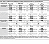 Table 1
Details of sample provenance for all DICER1- and DGCR8-mutated cases included in the different experiments
Genomic landscape of DICER1- and DGCR8-mutated BTLs and malignant thyroid lesions. We first performed WES on 18 samples harboring DICER1/DGCR8 mutations and integrated the data with published cases (12–14) and cases from The Cancer Genome Atlas Thyroid Cancer (TCGA-THCA) project (19). In total, 18 DICER1-mutated thyroid cases (5 benign, 13 malignant) and 10 DGCR8-mutated thyroid cases (6 benign, 4 malignant) were analyzed (Table 1 and Supplemental Table 3).
Thirteen out of the 18 (72.2%) DICER1-mutated cases followed the 2-hit mutational pattern of DICER1 (a DICER1 RNase IIIb hotspot mutation and a LoF variant or LOH at the DICER1 locus). In all instances, DICER1 changes were mutually exclusive from canonical MAPK gene changes, RET fusions, or TERT promoter changes (Figure 1A). While the 5 DICER1-benign lesions harbored mutations exclusively in DICER1, some cancers (4 out of 13, 30.7%) additionally harbored TP53 changes (Figure 1, A and B). Of these 4 (2 PDTCs, 1 FVPTC, and 1 invasive encapsulated FVPTC [IEFVPTC]), 3 presented aggressive features. Other variants in known thyroid cancer progression genes (PIK3CA, PTEN, ARID1A, ARID1B, KMT2C, KMT2D, EP300, ATM, and RBM10) were also observed in individual thyroid cancers (Figure 1A). Mutational co-occurrence with TP53 was confirmed by analyzing the GENIE cohort (v17.1) that contained 35 DICER1-hotspot-mutated thyroid tumors (28 primary tumors and 7 metastases) with TP53 changes found in 2 out of 9 primary PDTCs and 2 out of 4 PDTC metastases. In contrast with our IEFVPTC (sample 17), no TP53 change was found in the 22 well-differentiated thyroid cancers (19 primary tumors and 3 metastases), suggesting rather an association between TP53 changes and more aggressive thyroid cancers.
 Figure 1
Landscape of DICER1- and DGCR8-mutated thyroid lesions. (A) Molecular landscape of DICER1- and DGCR8-altered thyroid lesions showing MAPK pathway– or thyroid cancer progression–associated changes and recurrent copy number changes. Benign thyroid lesions (BTLs) harbored DICER1 or DGCR8 changes. In well-differentiated thyroid cancers (WDTCs) and poorly differentiated thyroid carcinomas (PDTCs), DICER1 mutations were mutually exclusive from MAPK gene changes, occasionally co-occurring with TP53 mutations. DGCR8-altered cancers co-occurred with MAPK pathway mutations. Both DICER1- and DGCR8-mutated thyroid cancers lacked TERT promoter changes as well as the most prevalent RET fusions (RET-PTC1 and RET-PTC3). All FVPTCs are invasive encapsulated FVPTCs (IEFVPTC) with the exception of TCGA-EL-A3D5-01, which showed follicular-patterned areas, but could not be classified as IEFVPTC or infiltrative FVPTC due to limited histological material. LoF, loss-of-function; LOH, loss of heterozygosity; VUS, variant of uncertain significance, TFND, follicular nodular disease; FTA, follicular adenoma; FTC, follicular thyroid cancer; FVPTC, follicular variant of papillary thyroid cancer; CPTC, classic papillary thyroid cancer. ‡, †, and § show sets of 2 different lesions from a single patient; #TFND with incidental microPTC; *Case IDs from Chernock et al. (12). (B) In the germline DICER1 context, a truncating variant is followed by a somatic RNase IIIb hotspot mutation, leading to BTL formation. In sporadic cases, a somatic RNase IIIb hotspot occurs first, followed by loss of the WT allele (truncating or LOH), sufficient for BTL development. In both contexts, BTLs progress to cancer in the absence of other changes and may progress to more aggressive cancer types (PDTC) via (a) TP53 mutation or (b) without further changes. (C) In the germline DGCR8 context, a missense c.1552G>A, p.E518K hotspot mutation is followed by LOH or truncation of the WT allele, forming a BTL. This BTL may acquire MAPK (BRAF, RAS) or PI3K (PIK3CA) mutations for malignant transformation. In sporadic cases, the same sequence of DGCR8 events occurs, followed by MAPK/PI3K changes during progression to cancer.
All 10 DGCR8 cases harbored the c.1552G>A, p.E518K hotspot variant, which was germline derived in 7 tumors (from 6 patients). All germline carriers developed a TFND; 1 carrier also developed a CPTC and another was found to have a micropapillary thyroid carcinoma (microPTC) within the TFND. In total, 7 out of 10 lesions lost the wild-type (WT) DGCR8 allele: 6 through LOH or full chromosome 22 deletion, 1 through a truncating DGCR8 variant, and another showed a low-VAF E518K variant with a copy number alteration (CNA) profile consistent with 22q LOH (Figure 1A and Supplemental Figure 2). In contrast with the DICER1 cancers, 4 out of 4 (100%) DGCR8-associated malignant lesions also harbored canonical thyroid cancer gene changes (NRAS hotspot mutations in the FTC and FVPTCs, and a BRAF V600E mutation in a CPTC), demonstrating the co-occurence of DGCR8 and MAPK gene changes (Figure 1, A–C). The DGCR8-benign lesions harbored DGCR8 mutations exclusively, highlighting a progression mechanism involving RAS pathway activation (Figure 1, A and C).
Both DICER1- and DGCR8-mutant samples exhibited a quiet genome with some recurrent CNAs, including full loss of chromosomes 17 and 22 in DICER1 thyroid cancers and in DGCR8 thyroid lesions, respectively (Figure 1A).
Impact of DICER1 and DGCR8 mutations on miRNA biogenesis. To assess the impact of DICER1 and DGCR8 mutations on miRNA biogenesis in the thyroid, we profiled the miRNA repertoire of DICER1- and DGCR8-mutated thyroid lesions compared to WT tumors. Unsupervised consensus clustering of miRNAs from 38 tumors (14 DICER1-mutated, 9 DGCR8-mutated, and 15 DICER1-/DGCR8-WT) identified 2 main clusters, including a WT group (cluster 1) and a DICER1-/DGCR8-mutant group (Figure 2A). The WT group comprised WT samples and 1 DGCR8-mutated TFND. The mutant group was further subdivided into 3 subclusters: (a) a DGCR8-mutant group (cluster 2), (b) a DICER1/DGCR8/WT mixed group (cluster 3), and (c) a DICER1-mutant group (cluster 4). The clusters were associated with mutation status and were independent of histology (Figure 2A).
 Figure 2
miRNA profiles of DICER1- and DGCR8-mutated thyroid lesions. (A) Unsupervised consensus clustering (1,000 repetitions) of miRNAs in 38 BTLs and malignant thyroid lesions identified: a WT group (cluster 1, n = 14) and a DICER1-/DGCR8-mutant group (n = 24), further subdivided into 3 subclusters: (a) a DGCR8-mutant group (cluster 2, n = 7), (b) a DICER1/DGCR8/WT mixed group (cluster 3, n = 7), and (c) a DICER1-mutant group (cluster 4, n = 10). The clusters were associated with mutation status and independent of histological diagnosis (benign vs. malignant). Of note, 1 DGCR8-mutated TFND clustered with the WT group and 2 WT samples clustered within the DICER1/DGCR8/WT mixed group. Consensus matrix using the most variable miRNAs with k set to 4 (elbow method). (B) All differentially expressed miRNAs (DEMs) in the DICER1 and DGCR8 BTLs (n = 3 and n = 12 DEMs, respectively) versus their respective malignant counterparts were persistently downregulated in the thyroid cancers (TCs) that harbored other DEMs, suggesting a linear model of progression. Two DEMs were shared by both DICER1- and DGCR8-mutated BTLs and TCs, while 34 DEMs were common to DICER1- and DGCR8-mutated TCs. No DEM was unique to DICER1-/DGCR8-mutant BTLs. Bar charts show the number of upregulated/downregulated 5p and 3p miRNAs.
miRNA profiling of benign and malignant lesions with impaired miRNA processing. Also, we defined the benign versus malignant profiles unique to each driver against each set of WT counterparts. In both entities, the differentially expressed miRNA (DEM) profiles confirmed that changes in miRNA processing leading to nodule development persist through the carcinoma process. We identified 3 DEMs (miR-30a-5p, miR-218-5p, and miR-324-5p), all of which were downregulated in DICER1-mutated BTLs compared with WT BTLs (Supplemental Table 4). These same 3 miRNAs and 38 extra downregulated DEMs and 1 upregulated DEM distinguished the cancer status (Figure 2B and Supplemental Table 5), showing an enrichment in certain miRNA families, including let-7 (the family with the most DEMs), miR-30, miR-135, miR-26, miR-221, miR-10, miR-15, and miR-181 (Supplemental Table 6). Following the same trend, between DGCR8-mutated and WT BTLs, we identified 12 miRNAs to be differentially expressed (Figure 2B and Supplemental Table 7), belonging to the miR-8, miR-30, and miR-135 families. In addition, 79 miRNAs were found to characterize the DGCR8 cancer status (Figure 2B and Supplemental Table 8), with an enrichment in several miRNA families (Supplemental Table 9). In all DICER1-mutant thyroid lesions (which were all RNase IIIb mutants), we observed a significant reduction in expression of 5p miRNAs, while a reduced expression of both 3p and 5p mature miRNAs was predominant in DGCR8-mutant cases, as expected according to previous studies in other tumor types (13, 15, 21, 27–36) (Figure 2B).
Shared miRNA defects in DICER1/DGCR8 lesions suggest linear thyroid tumor progression. Owing to the 2 genes’ shared involvement in miRNA biogenesis, we hypothesized that the common 5p miRNA deregulation observed in mutants of both genes is the key event in priming the cell context for the initiation of TFND. Two miRNAs (miR-218-5p and miR-30a-5p) were differentially expressed in both benign and malignant lesions and 34 additional DEMs were unique to mutant malignant thyroid lesions (Figure 2B), supporting an incremental miRNAome deregulation underlying a linear model of carcinoma progression. All 36 DEMs were downregulated, with 92% being 5p mature miRNAs (Figure 2B). Fifteen DEMs were selected for validations by quantitative PCR (Supplemental Figure 3 and Supplemental Table 10).
Spatially resolved transcriptomic profiling of benign nodules and malignant thyroid cancers with DICER1 or DGCR8 changes. We subsequently interrogated the transcriptomic differences between BTLs and malignant thyroid lesions at the spatial level in 2 different tumors individually (1 DICER1 sample [patient 1] and 1 DGCR8 sample [patient 2]) by interrogating (a) the follicular cell population expressing pan-cytokeratin (PanCK) and (b) vimentin-expressing (VIM-expressing) cells as a surrogate for enrichment of stromal populations (Figure 3A). We analyzed 4 histological components in patient 1: (a) normal thyroid, (b) a nonmutated non-neoplastic nodular area, (c) a DICER1-mutated PDTC, and (d) a capsular region between the PDTC and the normal thyroid tissue and non-neoplastic nodular area (Figure 3B). Gene set enrichment analysis (GSEA) revealed activation of the PI3K/AKT/mTOR pathway when comparing the PDTC with normal thyroid tissue at the region of interest (ROI) level (PanCK+VIM+) despite the lack of a mutation in RAS or PI3K/AKT/mTOR pathway genes (Figure 3C). In the epithelial component (PanCK+VIM–) of the PDTC, upregulation of oxidative phosphorylation (Figure 3C) and genes related to WNT signaling (Supplemental Figure 4) was observed. In the VIM-expressing cell population (PanCK–VIM+), we noted activation of cancer proliferation and progression signaling (PI3K/AKT/mTOR, KRAS, and WNT/β-catenin signaling), upregulation of developmental and stem cell pathways (Notch and Hedgehog signaling), involvement of metastasis and invasion processes (epithelial-mesenchymal transition [EMT] and TGF-β signaling), and upregulation of angiogenesis (Figure 3C). The angiogenic upregulation was further reflected by the larger proportion of endothelial cells in the PDTC compared with normal thyroid and the non-neoplastic nodular area, which was inferred from the deconvolution of the VIM+ cell populations (Figure 3D). Accordingly, the PDTC’s VIM+ component showed significant upregulation of genes involved in extracellular matrix (ECM) formation, angiogenic development, and endothelial function, thus supporting a strong remodeling of the ECM. Of note, the following ECM-related (COL3A1, COL4A1, COL4A2, FN1, and NID1) and angiogenesis-related (CDH5, FLT1, HSPG2, KDR, PLXND1, and SPARC) genes were direct targets of DEMs seen in the DICER1 cancers (Supplemental Figure 5).
 Figure 3
Transcriptomic profiles of a DICER1 thyroid lesion. (A) Immunofluorescent staining for pan-cytokeratin (PanCK) (green) and vimentin (VIM) (red) of a sporadic DICER1 case and a germline DGCR8 case. Enclosed areas represent selected regions of interest (ROIs). A minimum of 3 ROIs per histological regions was selected. Inset contains a representative image of the area of illumination (AOI) segmentation for PanCK and VIM. Original magnification, ×20 and ×200 (insets). (B) H&E staining of the DICER1 case showing the selected histological regions (normal thyroid tissue, non-neoplastic nodular area), poorly differentiated thyroid cancer, and the tumor capsule). Original magnification, ×20. (C) Dot plot of the significantly altered MSigDB hallmarks of cancer pathways in the poorly differentiated thyroid cancer (PDTC) compared with normal thyroid tissue (NT) at the ROI, PanCK+, and VIM+ levels. (D) Cellular deconvolution of the VIM+ component showing an enrichment in endothelial cells in the PDTC. NNNA (B), non-neoplastic nodular area (benign); PDTC (M), poorly differentiated thyroid cancer (malignant); Tcaps, tumor capsule.
Patient 2 harbored a germline DGCR8 p.E518K mutation and the sample consisted of 4 histological components: (a) normal thyroid, (b) non-neoplastic nodular areas, (c) a microPTC, and (d) normal adjacent thyroid (Figure 4A). Additionally, we captured VIM+ colloidal macrophages and CD45+ lymphocytes (Figure 4A). In contrast with the DICER1 sporadic case, we observed gradual changes in the transcriptomic profile from the normal thyroid to the benign non-neoplastic nodular areas to the microPTC at both the epithelial (PanCK+VIM–) and the stromal (PanCK–VIM+) levels. We hypothesized that these gradual changes reflected a germline E518K effect and not a tumor-adjacent effect, as no differences were observed between the distal normal thyroid tissue and the normal thyroid adjacent to the microPTC (data not shown). The microPTC was characterized by a remarkable upregulation of KRAS signaling and PI3K/AKT/mTOR signaling compared with the non-neoplastic nodular areas as well as by upregulation of metabolic reprogramming (oxidative phosphorylation, reactive oxygen species, and peroxisome) and of MYC targets (Figure 4B). Pathway analysis between the 3 main histological components (normal tissue, non-neoplastic nodular areas, and microPTC) uncovered a common immune pathway involvement (activation of TNF-α via NF-κB signaling, IL-6/JAK/STAT3 signaling, IL-2/STAT5 signaling, and inflammatory response) in both the non-neoplastic nodular areas and the microPTC compared with the normal tissue. The microPTC showed additional involvement of TGF-β and p53 signaling (Figure 4B). In line with these results, we observed specific upregulation of immune-related genes (Supplemental Figure 6, A and B). The DMKN gene was the top upregulated gene common to both nodular areas and microPTC at the ROI, the PanCK+, and the VIM+ levels and is a direct target of miR-93-5p, which was differentially expressed in the DGCR8 tumors.
 Figure 4
Transcriptomic profiles of DGCR8-mutated thyroid lesions. (A) H&E staining of the DGCR8 case showing the selected histological regions: normal thyroid tissue (NT), 3 non-neoplastic nodular areas (benign) [NNNA (B)], a micropapillary thyroid cancer (microPTC), and NT adjacent to the microPTC (NTadj). Original magnification, ×20. (B) Dot plot of the significantly altered MSigDB hallmarks of cancer pathways in the NNNAs and the microPTC at the region of interest (ROI) level. (C) Cellular deconvolution of the VIM+ component showing an enrichment in immune cells. M, macrophage; L, lymphocytes.
The immune pathway involvement underlying patient 2’s case was not present in the WT non-neoplastic nodular area from patient 1 and was further supported by the presence of different immune cell populations inferred in the VIM+ cells of the DGCR8 lesions (after cellular deconvolution) (Figure 4C). When comparing the normal tissue harboring the germline E518K mutation to the normal tissue from patient 1, the upregulation in inflammatory response, TGF-β signaling, and EMT processes were already detectable, thus supporting an effect of the germline change in the histologically normal thyroid tissue (Supplemental Figure 7).
Impact of the miRNA production impairment on the expression landscape of mutant BTLs and malignant thyroid lesions. We hypothesized that DGCR8 dysfunction promotes the inflammatory response observed in these thyroid tumors; thus, we investigated the transcriptional impact of the miRNA dysregulation in DGCR8-mutated cases. Integration of the 12 DEMs common to the DGCR8 BTLs and the DGCR8 thyroid cancers with the differentially expressed genes (DEGs) seen in the DGCR8 non-neoplastic nodular areas and the DGCR8 microPTC showed statistically significant associations with pathways linked to EMT, metabolic reprogramming (oxidative phosphorylation, reactive oxygen species, and peroxisome), and inflammation (IL-2/STAT5 signaling, IL-6/JAK/STAT3 signaling, IFN-γ response, and allograft rejection) (Supplemental Table 11). These results suggest that the first impact of the DGCR8 change results in an inflammatory process in the thyroid tissue that ignites cancer progression. Of note, the 79 DEMs unique to the DGCR8 thyroid cancers and the DEGs observed in the DGCR8 microPTC compared with DGCR8 non-neoplastic nodular areas were significantly associated with key thyroid cancer pathways (KRAS signaling, mTORC1 signaling, PI3K/AKT/mTOR signaling, p53 pathway, and TGF-β signaling) (Supplemental Table 12). miRNA-mRNA integration for the DICER1-mutated lesion was approximate, owing to unmatched dataset groups (Supplemental Methods and Supplemental Table 13).
miRNA-impaired thyroid lesions exhibit a distinct methylation profile. DICER1 thyroid tumors exhibit a wide range of histological subtypes, but DICER1-mutated CPTCs have only been reported in a handful of cases (n = 4) (37, 38). Consequently, DICER1 thyroid tumors have been classified as RAS-like (26) despite lacking RAS mutations. We explored this premise by interrogating the methylation profiles of DICER1- and DGCR8-mutated thyroid lesions compared to other subtypes.
We used 2,279 CpGs from the signature defined by Marczyk et al. (39) to categorize the lesions into follicular-like (RAS-associated) and PTC-like (BRAF-associated) entities. We benefitted from a publicly available dataset comprising 269 thyroid lesions (35 of which had available molecular data) and integrated it to our methylome data generated from 12 mutant thyroid samples. In total, methylome data from 34 mutant thyroid samples (26 DICER1 and 8 DGCR8) was available. Uniform manifold approximation and projection (UMAP) reduction dimensionality confirmed that thyroid lesions grouped based on histological tumor type, with normal thyroids and follicular-patterned thyroid lesions clustering near each other and away from PTC-like thyroid lesions (Figure 5A). Histological and molecular associations (PTC-like with BRAF-V600E and follicular-like with RAS mutations) were also confirmed (Figure 5B). This analysis revealed a distinct mutant cluster (n = 28) independent of the follicular-like and papillary-like lesions composed solely of DICER1-mutated BTLs, DGCR8-mutated BTLs, and DICER1 cancers (Figure 5B).
 Figure 5
Identification of distinct methylation clusters encompassing DICER1- and DGCR8-mutated thyroid lesions. Methylation clusters associated with histological diagnosis using the UMAP dimensionality reduction technique. (A) Samples are colored according to the methylation clusters observed by Marczyk et al. (39): papillary-like, follicular-like, and normal-like. (B) Samples are colored by histological subtype and shapes correspond to gene altered. One follicular-like cluster is made up of DICER1- and DGCR8-mutated thyroid lesions (DICER1-/DGCR8-mutated cluster). (C) Unsupervised hierarchical clustering (k = 9) revealed 3 main clusters (same as those defined by Marczyk et al.; ref. 39). Clusters A and B characterized the papillary-like group and were enriched in classic papillary thyroid cancers (CPTCs), but also contained tall cell PTCs (tcPTCs) and anaplastic thyroid cancers (ATCs). Clusters C and D characterized the normal-like histological group and were enriched in normal thyroid samples. Five clusters characterized the follicular-like group: cluster E, enriched in follicular thyroid cancers (FTCs); clusters F and G, stemming from a shared hierarchical node, were enriched in DICER1-/DGCR8-benign thyroid lesions and composed exclusively of DICER1-mutated samples (enriched in thyroid cancers), respectively; and clusters H and I, also stemming from a common node, were enriched in oncocytic thyroid lesions (OA and OC) and benign thyroid lesions, respectively.
Unsupervised hierarchical clustering of the 250 most differentially methylated probes supported the presence of the 3 main histological groups defined by Marczyk et al. (39): PTC-like (clusters A and B), normal thyroid (clusters C and D), and follicular-like (clusters E–I). Within the follicular-like group, clusters E, H, and I were enriched in thyroid cancers, oncocytic lesions, and benign thyroid lesions, respectively. Additionally, 32 out of 34 (94%) of DICER1-/DGCR8-mutated thyroid lesions also belonged to the follicular-like group (Figure 5C). Although the mutant thyroid lesions exhibited a closer hierarchy to follicular-patterned tumors, the unsupervised analysis identified 2 mutant groups stemming from the same hierarchical node corresponding to the DICER1/DGCR8 thyroid lesions highlighted in the UMAP (Figure 5B). On one hand, cluster F was a group strongly enriched in DICER1 and DGCR8 BTLs (D1/D8-B hereafter) and cluster G was a group composed solely of DICER1 lesions, primarily DICER1 cancers (D1-M hereafter). On the other hand, 2 DGCR8 cancers (both also harboring a NRAS mutation) clustered within a follicular-like cluster containing RAS-mutated thyroid cancers (cluster E, Figure 5C). Additionally, 2 mutant TFND samples clustered with the normal thyroid samples. Upon review of the pathological and molecular data, we found that (a) the germline DGCR8-mutated TFND sample was highly enriched in normal thyroid tissue and was the only DGCR8 case retaining a WT copy of DGCR8, and (b) the DICER1 TFND, although harboring a DICER1 truncating variant, lacked a DICER1 hotspot mutation. Two DICER1 cases (1 PDTC and 1 TFND) grouped with the 2 DGCR8 cancers, found in cluster E, which was enriched in RAS tumors. Altogether, these results indicate that DICER1/DGCR8 lesions share molecular features with other follicular-derived tumors but display a particular epi-signature that can showcase DICER1 cancers.
DICER1- and DGCR8-mutated thyroid lesions are characterized by hypomethylation of miRNA-encoding genes. Differentiated papillary thyroid tumors are characterized by a general hypomethylation, while follicular-like thyroid tumors exhibit a higher proportion of hypermethylated probes compared with normal thyroid tissue (39–41). The miRNA-impaired tumors, although histologically mainly follicular-patterned and clustering with the follicular-like tumors, exhibited a higher proportion of hypomethylated CpGs (located in the open sea), thus behaving rather similarly to papillary thyroid cancers (Table 2 and Supplemental Figure 8).
 Table 2
Differentially methylated genomic positions and regions in comparison to the normal-like cluster
We then interrogated how the miRNA-impaired tumors (clusters F and G) differed from the rest. Compared with non–DICER1-/non–DGCR8-mutated follicular-like thyroid lesions (clusters E, H, and I), the miRNA-impaired lesions (clusters F for D1/D8-B and G for D1-M) had 345 and 436 differentially methylated genes (DMGs), respectively (Supplemental Tables 14 and 15). GSEA showed a common significant enrichment of hypomethylated genes corresponding to Gene Ontology (GO) processes related to miRNA-mediated gene-silencing mechanisms (Supplemental Tables 16 and 17). This finding was further validated by an enrichment of individual miRNA genes within the individual hypomethylated genes (Supplemental Tables 14 and 15). Hypomethylation affected mostly the miRNA gene promoters. Likewise, enrichment in GO terms related to miRNA-mediated gene silencing was observed in miRNA-mutated cases compared with papillary-like lesions, supporting this hypomethylation signature as a feature of miRNA-mutated samples (Supplemental Tables 18 and 19).
Evolving methylation profile of DICER1 thyroid cancers compared to mutant benign lesions. DICER1 benign and malignant lesions had subtle differences in their mutational profiles, with DICER1/DGCR8 genes equally mutated in both; however, deficiencies in miRNA production were severely pronounced in the cancers. Since DICER1-/DGCR8-mutant BTLs exhibited a lower global DNA methylation level than cancers (at the general level and when focusing on differentially methylation probes [DMPs]) (Table 2 and Supplemental Figure 9), we interrogated methylation profiles possibly associated with carcinoma progression. We identified 322 DMGs between the mutant malignant (D1-M) and benign (D1/D8-B) groups (Supplemental Table 20). Although DICER1-/DGCR8-mutant lesions were characterized by hypomethylation of miRNA-mediated gene-silencing processes, these processes were altered to a lesser extent in DICER1-driven cancers (Figure 6 and Supplemental Table 21). In fact, when looking at individual miRNA genes, benign lesions showed twice the number of hypomethylated miRNA genes compared with malignant lesions (40 vs. 24 DMGs compared with normal thyroid, respectively) (Figure 6 and Supplemental Tables 22 and 23). Five tumor suppressor miRNA genes (MIR517A, MIR519D, MIR1266, MIR548C, and MIR711) were specifically hypermethylated in the mutant cancers compared with the benign forms; of note, MIR1266 and MIR548C are associated with tumor growth and metastasis (42, 43).
 Figure 6
Scheme of epigenetic miRNA compensation mechanism in benign and malignant mutant lesions. We propose a compensatory mechanism involving regulation of miRNA genes at the epigenetic level where both benign and malignant mutant thyroid lesions activate gene expression of miRNA genes in response to lack of miRNA production, in contrast with gene repression of miRNA genes by DNA methylation in the context of normal miRNA production in normal thyroid. This compensatory mechanism was more active in the mutant benign thyroid lesions, supporting successful rescue of some miRNA processing exemplified by a less prominent loss of miRNAs. In the mutant thyroid cancers, we observed a decreased activity of this compensatory mechanism, supported by their greater loss in miRNAs.
As a feature, DICER1 cancers (D1-M) showed an enrichment of hypermethylated genes in biological processes related to immune response, cell signaling, and migration, which might underlie a putative silencing mechanism leading to immune evasion and impaired tissue remodeling. The gene enrichment processes with a marked hypomethylation involved mechanisms of adaptation to cellular stress such as enhanced metabolic pathways and DNA repair, proper to cancer processes (Supplemental Table 15). Looking at specific genes, we noted that DICER1-mutant thyroid cancers displayed differential hypomethylation in the promoters of MIRLET7G (a top DEM in the cancers), KRT19, CTPBP1, CXXC5, PRAME, THRSP, and FERMT1, as well as hypermethylation in the promoters of ETS1, TGFRB1, EYA1, FGF2, and SERPINF1, all of which are involved in thyroid cancer progression (21, 44–60). We hypothesized that these methylation changes might result from loss of miRNA regulation of methylation enzymes. Analysis of cancer-specific DEMs identified 24 targeting DNMT1/3A/3B and TET1/2/3 genes. Evaluation of the spatial transcriptomic data revealed significant upregulation of DNMT3B and lower expression of the eraser TET2 in the epithelial component of the DICER1 PDTC compared with the corresponding normal tissue (P = 0.0121 and P = 0.0242, respectively).


