
Research ArticleBone biologyInflammation
Open Access |  10.1172/jci.insight.193632
Activation of SLIT2/ROBO1/LRP6 axis aggravates cartilage degradation via β-catenin signaling in TMJOA
Guan Luo,1 Baoyi Chen,1 Wenjun Chen,1 Huiyi Lin,1,2 Weiqi Guo,1,3 Qingbin Zhang,1 Jiang Li,1 Lijing Wang,4 Janak Lal Pathak,1 Yuhui Yang,5 Weijun Zhang,1 Xiaoyu Zhang,1 Beining Zheng,1 Ziyi Wang,1 Shiting Wei,1 Jiaxin He,1 Wei-Jie Zhou,6 and Chang Liu1
1Department of Orthodontics, School and Hospital of Stomatology, Guangdong Engineering Research Center of Oral Restoration and Reconstruction, Guangzhou Key Laboratory of Basic and Applied Research of Oral Regenerative Medicine, Guangzhou Medical University, Guangzhou, Guangdong, China.
2Department of Orthodontics, Jiangmen Municipal Stomatology Hospital, Jiangmen, Guangdong, China.
3Department of Stomatology, The First Affiliated Hospital of Guangdong Pharmaceutical University, Guangzhou, Guangdong, China.
4Vascular Biology Research Institute, School of Life Sciences and Biopharmaceutics, Guangdong Pharmaceutical University, Guangzhou, Guangdong, China.
5Department of Orthopedics, Guangdong Provincial People’s Hospital (Guangdong Academy of Medical Sciences), Southern Medical University, Guangzhou, Guangdong, China.
6Department of General Surgery, Guangdong Provincial Key Laboratory of Precision Medicine for Gastrointestinal Tumor, State Key Laboratory of Multi-organ Injury Prevention and Treatment, Cancer Research Institute, School of Basic Medical Sciences, Nanfang Hospital, Southern Medical University, Guangzhou, Guangdong, China.
Address correspondence to: Chang Liu, Department of Orthodontics, School and Hospital of Stomatology, Guangdong Engineering Research Center of Oral Restoration and Reconstruction & Guangzhou Key Laboratory of Basic and Applied Research of Oral Regenerative Medicine, Guangzhou Medical University, Guangzhou, Guangdong, China. Phone: 8613710210667; Email: changliudentist@gzhmu.edu.cn. Or to: Wei-Jie Zhou, Department of General Surgery, Guangdong Provincial Key Laboratory of Precision Medicine for Gastrointestinal Tumor, State Key Laboratory of Multi-organ Injury Prevention and Treatment, Cancer Research Institute, School of Basic Medical Sciences, Nanfang Hospital, Southern Medical University, Guangzhou, Guangdong, China. Phone: 8613925005634; Email: weijiezhouum@163.com.
Authorship note: GL, BC, and WC contributed equally to this work and are co–first authors.
Find articles by Luo, G. in: PubMed | Google Scholar
1Department of Orthodontics, School and Hospital of Stomatology, Guangdong Engineering Research Center of Oral Restoration and Reconstruction, Guangzhou Key Laboratory of Basic and Applied Research of Oral Regenerative Medicine, Guangzhou Medical University, Guangzhou, Guangdong, China.
2Department of Orthodontics, Jiangmen Municipal Stomatology Hospital, Jiangmen, Guangdong, China.
3Department of Stomatology, The First Affiliated Hospital of Guangdong Pharmaceutical University, Guangzhou, Guangdong, China.
4Vascular Biology Research Institute, School of Life Sciences and Biopharmaceutics, Guangdong Pharmaceutical University, Guangzhou, Guangdong, China.
5Department of Orthopedics, Guangdong Provincial People’s Hospital (Guangdong Academy of Medical Sciences), Southern Medical University, Guangzhou, Guangdong, China.
6Department of General Surgery, Guangdong Provincial Key Laboratory of Precision Medicine for Gastrointestinal Tumor, State Key Laboratory of Multi-organ Injury Prevention and Treatment, Cancer Research Institute, School of Basic Medical Sciences, Nanfang Hospital, Southern Medical University, Guangzhou, Guangdong, China.
Address correspondence to: Chang Liu, Department of Orthodontics, School and Hospital of Stomatology, Guangdong Engineering Research Center of Oral Restoration and Reconstruction & Guangzhou Key Laboratory of Basic and Applied Research of Oral Regenerative Medicine, Guangzhou Medical University, Guangzhou, Guangdong, China. Phone: 8613710210667; Email: changliudentist@gzhmu.edu.cn. Or to: Wei-Jie Zhou, Department of General Surgery, Guangdong Provincial Key Laboratory of Precision Medicine for Gastrointestinal Tumor, State Key Laboratory of Multi-organ Injury Prevention and Treatment, Cancer Research Institute, School of Basic Medical Sciences, Nanfang Hospital, Southern Medical University, Guangzhou, Guangdong, China. Phone: 8613925005634; Email: weijiezhouum@163.com.
Authorship note: GL, BC, and WC contributed equally to this work and are co–first authors.
Find articles by Chen, B. in: PubMed | Google Scholar
1Department of Orthodontics, School and Hospital of Stomatology, Guangdong Engineering Research Center of Oral Restoration and Reconstruction, Guangzhou Key Laboratory of Basic and Applied Research of Oral Regenerative Medicine, Guangzhou Medical University, Guangzhou, Guangdong, China.
2Department of Orthodontics, Jiangmen Municipal Stomatology Hospital, Jiangmen, Guangdong, China.
3Department of Stomatology, The First Affiliated Hospital of Guangdong Pharmaceutical University, Guangzhou, Guangdong, China.
4Vascular Biology Research Institute, School of Life Sciences and Biopharmaceutics, Guangdong Pharmaceutical University, Guangzhou, Guangdong, China.
5Department of Orthopedics, Guangdong Provincial People’s Hospital (Guangdong Academy of Medical Sciences), Southern Medical University, Guangzhou, Guangdong, China.
6Department of General Surgery, Guangdong Provincial Key Laboratory of Precision Medicine for Gastrointestinal Tumor, State Key Laboratory of Multi-organ Injury Prevention and Treatment, Cancer Research Institute, School of Basic Medical Sciences, Nanfang Hospital, Southern Medical University, Guangzhou, Guangdong, China.
Address correspondence to: Chang Liu, Department of Orthodontics, School and Hospital of Stomatology, Guangdong Engineering Research Center of Oral Restoration and Reconstruction & Guangzhou Key Laboratory of Basic and Applied Research of Oral Regenerative Medicine, Guangzhou Medical University, Guangzhou, Guangdong, China. Phone: 8613710210667; Email: changliudentist@gzhmu.edu.cn. Or to: Wei-Jie Zhou, Department of General Surgery, Guangdong Provincial Key Laboratory of Precision Medicine for Gastrointestinal Tumor, State Key Laboratory of Multi-organ Injury Prevention and Treatment, Cancer Research Institute, School of Basic Medical Sciences, Nanfang Hospital, Southern Medical University, Guangzhou, Guangdong, China. Phone: 8613925005634; Email: weijiezhouum@163.com.
Authorship note: GL, BC, and WC contributed equally to this work and are co–first authors.
Find articles by Chen, W. in: PubMed | Google Scholar
1Department of Orthodontics, School and Hospital of Stomatology, Guangdong Engineering Research Center of Oral Restoration and Reconstruction, Guangzhou Key Laboratory of Basic and Applied Research of Oral Regenerative Medicine, Guangzhou Medical University, Guangzhou, Guangdong, China.
2Department of Orthodontics, Jiangmen Municipal Stomatology Hospital, Jiangmen, Guangdong, China.
3Department of Stomatology, The First Affiliated Hospital of Guangdong Pharmaceutical University, Guangzhou, Guangdong, China.
4Vascular Biology Research Institute, School of Life Sciences and Biopharmaceutics, Guangdong Pharmaceutical University, Guangzhou, Guangdong, China.
5Department of Orthopedics, Guangdong Provincial People’s Hospital (Guangdong Academy of Medical Sciences), Southern Medical University, Guangzhou, Guangdong, China.
6Department of General Surgery, Guangdong Provincial Key Laboratory of Precision Medicine for Gastrointestinal Tumor, State Key Laboratory of Multi-organ Injury Prevention and Treatment, Cancer Research Institute, School of Basic Medical Sciences, Nanfang Hospital, Southern Medical University, Guangzhou, Guangdong, China.
Address correspondence to: Chang Liu, Department of Orthodontics, School and Hospital of Stomatology, Guangdong Engineering Research Center of Oral Restoration and Reconstruction & Guangzhou Key Laboratory of Basic and Applied Research of Oral Regenerative Medicine, Guangzhou Medical University, Guangzhou, Guangdong, China. Phone: 8613710210667; Email: changliudentist@gzhmu.edu.cn. Or to: Wei-Jie Zhou, Department of General Surgery, Guangdong Provincial Key Laboratory of Precision Medicine for Gastrointestinal Tumor, State Key Laboratory of Multi-organ Injury Prevention and Treatment, Cancer Research Institute, School of Basic Medical Sciences, Nanfang Hospital, Southern Medical University, Guangzhou, Guangdong, China. Phone: 8613925005634; Email: weijiezhouum@163.com.
Authorship note: GL, BC, and WC contributed equally to this work and are co–first authors.
Find articles by Lin, H. in: PubMed | Google Scholar
1Department of Orthodontics, School and Hospital of Stomatology, Guangdong Engineering Research Center of Oral Restoration and Reconstruction, Guangzhou Key Laboratory of Basic and Applied Research of Oral Regenerative Medicine, Guangzhou Medical University, Guangzhou, Guangdong, China.
2Department of Orthodontics, Jiangmen Municipal Stomatology Hospital, Jiangmen, Guangdong, China.
3Department of Stomatology, The First Affiliated Hospital of Guangdong Pharmaceutical University, Guangzhou, Guangdong, China.
4Vascular Biology Research Institute, School of Life Sciences and Biopharmaceutics, Guangdong Pharmaceutical University, Guangzhou, Guangdong, China.
5Department of Orthopedics, Guangdong Provincial People’s Hospital (Guangdong Academy of Medical Sciences), Southern Medical University, Guangzhou, Guangdong, China.
6Department of General Surgery, Guangdong Provincial Key Laboratory of Precision Medicine for Gastrointestinal Tumor, State Key Laboratory of Multi-organ Injury Prevention and Treatment, Cancer Research Institute, School of Basic Medical Sciences, Nanfang Hospital, Southern Medical University, Guangzhou, Guangdong, China.
Address correspondence to: Chang Liu, Department of Orthodontics, School and Hospital of Stomatology, Guangdong Engineering Research Center of Oral Restoration and Reconstruction & Guangzhou Key Laboratory of Basic and Applied Research of Oral Regenerative Medicine, Guangzhou Medical University, Guangzhou, Guangdong, China. Phone: 8613710210667; Email: changliudentist@gzhmu.edu.cn. Or to: Wei-Jie Zhou, Department of General Surgery, Guangdong Provincial Key Laboratory of Precision Medicine for Gastrointestinal Tumor, State Key Laboratory of Multi-organ Injury Prevention and Treatment, Cancer Research Institute, School of Basic Medical Sciences, Nanfang Hospital, Southern Medical University, Guangzhou, Guangdong, China. Phone: 8613925005634; Email: weijiezhouum@163.com.
Authorship note: GL, BC, and WC contributed equally to this work and are co–first authors.
Find articles by Guo, W. in: PubMed | Google Scholar
1Department of Orthodontics, School and Hospital of Stomatology, Guangdong Engineering Research Center of Oral Restoration and Reconstruction, Guangzhou Key Laboratory of Basic and Applied Research of Oral Regenerative Medicine, Guangzhou Medical University, Guangzhou, Guangdong, China.
2Department of Orthodontics, Jiangmen Municipal Stomatology Hospital, Jiangmen, Guangdong, China.
3Department of Stomatology, The First Affiliated Hospital of Guangdong Pharmaceutical University, Guangzhou, Guangdong, China.
4Vascular Biology Research Institute, School of Life Sciences and Biopharmaceutics, Guangdong Pharmaceutical University, Guangzhou, Guangdong, China.
5Department of Orthopedics, Guangdong Provincial People’s Hospital (Guangdong Academy of Medical Sciences), Southern Medical University, Guangzhou, Guangdong, China.
6Department of General Surgery, Guangdong Provincial Key Laboratory of Precision Medicine for Gastrointestinal Tumor, State Key Laboratory of Multi-organ Injury Prevention and Treatment, Cancer Research Institute, School of Basic Medical Sciences, Nanfang Hospital, Southern Medical University, Guangzhou, Guangdong, China.
Address correspondence to: Chang Liu, Department of Orthodontics, School and Hospital of Stomatology, Guangdong Engineering Research Center of Oral Restoration and Reconstruction & Guangzhou Key Laboratory of Basic and Applied Research of Oral Regenerative Medicine, Guangzhou Medical University, Guangzhou, Guangdong, China. Phone: 8613710210667; Email: changliudentist@gzhmu.edu.cn. Or to: Wei-Jie Zhou, Department of General Surgery, Guangdong Provincial Key Laboratory of Precision Medicine for Gastrointestinal Tumor, State Key Laboratory of Multi-organ Injury Prevention and Treatment, Cancer Research Institute, School of Basic Medical Sciences, Nanfang Hospital, Southern Medical University, Guangzhou, Guangdong, China. Phone: 8613925005634; Email: weijiezhouum@163.com.
Authorship note: GL, BC, and WC contributed equally to this work and are co–first authors.
Find articles by Zhang, Q. in: PubMed | Google Scholar
1Department of Orthodontics, School and Hospital of Stomatology, Guangdong Engineering Research Center of Oral Restoration and Reconstruction, Guangzhou Key Laboratory of Basic and Applied Research of Oral Regenerative Medicine, Guangzhou Medical University, Guangzhou, Guangdong, China.
2Department of Orthodontics, Jiangmen Municipal Stomatology Hospital, Jiangmen, Guangdong, China.
3Department of Stomatology, The First Affiliated Hospital of Guangdong Pharmaceutical University, Guangzhou, Guangdong, China.
4Vascular Biology Research Institute, School of Life Sciences and Biopharmaceutics, Guangdong Pharmaceutical University, Guangzhou, Guangdong, China.
5Department of Orthopedics, Guangdong Provincial People’s Hospital (Guangdong Academy of Medical Sciences), Southern Medical University, Guangzhou, Guangdong, China.
6Department of General Surgery, Guangdong Provincial Key Laboratory of Precision Medicine for Gastrointestinal Tumor, State Key Laboratory of Multi-organ Injury Prevention and Treatment, Cancer Research Institute, School of Basic Medical Sciences, Nanfang Hospital, Southern Medical University, Guangzhou, Guangdong, China.
Address correspondence to: Chang Liu, Department of Orthodontics, School and Hospital of Stomatology, Guangdong Engineering Research Center of Oral Restoration and Reconstruction & Guangzhou Key Laboratory of Basic and Applied Research of Oral Regenerative Medicine, Guangzhou Medical University, Guangzhou, Guangdong, China. Phone: 8613710210667; Email: changliudentist@gzhmu.edu.cn. Or to: Wei-Jie Zhou, Department of General Surgery, Guangdong Provincial Key Laboratory of Precision Medicine for Gastrointestinal Tumor, State Key Laboratory of Multi-organ Injury Prevention and Treatment, Cancer Research Institute, School of Basic Medical Sciences, Nanfang Hospital, Southern Medical University, Guangzhou, Guangdong, China. Phone: 8613925005634; Email: weijiezhouum@163.com.
Authorship note: GL, BC, and WC contributed equally to this work and are co–first authors.
Find articles by Li, J. in: PubMed | Google Scholar
1Department of Orthodontics, School and Hospital of Stomatology, Guangdong Engineering Research Center of Oral Restoration and Reconstruction, Guangzhou Key Laboratory of Basic and Applied Research of Oral Regenerative Medicine, Guangzhou Medical University, Guangzhou, Guangdong, China.
2Department of Orthodontics, Jiangmen Municipal Stomatology Hospital, Jiangmen, Guangdong, China.
3Department of Stomatology, The First Affiliated Hospital of Guangdong Pharmaceutical University, Guangzhou, Guangdong, China.
4Vascular Biology Research Institute, School of Life Sciences and Biopharmaceutics, Guangdong Pharmaceutical University, Guangzhou, Guangdong, China.
5Department of Orthopedics, Guangdong Provincial People’s Hospital (Guangdong Academy of Medical Sciences), Southern Medical University, Guangzhou, Guangdong, China.
6Department of General Surgery, Guangdong Provincial Key Laboratory of Precision Medicine for Gastrointestinal Tumor, State Key Laboratory of Multi-organ Injury Prevention and Treatment, Cancer Research Institute, School of Basic Medical Sciences, Nanfang Hospital, Southern Medical University, Guangzhou, Guangdong, China.
Address correspondence to: Chang Liu, Department of Orthodontics, School and Hospital of Stomatology, Guangdong Engineering Research Center of Oral Restoration and Reconstruction & Guangzhou Key Laboratory of Basic and Applied Research of Oral Regenerative Medicine, Guangzhou Medical University, Guangzhou, Guangdong, China. Phone: 8613710210667; Email: changliudentist@gzhmu.edu.cn. Or to: Wei-Jie Zhou, Department of General Surgery, Guangdong Provincial Key Laboratory of Precision Medicine for Gastrointestinal Tumor, State Key Laboratory of Multi-organ Injury Prevention and Treatment, Cancer Research Institute, School of Basic Medical Sciences, Nanfang Hospital, Southern Medical University, Guangzhou, Guangdong, China. Phone: 8613925005634; Email: weijiezhouum@163.com.
Authorship note: GL, BC, and WC contributed equally to this work and are co–first authors.
Find articles by Wang, L. in: PubMed | Google Scholar
1Department of Orthodontics, School and Hospital of Stomatology, Guangdong Engineering Research Center of Oral Restoration and Reconstruction, Guangzhou Key Laboratory of Basic and Applied Research of Oral Regenerative Medicine, Guangzhou Medical University, Guangzhou, Guangdong, China.
2Department of Orthodontics, Jiangmen Municipal Stomatology Hospital, Jiangmen, Guangdong, China.
3Department of Stomatology, The First Affiliated Hospital of Guangdong Pharmaceutical University, Guangzhou, Guangdong, China.
4Vascular Biology Research Institute, School of Life Sciences and Biopharmaceutics, Guangdong Pharmaceutical University, Guangzhou, Guangdong, China.
5Department of Orthopedics, Guangdong Provincial People’s Hospital (Guangdong Academy of Medical Sciences), Southern Medical University, Guangzhou, Guangdong, China.
6Department of General Surgery, Guangdong Provincial Key Laboratory of Precision Medicine for Gastrointestinal Tumor, State Key Laboratory of Multi-organ Injury Prevention and Treatment, Cancer Research Institute, School of Basic Medical Sciences, Nanfang Hospital, Southern Medical University, Guangzhou, Guangdong, China.
Address correspondence to: Chang Liu, Department of Orthodontics, School and Hospital of Stomatology, Guangdong Engineering Research Center of Oral Restoration and Reconstruction & Guangzhou Key Laboratory of Basic and Applied Research of Oral Regenerative Medicine, Guangzhou Medical University, Guangzhou, Guangdong, China. Phone: 8613710210667; Email: changliudentist@gzhmu.edu.cn. Or to: Wei-Jie Zhou, Department of General Surgery, Guangdong Provincial Key Laboratory of Precision Medicine for Gastrointestinal Tumor, State Key Laboratory of Multi-organ Injury Prevention and Treatment, Cancer Research Institute, School of Basic Medical Sciences, Nanfang Hospital, Southern Medical University, Guangzhou, Guangdong, China. Phone: 8613925005634; Email: weijiezhouum@163.com.
Authorship note: GL, BC, and WC contributed equally to this work and are co–first authors.
Find articles by Pathak, J. in: PubMed | Google Scholar
1Department of Orthodontics, School and Hospital of Stomatology, Guangdong Engineering Research Center of Oral Restoration and Reconstruction, Guangzhou Key Laboratory of Basic and Applied Research of Oral Regenerative Medicine, Guangzhou Medical University, Guangzhou, Guangdong, China.
2Department of Orthodontics, Jiangmen Municipal Stomatology Hospital, Jiangmen, Guangdong, China.
3Department of Stomatology, The First Affiliated Hospital of Guangdong Pharmaceutical University, Guangzhou, Guangdong, China.
4Vascular Biology Research Institute, School of Life Sciences and Biopharmaceutics, Guangdong Pharmaceutical University, Guangzhou, Guangdong, China.
5Department of Orthopedics, Guangdong Provincial People’s Hospital (Guangdong Academy of Medical Sciences), Southern Medical University, Guangzhou, Guangdong, China.
6Department of General Surgery, Guangdong Provincial Key Laboratory of Precision Medicine for Gastrointestinal Tumor, State Key Laboratory of Multi-organ Injury Prevention and Treatment, Cancer Research Institute, School of Basic Medical Sciences, Nanfang Hospital, Southern Medical University, Guangzhou, Guangdong, China.
Address correspondence to: Chang Liu, Department of Orthodontics, School and Hospital of Stomatology, Guangdong Engineering Research Center of Oral Restoration and Reconstruction & Guangzhou Key Laboratory of Basic and Applied Research of Oral Regenerative Medicine, Guangzhou Medical University, Guangzhou, Guangdong, China. Phone: 8613710210667; Email: changliudentist@gzhmu.edu.cn. Or to: Wei-Jie Zhou, Department of General Surgery, Guangdong Provincial Key Laboratory of Precision Medicine for Gastrointestinal Tumor, State Key Laboratory of Multi-organ Injury Prevention and Treatment, Cancer Research Institute, School of Basic Medical Sciences, Nanfang Hospital, Southern Medical University, Guangzhou, Guangdong, China. Phone: 8613925005634; Email: weijiezhouum@163.com.
Authorship note: GL, BC, and WC contributed equally to this work and are co–first authors.
Find articles by Yang, Y. in: PubMed | Google Scholar
1Department of Orthodontics, School and Hospital of Stomatology, Guangdong Engineering Research Center of Oral Restoration and Reconstruction, Guangzhou Key Laboratory of Basic and Applied Research of Oral Regenerative Medicine, Guangzhou Medical University, Guangzhou, Guangdong, China.
2Department of Orthodontics, Jiangmen Municipal Stomatology Hospital, Jiangmen, Guangdong, China.
3Department of Stomatology, The First Affiliated Hospital of Guangdong Pharmaceutical University, Guangzhou, Guangdong, China.
4Vascular Biology Research Institute, School of Life Sciences and Biopharmaceutics, Guangdong Pharmaceutical University, Guangzhou, Guangdong, China.
5Department of Orthopedics, Guangdong Provincial People’s Hospital (Guangdong Academy of Medical Sciences), Southern Medical University, Guangzhou, Guangdong, China.
6Department of General Surgery, Guangdong Provincial Key Laboratory of Precision Medicine for Gastrointestinal Tumor, State Key Laboratory of Multi-organ Injury Prevention and Treatment, Cancer Research Institute, School of Basic Medical Sciences, Nanfang Hospital, Southern Medical University, Guangzhou, Guangdong, China.
Address correspondence to: Chang Liu, Department of Orthodontics, School and Hospital of Stomatology, Guangdong Engineering Research Center of Oral Restoration and Reconstruction & Guangzhou Key Laboratory of Basic and Applied Research of Oral Regenerative Medicine, Guangzhou Medical University, Guangzhou, Guangdong, China. Phone: 8613710210667; Email: changliudentist@gzhmu.edu.cn. Or to: Wei-Jie Zhou, Department of General Surgery, Guangdong Provincial Key Laboratory of Precision Medicine for Gastrointestinal Tumor, State Key Laboratory of Multi-organ Injury Prevention and Treatment, Cancer Research Institute, School of Basic Medical Sciences, Nanfang Hospital, Southern Medical University, Guangzhou, Guangdong, China. Phone: 8613925005634; Email: weijiezhouum@163.com.
Authorship note: GL, BC, and WC contributed equally to this work and are co–first authors.
Find articles by Zhang, W. in: PubMed | Google Scholar
1Department of Orthodontics, School and Hospital of Stomatology, Guangdong Engineering Research Center of Oral Restoration and Reconstruction, Guangzhou Key Laboratory of Basic and Applied Research of Oral Regenerative Medicine, Guangzhou Medical University, Guangzhou, Guangdong, China.
2Department of Orthodontics, Jiangmen Municipal Stomatology Hospital, Jiangmen, Guangdong, China.
3Department of Stomatology, The First Affiliated Hospital of Guangdong Pharmaceutical University, Guangzhou, Guangdong, China.
4Vascular Biology Research Institute, School of Life Sciences and Biopharmaceutics, Guangdong Pharmaceutical University, Guangzhou, Guangdong, China.
5Department of Orthopedics, Guangdong Provincial People’s Hospital (Guangdong Academy of Medical Sciences), Southern Medical University, Guangzhou, Guangdong, China.
6Department of General Surgery, Guangdong Provincial Key Laboratory of Precision Medicine for Gastrointestinal Tumor, State Key Laboratory of Multi-organ Injury Prevention and Treatment, Cancer Research Institute, School of Basic Medical Sciences, Nanfang Hospital, Southern Medical University, Guangzhou, Guangdong, China.
Address correspondence to: Chang Liu, Department of Orthodontics, School and Hospital of Stomatology, Guangdong Engineering Research Center of Oral Restoration and Reconstruction & Guangzhou Key Laboratory of Basic and Applied Research of Oral Regenerative Medicine, Guangzhou Medical University, Guangzhou, Guangdong, China. Phone: 8613710210667; Email: changliudentist@gzhmu.edu.cn. Or to: Wei-Jie Zhou, Department of General Surgery, Guangdong Provincial Key Laboratory of Precision Medicine for Gastrointestinal Tumor, State Key Laboratory of Multi-organ Injury Prevention and Treatment, Cancer Research Institute, School of Basic Medical Sciences, Nanfang Hospital, Southern Medical University, Guangzhou, Guangdong, China. Phone: 8613925005634; Email: weijiezhouum@163.com.
Authorship note: GL, BC, and WC contributed equally to this work and are co–first authors.
Find articles by Zhang, X. in: PubMed | Google Scholar
1Department of Orthodontics, School and Hospital of Stomatology, Guangdong Engineering Research Center of Oral Restoration and Reconstruction, Guangzhou Key Laboratory of Basic and Applied Research of Oral Regenerative Medicine, Guangzhou Medical University, Guangzhou, Guangdong, China.
2Department of Orthodontics, Jiangmen Municipal Stomatology Hospital, Jiangmen, Guangdong, China.
3Department of Stomatology, The First Affiliated Hospital of Guangdong Pharmaceutical University, Guangzhou, Guangdong, China.
4Vascular Biology Research Institute, School of Life Sciences and Biopharmaceutics, Guangdong Pharmaceutical University, Guangzhou, Guangdong, China.
5Department of Orthopedics, Guangdong Provincial People’s Hospital (Guangdong Academy of Medical Sciences), Southern Medical University, Guangzhou, Guangdong, China.
6Department of General Surgery, Guangdong Provincial Key Laboratory of Precision Medicine for Gastrointestinal Tumor, State Key Laboratory of Multi-organ Injury Prevention and Treatment, Cancer Research Institute, School of Basic Medical Sciences, Nanfang Hospital, Southern Medical University, Guangzhou, Guangdong, China.
Address correspondence to: Chang Liu, Department of Orthodontics, School and Hospital of Stomatology, Guangdong Engineering Research Center of Oral Restoration and Reconstruction & Guangzhou Key Laboratory of Basic and Applied Research of Oral Regenerative Medicine, Guangzhou Medical University, Guangzhou, Guangdong, China. Phone: 8613710210667; Email: changliudentist@gzhmu.edu.cn. Or to: Wei-Jie Zhou, Department of General Surgery, Guangdong Provincial Key Laboratory of Precision Medicine for Gastrointestinal Tumor, State Key Laboratory of Multi-organ Injury Prevention and Treatment, Cancer Research Institute, School of Basic Medical Sciences, Nanfang Hospital, Southern Medical University, Guangzhou, Guangdong, China. Phone: 8613925005634; Email: weijiezhouum@163.com.
Authorship note: GL, BC, and WC contributed equally to this work and are co–first authors.
Find articles by Zheng, B. in: PubMed | Google Scholar
1Department of Orthodontics, School and Hospital of Stomatology, Guangdong Engineering Research Center of Oral Restoration and Reconstruction, Guangzhou Key Laboratory of Basic and Applied Research of Oral Regenerative Medicine, Guangzhou Medical University, Guangzhou, Guangdong, China.
2Department of Orthodontics, Jiangmen Municipal Stomatology Hospital, Jiangmen, Guangdong, China.
3Department of Stomatology, The First Affiliated Hospital of Guangdong Pharmaceutical University, Guangzhou, Guangdong, China.
4Vascular Biology Research Institute, School of Life Sciences and Biopharmaceutics, Guangdong Pharmaceutical University, Guangzhou, Guangdong, China.
5Department of Orthopedics, Guangdong Provincial People’s Hospital (Guangdong Academy of Medical Sciences), Southern Medical University, Guangzhou, Guangdong, China.
6Department of General Surgery, Guangdong Provincial Key Laboratory of Precision Medicine for Gastrointestinal Tumor, State Key Laboratory of Multi-organ Injury Prevention and Treatment, Cancer Research Institute, School of Basic Medical Sciences, Nanfang Hospital, Southern Medical University, Guangzhou, Guangdong, China.
Address correspondence to: Chang Liu, Department of Orthodontics, School and Hospital of Stomatology, Guangdong Engineering Research Center of Oral Restoration and Reconstruction & Guangzhou Key Laboratory of Basic and Applied Research of Oral Regenerative Medicine, Guangzhou Medical University, Guangzhou, Guangdong, China. Phone: 8613710210667; Email: changliudentist@gzhmu.edu.cn. Or to: Wei-Jie Zhou, Department of General Surgery, Guangdong Provincial Key Laboratory of Precision Medicine for Gastrointestinal Tumor, State Key Laboratory of Multi-organ Injury Prevention and Treatment, Cancer Research Institute, School of Basic Medical Sciences, Nanfang Hospital, Southern Medical University, Guangzhou, Guangdong, China. Phone: 8613925005634; Email: weijiezhouum@163.com.
Authorship note: GL, BC, and WC contributed equally to this work and are co–first authors.
Find articles by Wang, Z. in: PubMed | Google Scholar
1Department of Orthodontics, School and Hospital of Stomatology, Guangdong Engineering Research Center of Oral Restoration and Reconstruction, Guangzhou Key Laboratory of Basic and Applied Research of Oral Regenerative Medicine, Guangzhou Medical University, Guangzhou, Guangdong, China.
2Department of Orthodontics, Jiangmen Municipal Stomatology Hospital, Jiangmen, Guangdong, China.
3Department of Stomatology, The First Affiliated Hospital of Guangdong Pharmaceutical University, Guangzhou, Guangdong, China.
4Vascular Biology Research Institute, School of Life Sciences and Biopharmaceutics, Guangdong Pharmaceutical University, Guangzhou, Guangdong, China.
5Department of Orthopedics, Guangdong Provincial People’s Hospital (Guangdong Academy of Medical Sciences), Southern Medical University, Guangzhou, Guangdong, China.
6Department of General Surgery, Guangdong Provincial Key Laboratory of Precision Medicine for Gastrointestinal Tumor, State Key Laboratory of Multi-organ Injury Prevention and Treatment, Cancer Research Institute, School of Basic Medical Sciences, Nanfang Hospital, Southern Medical University, Guangzhou, Guangdong, China.
Address correspondence to: Chang Liu, Department of Orthodontics, School and Hospital of Stomatology, Guangdong Engineering Research Center of Oral Restoration and Reconstruction & Guangzhou Key Laboratory of Basic and Applied Research of Oral Regenerative Medicine, Guangzhou Medical University, Guangzhou, Guangdong, China. Phone: 8613710210667; Email: changliudentist@gzhmu.edu.cn. Or to: Wei-Jie Zhou, Department of General Surgery, Guangdong Provincial Key Laboratory of Precision Medicine for Gastrointestinal Tumor, State Key Laboratory of Multi-organ Injury Prevention and Treatment, Cancer Research Institute, School of Basic Medical Sciences, Nanfang Hospital, Southern Medical University, Guangzhou, Guangdong, China. Phone: 8613925005634; Email: weijiezhouum@163.com.
Authorship note: GL, BC, and WC contributed equally to this work and are co–first authors.
Find articles by Wei, S. in: PubMed | Google Scholar
1Department of Orthodontics, School and Hospital of Stomatology, Guangdong Engineering Research Center of Oral Restoration and Reconstruction, Guangzhou Key Laboratory of Basic and Applied Research of Oral Regenerative Medicine, Guangzhou Medical University, Guangzhou, Guangdong, China.
2Department of Orthodontics, Jiangmen Municipal Stomatology Hospital, Jiangmen, Guangdong, China.
3Department of Stomatology, The First Affiliated Hospital of Guangdong Pharmaceutical University, Guangzhou, Guangdong, China.
4Vascular Biology Research Institute, School of Life Sciences and Biopharmaceutics, Guangdong Pharmaceutical University, Guangzhou, Guangdong, China.
5Department of Orthopedics, Guangdong Provincial People’s Hospital (Guangdong Academy of Medical Sciences), Southern Medical University, Guangzhou, Guangdong, China.
6Department of General Surgery, Guangdong Provincial Key Laboratory of Precision Medicine for Gastrointestinal Tumor, State Key Laboratory of Multi-organ Injury Prevention and Treatment, Cancer Research Institute, School of Basic Medical Sciences, Nanfang Hospital, Southern Medical University, Guangzhou, Guangdong, China.
Address correspondence to: Chang Liu, Department of Orthodontics, School and Hospital of Stomatology, Guangdong Engineering Research Center of Oral Restoration and Reconstruction & Guangzhou Key Laboratory of Basic and Applied Research of Oral Regenerative Medicine, Guangzhou Medical University, Guangzhou, Guangdong, China. Phone: 8613710210667; Email: changliudentist@gzhmu.edu.cn. Or to: Wei-Jie Zhou, Department of General Surgery, Guangdong Provincial Key Laboratory of Precision Medicine for Gastrointestinal Tumor, State Key Laboratory of Multi-organ Injury Prevention and Treatment, Cancer Research Institute, School of Basic Medical Sciences, Nanfang Hospital, Southern Medical University, Guangzhou, Guangdong, China. Phone: 8613925005634; Email: weijiezhouum@163.com.
Authorship note: GL, BC, and WC contributed equally to this work and are co–first authors.
Find articles by He, J. in: PubMed | Google Scholar
1Department of Orthodontics, School and Hospital of Stomatology, Guangdong Engineering Research Center of Oral Restoration and Reconstruction, Guangzhou Key Laboratory of Basic and Applied Research of Oral Regenerative Medicine, Guangzhou Medical University, Guangzhou, Guangdong, China.
2Department of Orthodontics, Jiangmen Municipal Stomatology Hospital, Jiangmen, Guangdong, China.
3Department of Stomatology, The First Affiliated Hospital of Guangdong Pharmaceutical University, Guangzhou, Guangdong, China.
4Vascular Biology Research Institute, School of Life Sciences and Biopharmaceutics, Guangdong Pharmaceutical University, Guangzhou, Guangdong, China.
5Department of Orthopedics, Guangdong Provincial People’s Hospital (Guangdong Academy of Medical Sciences), Southern Medical University, Guangzhou, Guangdong, China.
6Department of General Surgery, Guangdong Provincial Key Laboratory of Precision Medicine for Gastrointestinal Tumor, State Key Laboratory of Multi-organ Injury Prevention and Treatment, Cancer Research Institute, School of Basic Medical Sciences, Nanfang Hospital, Southern Medical University, Guangzhou, Guangdong, China.
Address correspondence to: Chang Liu, Department of Orthodontics, School and Hospital of Stomatology, Guangdong Engineering Research Center of Oral Restoration and Reconstruction & Guangzhou Key Laboratory of Basic and Applied Research of Oral Regenerative Medicine, Guangzhou Medical University, Guangzhou, Guangdong, China. Phone: 8613710210667; Email: changliudentist@gzhmu.edu.cn. Or to: Wei-Jie Zhou, Department of General Surgery, Guangdong Provincial Key Laboratory of Precision Medicine for Gastrointestinal Tumor, State Key Laboratory of Multi-organ Injury Prevention and Treatment, Cancer Research Institute, School of Basic Medical Sciences, Nanfang Hospital, Southern Medical University, Guangzhou, Guangdong, China. Phone: 8613925005634; Email: weijiezhouum@163.com.
Authorship note: GL, BC, and WC contributed equally to this work and are co–first authors.
Find articles by Zhou, W. in: PubMed | Google Scholar
1Department of Orthodontics, School and Hospital of Stomatology, Guangdong Engineering Research Center of Oral Restoration and Reconstruction, Guangzhou Key Laboratory of Basic and Applied Research of Oral Regenerative Medicine, Guangzhou Medical University, Guangzhou, Guangdong, China.
2Department of Orthodontics, Jiangmen Municipal Stomatology Hospital, Jiangmen, Guangdong, China.
3Department of Stomatology, The First Affiliated Hospital of Guangdong Pharmaceutical University, Guangzhou, Guangdong, China.
4Vascular Biology Research Institute, School of Life Sciences and Biopharmaceutics, Guangdong Pharmaceutical University, Guangzhou, Guangdong, China.
5Department of Orthopedics, Guangdong Provincial People’s Hospital (Guangdong Academy of Medical Sciences), Southern Medical University, Guangzhou, Guangdong, China.
6Department of General Surgery, Guangdong Provincial Key Laboratory of Precision Medicine for Gastrointestinal Tumor, State Key Laboratory of Multi-organ Injury Prevention and Treatment, Cancer Research Institute, School of Basic Medical Sciences, Nanfang Hospital, Southern Medical University, Guangzhou, Guangdong, China.
Address correspondence to: Chang Liu, Department of Orthodontics, School and Hospital of Stomatology, Guangdong Engineering Research Center of Oral Restoration and Reconstruction & Guangzhou Key Laboratory of Basic and Applied Research of Oral Regenerative Medicine, Guangzhou Medical University, Guangzhou, Guangdong, China. Phone: 8613710210667; Email: changliudentist@gzhmu.edu.cn. Or to: Wei-Jie Zhou, Department of General Surgery, Guangdong Provincial Key Laboratory of Precision Medicine for Gastrointestinal Tumor, State Key Laboratory of Multi-organ Injury Prevention and Treatment, Cancer Research Institute, School of Basic Medical Sciences, Nanfang Hospital, Southern Medical University, Guangzhou, Guangdong, China. Phone: 8613925005634; Email: weijiezhouum@163.com.
Authorship note: GL, BC, and WC contributed equally to this work and are co–first authors.
Find articles by Liu, C. in: PubMed | Google Scholar
Published April 8, 2026 - More info
JCI Insight. 2026;11(7):e193632. https://doi.org/10.1172/jci.insight.193632.
© 2026 Luo et al. This work is licensed under the Creative Commons Attribution 4.0 International License. To view a copy of this license, visit http://creativecommons.org/licenses/by/4.0/.
Received: March 24, 2025; Accepted: February 12, 2026


-
Results
Upregulation of SLIT2 expression in joint tissues in OA. Mice subjected to UAC treatment displayed pronounced degenerative changes in condylar cartilage, characterized by thinning of both the superficial and deep zones, a reduction in overall thickness (Figure 1, A and B), and increased matrix degradation (Figure 1, C and D), consistent with prior studies (31–33). In mice with UAC-induced TMJOA, Slit2 expression was markedly upregulated in the TMJ, while Slit1 and Slit3 expression was not (Figure 1E). Enzyme-linked immunosorbent assay (ELISA) analysis of synovial fluid from patients with TMJOA revealed that SLIT2 levels were elevated in the pretreatment group compared with the posttreatment group (Figure 1F). Additionally, SLIT2 levels in synovial fluid were positively correlated with the severity of TMJOA (Figure 1G). Consistently, SLIT2 expression was increased in synovial tissues from TMJOA samples (Supplemental Figure 2, A and B; supplemental material available online with this article; https://doi.org/10.1172/jci.insight.193632DS1). Analysis of the NCBI Gene Expression Omnibus (GEO) single-cell RNA sequencing (scRNA-seq) dataset GSE216651 from knee OA revealed that SLIT2 is primarily expressed in intermediate and lining-layer fibroblasts, with its expression markedly increased under OA conditions (Supplemental Figure 2, C–E). These results offered a possible reason for the elevated levels of SLIT2 in TMJ synovial fluid. We then examined SLIT2 expression in cartilage. Immunofluorescent (IF) staining revealed that SLIT2 was predominantly localized in deep-zone chondrocytes, with minimal expression in the superficial zone (SZ) (Figure 1H), a finding further validated by reanalysis of dataset GSE162823 (Supplemental Figure 2F). In addition, immunohistochemistry (IHC) confirmed higher SLIT2 levels in UAC-induced TMJOA cartilage compared with Sham controls (Figure 1, I and J). Primary chondrocytes were isolated from mouse condylar cartilage (Supplemental Figure 2G) and subsequently treated with IL-1β to mimic inflammatory conditions, which resulted in a marked upregulation of SLIT2 expression (Figure 1, K and L). Human knee OA cartilage samples were collected, and Osteoarthritis Research Society International (OARSI) scoring was performed using safranin O/Fast Green (S&F) staining, as previously described (34). The results demonstrated a positive correlation between SLIT2 levels and the severity of cartilage damage (Supplemental Figure 2, H and I). To identify chondrocyte subpopulations expressing SLIT2 in knee OA, the scRNA-seq dataset GSE255460 was reanalyzed. This analysis revealed that SLIT2 is enriched in fibrocartilage chondrocytes and is upregulated in knee OA cartilage lesions (Supplemental Figure 2, J and K). Additionally, a reanalysis of dataset GSE51588 showed higher SLIT2 expression in subchondral bone associated with damaged cartilage compared with normal tissue (Supplemental Figure 2L).
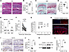 Figure 1
Upregulation of SLIT2 expression in joint tissues in OA. (A and B) Histological images of condylar cartilage stained with H&E from TMJ tissues of Sham or UAC group, and quantitative analysis of images (n = 6). The black dashed lines mark the upper and lower boundaries of the condylar cartilage. (C and D) Histological images of condylar cartilage stained with S&F from TMJ tissues of Sham or UAC group, and quantitative analysis of images (n = 6). (E) qRT-PCR analysis of Slit family gene expression (n = 3) for mouse TMJ tissues, including the disc, articular cartilage, subchondral bone, and synovial tissues. (F) ELISA analysis of SLIT2 concentrations in joint lavage fluid from patients with TMJOA before and after treatment (n = 39). TMJOA-pre, TMJOA pretreatment; TMJOA-post, TMJOA posttreatment. (G) Correlation analysis between radiographic imaging score of TMJOA and the concentration of SLIT2 in TMJOA-pre (n = 39). (H) IF staining showing SLIT2 localization in the condylar cartilage of 10-week-old mice. (I and J) Histological images of condylar cartilage stained with IHC for SLIT2 expression from TMJ tissues of Sham or UAC group, and quantitative analysis of images (n = 6). (K and L) Western blotting analysis of SLIT2 expression (n = 3). Scale bars: 50 μm (A, C, H, and I [right]) and 100 μm (I [left]). All data are shown as mean ± SD. Two-tailed Student’s t test was used for 2-group comparisons, and Spearman’s rank correlation was applied for correlation analysis. NS, not significant. ***P < 0.001.
SLIT2 overexpression aggravated cartilage degradation in UAC-induced and age-induced TMJOA mice. To investigate the role of SLIT2 in TMJOA progression, SLIT2-transgenic (SLIT2-Tg) mice with constitutive SLIT2 overexpression (Supplemental Figure 3, A and B) and C57BL/6J (C57) mice as controls were used. Two experimental models were established: UAC-induced TMJOA and age-associated TMJOA (Figure 2A). In the SLIT2-Tg Sham group, marked degenerative changes were observed compared with the C57 Sham controls, including a reduction in the thickness of the SZ and proliferative zone, decreased overall cartilage thickness, an increased proportion of the deep zone, a higher prevalence of hypertrophic chondrocytes, and more severe cartilage matrix degradation. Cartilage destruction was more severe in the SLIT2-Tg UAC group than in the C57 UAC group. After SLIT2 overexpression, COL2A1-positive areas in the condylar cartilage decreased, while the number of MMP13-positive cells increased, and this trend was more pronounced under UAC induction than in the Sham group (Figure 2, B and C). Meanwhile, the synovium of SLIT2-Tg mice exhibited marked thickening and increased inflammatory cell infiltration compared with that of C57 mice. (Supplemental Figure 3, C and D). The incidence of TMJOA increases with age, and aged mice spontaneously develop TMJOA-like pathological changes, which have been observed at 24 and 48 weeks of age (35–38). Consistent with previous reports, our hematoxylin and eosin (H&E) staining revealed that in aging C57 mice, the condylar cartilage surface exhibited fibrosis, irregular chondrocyte organization, acellular regions, and chondrocyte clustering, accompanied by a reduction in S&F-positive cartilage areas. In SLIT2-Tg mice, a pronounced loss of proteoglycan staining and the formation of vertical fissures were already evident by 24 weeks of age. By 48 weeks, these mice displayed further surface irregularities, multiple fissures, matrix delamination, marked proteoglycan depletion, and chondrocyte clustering — changes more severe than in C57 mice (Figure 2, D and E). Moreover, MMP13 expression remained higher in SLIT2-Tg mice than in C57 mice at 24 and 48 weeks (Supplemental Figure 3, E and F), whereas COL2A1 expression was markedly reduced in SLIT2-Tg mice at 24 and 48 weeks (Supplemental Figure 3, G and H).
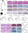 Figure 2
SLIT2 overexpression aggravated cartilage degradation in UAC-induced and age-induced TMJOA mice. (A) Flowchart of experimental procedure in vivo. (B) Histological images of condylar cartilage stained with H&E, S&F, and IHC for COL2A1 and MMP13. The black dashed lines mark the upper and lower boundaries of the condylar cartilage. The superficial layer (SL) is marked by solid yellow lines. The deep layer (DL) is marked by solid green lines. (C) Quantitative analysis of cartilage thickness and S&F-positive area (n = 6), and quantitative analysis of COL2A1-positive area (n = 4) and percentage of MMP13-positive cells (n = 6). (D) H&E and S&F staining of 2-week, 24-week, and 48-week condylar cartilage. (E) Quantitative analysis of the S&F-positive area and OARSI score (n = 6). Scale bars: 50 μm. All data are shown as mean ± SD. Statistical significance was assessed by 2-way ANOVA with Šidák’s post hoc analysis. NS, not significant. *P < 0.05; **P < 0.01; ***P < 0.001.
SLIT2 overexpression exacerbated subchondral bone loss in UAC-induced TMJOA mice. Next, we examined TMJOA cartilage morphology and subchondral bone formation after SLIT2 overexpression. Under a medical microscope, normal condylar cartilage surfaces appeared clear and undamaged. After UAC treatment, slight redness appeared on the surface, with more pronounced redness and higher OARSI scores observed in the SLIT2-Tg UAC group (Figure 3, A and B). We then performed tartrate-resistant acid phosphatase (TRAP) staining to assess osteoclast activity, which is crucial for evaluating subchondral bone resorption (39). A marked increase in TRAP-positive cells was noted in the subchondral bone of SLIT2-Tg mice relative to C57 mice, with a more pronounced elevation observed after UAC induction compared with Sham-operated controls (Figure 3, C and D). Micro-computed tomography (micro-CT) analysis revealed that the bone surface was smooth in the Sham group, whereas UAC induction led to evident surface damage. The extent of bone surface deterioration was more severe in the SLIT2-Tg UAC group than in the C57 UAC group (Figure 3E). Quantitative measurements demonstrated markedly lower bone volume fraction (BV/TV) and trabecular thickness (Tb.Th), along with higher bone surface fraction (BS/BV) and trabecular separation (Tb.Sp) in the SLIT2-Tg UAC compared with the C57 UAC group (Figure 3F). These findings suggest that transgenic overexpression of SLIT2 in mice leads to the destruction of subchondral bone, exacerbating the progression of TMJOA.
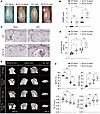 Figure 3
SLIT2 overexpression exacerbated subchondral bone loss in UAC-induced TMJOA mice. (A and B) Gross appearance of TMJ condyles and morphological scoring based on OARSI criteria (n = 6). (C and D) TRAP staining of the TMJ subchondral bone, and quantitative analysis of TRAP-positive cells per unit of bone perimeter (TRAP+/B.pm) (n = 6). The black dashed lines denote the upper boundary of the subchondral bone. Scale bar: 50 μm. (E and F) Two-dimensional and 3-dimensional micro-CT images illustrating the microstructure of subchondral bone, and quantitative analysis using bone structural parameters (n = 6). BV/TV, bone volume fraction; BS/BV, bone surface fraction; Tb.Th, trabecular thickness; Tb.Sp, trabecular separation. All data are shown as mean ± SD. Statistical significance was assessed using 2-way ANOVA with Šidák’s post hoc test. NS, not significant. *P < 0.05; **P < 0.01; ***P < 0.001.
SLIT2 led to an inflammatory response and catabolism in chondrocytes. The primary mouse chondrocytes from SLIT2-Tg mice exhibited OA-like changes, characterized by reduced COL2A1 protein levels and increased MMP13 protein levels compared with the C57 mice. The expression of the inflammatory factor IL-1β was also upregulated (Figure 4, A and B). Consistent with these protein level changes, Col2a1 expression was reduced, whereas Mmp13 expression was increased in SLIT2-overexpressing chondrocytes (Figure 4C). After treating chondrocytes with 10 ng/mL IL-1β, we observed an increase in the expression of Mmp3 and Mmp13, along with a decrease in the expression of Col2a1 in primary chondrocytes. The knockdown of SLIT2 reversed these effects (Supplemental Figure 4A). We further verified the regulation of SLIT2 expression in chondrocytes in vitro using the human chondrocyte cell line, SW1353. We treated SW1353 cells with 0, 10, 20, 50, and 100 ng/mL recombinant human SLIT2 (rhSLIT2) and performed Western blot analysis after 48 hours. Compared with the 0 ng/mL group, the other treatment groups showed decreased expression of COL2A1 and SOX9 and increased expression of MMP13 and MMP3 (Figure 4, D and E). Knockdown of SLIT2 in chondrocytes decreased the expression of MMP3 and MMP13 and enhanced the expression of SOX9 compared with the negative control (NC) group (Supplemental Figure 4, B and C, and Figure 4, F, and G). qRT-PCR analysis revealed an increase in ACAN, COL2A1, and SOX9 expression, and a decrease in MMP3 and MMP13 expression (Figure 4H). RNA-seq analysis revealed that markers of inflammation and catabolism in chondrocytes were downregulated following SLIT2 knockdown (Figure 4I). Gene Ontology (GO) analysis highlighted cellular processes related to the inflammatory response, protein binding, and other associated pathways (Figure 4J).
 Figure 4
SLIT2 led to an inflammatory response and catabolism in chondrocytes. (A) Western blot detection of the effects of SLIT2 overexpression in primary chondrocytes from 3-week-old C57 and SLIT2-Tg mice. Primary chondrocytes for each independent isolation batch were pooled from 3 mice, and the process was repeated 3 times. (B) Relative quantification of COL2A1, MMP13, and IL-1β proteins. (C) qRT-PCR analysis of the effect of SLIT2 overexpression in primary chondrocytes from 3-week-old C57 or SLIT2-Tg mice. (D) Western blot detection of COL2A1, MMP13, and MMP3 after 48 hours of treatment with rhSLIT2 at 0, 10, 20, 50, and 100 ng/mL in SW1353 cells. (E) Relative quantification of COL2A1, MMP3, MMP13 and SOX9 proteins. #P < 0.05, ##P < 0.01, ###P < 0.001 compared with the 0 ng/mL group. (F) Western blot detection of the effect of SLIT2 knockdown on human chondrocytes treated with NC or Si-SLIT2 sequence for 48 hours. (G) Relative quantification of MMP3, MMP13, and SOX9 proteins. (H) qRT-PCR detection of the effect of SLIT2 knockdown on SW1353 cells treated with NC or Si-SLIT2 sequence for 24 hours. (I) A heatmap illustrating differentially expressed genes related to TMJOA from RNA-seq analysis between the NC and Si-SLIT2 groups. (J) GO analysis of 12 key terms from the top terms. All data are shown as mean ± SD. Statistical significance was assessed by 2-tailed Student’s t test (B, C, G, and H) and 1-way ANOVA with Dunnett’s multiple-comparison test (E). *P < 0.05; **P < 0.01; ***P < 0.001.
Knocking down ROBO1 alleviated cartilage degradation in TMJOA. Having established that SLIT2 accelerates TMJOA progression, we next sought to elucidate the underlying molecular mechanism. Given that the canonical physiological roles of SLIT2, ranging from axon guidance and contact-mediated repulsion to angiogenesis regulation and organ formation, strictly depend on its binding to the ROBO receptor (40, 41), we proceeded to dissect the functional interactions between SLIT2 and the ROBO family in the context of TMJOA pathogenesis. Robo1 was the most abundantly expressed receptor and exhibited markedly greater upregulation compared with other receptors in the TMJOA cartilage (Figure 5A). Knockdown of ROBO1 reversed the SLIT2-mediated downregulation of COL2A1 and SOX9 protein expression and upregulation of MMP13 in chondrocytes, whereas knockdown of ROBO2–4 receptors had no apparent effect (Figure 5, B–F). ROBO1 was widely expressed across all cartilage layers and various chondrocyte subpopulations (Figure 5G and Supplemental Figure 5A). Notably, ROBO1 expression was upregulated in TMJOA cartilage (Figure 5, H and I) and also showed a pronounced increase in knee OA, mirroring the expression pattern of SLIT2 (Supplemental Figure 5B). These results suggest that SLIT2 primarily promotes cartilage degradation via a ROBO1-mediated mechanism. The regulatory effect of ROBO1 on TMJOA was then further explored in vivo. To circumvent the embryonic lethality associated with complete or partial Robo1 deletion (29, 42), we utilized Robo1+/–Robo2+/– (Robo1/2+/–) mice, which feature partial deletion of both ROBO1 and ROBO2 receptors, to investigate the role of ROBO1 in TMJOA. Due to the low expression level of ROBO2 in cartilage (Figure 5A and Supplemental Figure 5C) and the fact that its knockdown does not reverse SLIT2-mediated cartilage degradation (Figure 5, B–F), partial ROBO2 deletion has minimal impact on cartilage. Therefore, the Robo1/2+/– model effectively mimics ROBO1 knockdown in vivo. S&F staining revealed no apparent differences in cartilage matrix between the wild-type (WT) and Robo1/2+/– groups. However, Robo1/2+/– mice exhibited a mitigated loss of cartilage matrix induced by UAC compared with WT littermates (Figure 5, J and K). Micro-CT analyses further demonstrated reduced subchondral bone destruction in the Robo1/2+/– UAC group compared with the WT UAC group (Figure 5L). Specifically, the Robo1/2+/– UAC group exhibited increased BV/TV and decreased BS/BV relative to the WT UAC group (Figure 5M). The knockdown of ROBO1 in SW1353 cells induced the downregulation of MMP3 and MMP13 expression while upregulating SOX9 expression in vitro (Figure 5, N and O). These results suggest that ROBO1 acts as the main receptor in regulating SLIT2-induced cartilage degradation.
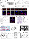 Figure 5
Knocking down ROBO1 alleviated cartilage degradation in TMJOA. (A) Robo family gene expression levels in cartilage measured by qRT-PCR. (B–D) Detection of the effect of ROBO1 knockdown in chondrocytes treated with 10 ng/mL rhSLIT2, and relative quantification of the expression of COL2A1 and SOX9 proteins. R1, Si-ROBO1; R2, Si-ROBO2; R3, Si-ROBO3; R4, Si-ROBO4. (E and F) IF staining showing the effect of ROBO family protein knockdown in chondrocytes treated with 10 ng/mL rhSLIT2, and relative fluorescence intensity of MMP13. (G) IF staining showing ROBO1 localization in the condylar cartilage of 10-week-old mice. (H and I) IHC images of ROBO1 in the condylar cartilage, and quantitative analysis of the percentage of ROBO1-positive cells (n = 4). (J and K) Histological images of condylar cartilage from TMJ tissues stained with S&F, and quantitative analysis of images (n = 6). (L and M) Two-dimensional images illustrating the microstructure of subchondral bone, and quantitative analysis using bone structural parameters (n = 6). (N and O) Detection of the effect of ROBO1 knockdown in chondrocytes, and relative quantification of the expression of ROBO1, MMP3, MMP13, and SOX9 proteins. Scale bars: 50 μm (E and J), 25 μm (G), and 500 μm (L). All data are shown as mean ± SD. Statistical significance was assessed by 2-tailed Student’s t test (A, I, and O) and 1-way (C, D, and F) or 2-way ANOVA (K and M) with Šidák post hoc analysis. NS, not significant. *P < 0.05; **P < 0.01; ***P < 0.001.
SLIT2/ROBO1 signaling–induced cartilage degradation was regulated by LRP6-mediated β-catenin signaling. Gene set enrichment analysis (GSEA) revealed suppression of Wnt/β-catenin signaling following SLIT2 knockdown (Figure 6A). IHC in SLIT2-Tg mice showed increased nuclear translocation of active β-catenin in chondrocytes (Figure 6, B and C), along with elevated levels of phosphorylated LRP6 (p-LRP6) (Figure 6, D and E). Phosphorylated GSK-3β (p-GSK-3β) represents an inactive state that reduces the ability to degrade β-catenin (43). Following ROBO1 knockdown, total GSK-3β (t-GSK-3β) protein levels remained unchanged; however, its phosphorylation was markedly reduced, concomitant with a decrease in active β-catenin levels (Figure 6, F and G). IF staining further showed that treatment with rhSLIT2 increased β-catenin expression and enhanced its nuclear translocation, effects that were reversed by ROBO1 knockdown in chondrocytes (Figure 6, H and I). LRP6 is a key upstream receptor of β-catenin (44). The rhSLIT2-induced increase in MMP3 and MMP13 and the decrease in SOX9 were reversed upon LRP6 knockdown (Figure 6, J and K, and Supplemental Figure 5D). Treatment with rhSLIT2 promoted nuclear translocation of β-catenin; however, this effect was reversed by LRP6 knockdown (Figure 6, L and M). These results suggest that SLIT2/ROBO1 signaling–induced cartilage degradation is regulated by LRP6-mediated β-catenin signaling.
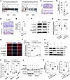 Figure 6
SLIT2/ROBO1 signaling–induced cartilage degradation is regulated by LRP6-mediated β-catenin signaling. (A) GSEA was performed using gene sets related to the Wnt/β-catenin pathway from the GO and KEGG databases to assess differences between the NC and Si-SLIT2 groups. (B and C) IHC images of active β-catenin in condylar cartilage, and quantitative analysis of the percentage of active β-catenin–positive cells (n = 4). (D and E) IHC images of p-LRP6 in condylar cartilage, and quantitative analysis of the percentage of p-LRP6–positive cells (n = 4). (F and G) Western blot detection of the effect of ROBO1 knockdown for GSK-3β and active β-catenin proteins in SW1353 chondrocytes, and relative quantification of p-GSK-3β/t-GSK-3β and active β-catenin proteins. (H and I) IF staining images of β-catenin proteins after being treated with rhSLIT2 and/or Si-ROBO1, and relative quantification of fluorescence intensity of β-catenin proteins. (J and K) Western blot analysis assessing the effect of LRP6 knockdown on rhSLIT2-induced catabolism in SW1353 chondrocytes, and relative quantification of MMP3, MMP13, and SOX9 proteins. (L and M) Western blot analysis showing the impact of LRP6 knockdown on rhSLIT2-induced nuclear translocation of β-catenin in SW1353 cells, and relative quantification of β-catenin proteins in the cytoplasm and nucleus. Scale bars: 50 μm. All data are shown as mean ± SD. Statistical significance was assessed by 2-tailed Student’s t test (C, E, and G), 2-way ANOVA with Šidák’s post hoc analysis (I and K), and 1-way ANOVA with Dunnett’s multiple-comparison test (M). *P < 0.05; **P < 0.01; ***P < 0.001.
SLIT2 enhanced ROBO1-LRP6 complex formation and LRP6 phosphorylation. We next examined the relationship between SLIT2/ROBO1 and LRP6-associated β-catenin signaling. Western blot analysis indicated that rhSLIT2 enhanced LRP6 phosphorylation, an effect reversed by ROBO1 knockdown (Figure 7, A and B). IF staining also showed that rhSLIT2 increased the fluorescence intensity of p-LRP6; however, this increase was attenuated by ROBO1 knockdown (Figure 7, C and D). ROBO1 expression remained unchanged following LRP6 knockdown (Figure 7, E and F). These results indicate that SLIT2/ROBO1 promotes LRP6 phosphorylation, thereby activating β-catenin signaling. SLIT2 binding induces a conformational change in ROBO1, leading to the recruitment of kinases and activation of downstream signaling pathways (9, 45). Similarly, Wnt binding to Frizzled induces a conformational change that promotes the interaction between Frizzled and LRP6 and recruits intracellular signaling proteins, thereby facilitating LRP6 phosphorylation (46, 47). Based on these parallels, we hypothesized that ROBO1 and LRP6 may form a complex through a mechanism analogous to that of the Frizzled-LRP6 interaction. To explore this possibility, we analyzed the GEPIA platform database, which revealed a positive correlation between ROBO1 and LRP6 expression (Supplemental Figure 6A). Bioinformatics analysis showed the binding free energy of ROBO1-LRP6 complex was –322.69 kcal/mol (Supplemental Table 5), with multiple amino acid clusters identified at both extracellular and intracellular binding sites (Figure 7G). To validate these findings experimentally, we performed co-IP assays in SW1353 chondrocytes. ROBO1 was detected in the LRP6-immunoprecipitated complex (Figure 7H). Furthermore, SW1353 chondrocytes were transfected with FLAG-tagged ROBO1 or Myc-tagged LRP6 expression constructs (Supplemental Figure 6B). Co-IP assays revealed that Myc-tagged LRP6 was detected in FLAG-tagged ROBO1 immunoprecipitates isolated using anti-FLAG nanobody agarose beads (Figure 7I). Reciprocally, FLAG-tagged ROBO1 was detected in complexes pulled down with Myc-tagged LRP6 (Figure 7J). Following rhSLIT2 treatment, co-IP analysis revealed increased enrichment of FLAG-tagged ROBO1 in Myc-tagged LRP6 immunoprecipitates (Figure 7K). Consistently, IF staining demonstrated that treatment with rhSLIT2 enhanced the colocalization of ROBO1 and LRP6 in chondrocytes (Figure 7, L–N).
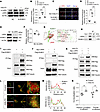 Figure 7
SLIT2 enhanced ROBO1-LRP6 complex formation and LRP6 phosphorylation. (A and B) Western blot analysis of p-LRP6 with the effect of rhSLIT2 and/or Si-ROBO1 in SW1353 cells, and relative quantification of p-LRP6 proteins. (C and D) IF staining images of the effect of rhSLIT2 and Si-ROBO1 on the expression of p-LRP6 in SW1353 cells, and relative quantification of fluorescence intensity of p-LRP6 proteins. Scale bar: 50 μm. (E and F) Western blot analysis examining the effect of LRP6 knockdown on ROBO1 expression in SW1353 cells, and relative quantification of ROBO1 proteins. (G) Visualization of representative potential binding sites between LRP6 and ROBO1 using PyMOL. (H) Co-IP experiments detected the interaction between ROBO1 and LRP6 in SW1353 cells. (I and J) Co-IP experiments using overexpression plasmids for LRP6 and ROBO1 to validate their interaction in SW1353 chondrocytes. (K) Co-IP analysis of the effect of rhSLIT2 on the interaction between LRP6 and ROBO1 in SW1353 chondrocytes transfected with overexpression plasmids. (L) IF staining showing the effect of rhSLIT2 on the colocalization of ROBO1 and LRP6 in SW1353 cells. Scale bars: 10 μm (left) and 1 μm (right). (M) IF staining intensity curve of ROBO1 and LRP6 in IF images. (N) Quantification of colocalization using Pearson’s correlation and overlap coefficients. All data are shown as mean ± SD. Statistical significance was assessed by 1-way ANOVA with Dunnett’s multiple-comparison test (B and D) and 2-tailed Student’s t test (F and N). NS, not significant. **P < 0.01; ***P < 0.001.


