
Research ArticleClinical ResearchHematologyOncology
Open Access |  10.1172/jci.insight.209042
Plasma proteins associated with chronic graft-versus-host disease organ involvement
Stephanie J. Lee,1 Corey Cutler,2 Ningxin Ma,1 Timothy W. Randolph,1 George L. Chen,3 Joseph Pidala,4 Betty K. Hamilton,5 Carrie L. Kitko,6 Sally Arai,7 Najla El Jurdi,8 Lynn Onstad,1 Motoko Koyama,1 Catherine J. Lee,1 Sophie Paczesny,9 and Geoffrey R. Hill1
1Fred Hutchinson Cancer Center, Seattle, Washington, USA.
2Division of Transplantation and Cellular Therapy, Dana-Farber Cancer Institute, Boston, Massachusetts, USA.
3The University of Texas MD Anderson Cancer Center, Houston, Texas, USA.
4Department of Blood and Marrow Transplantation and Cellular Immunotherapy, Moffitt Cancer Center, Tampa, Florida, USA.
5Taussig Cancer Institute, Cleveland Clinic, Cleveland, Ohio, USA.
6Vanderbilt University Medical Center, Nashville, Tennessee, USA.
7Stanford Blood and Marrow Transplantation and Cellular Therapy Division, Stanford School of Medicine, Stanford University, Stanford, California, USA.
8Immune Deficiency Cellular Therapy Program, Center for Cancer Research, National Cancer Institute, NIH, Bethesda, Maryland, USA.
9Hollings Cancer Center and Department of Pharmacology and Immunology, Medical University of South Carolina, Charleston, South Carolina, USA.ZZZZÆ
Address correspondence to: Stephanie J. Lee, Fred Hutchinson Cancer Center, 1100 Fairview Ave. N, M2-B230, Seattle, Washington, 98109, USA. Phone: 206.667.6190; Email: sjlee@fredhutch.org.
Authorship note: SP and GRH are co-senior authors.
Find articles by
Lee, S.
in:
PubMed
|
Google Scholar
|

1Fred Hutchinson Cancer Center, Seattle, Washington, USA.
2Division of Transplantation and Cellular Therapy, Dana-Farber Cancer Institute, Boston, Massachusetts, USA.
3The University of Texas MD Anderson Cancer Center, Houston, Texas, USA.
4Department of Blood and Marrow Transplantation and Cellular Immunotherapy, Moffitt Cancer Center, Tampa, Florida, USA.
5Taussig Cancer Institute, Cleveland Clinic, Cleveland, Ohio, USA.
6Vanderbilt University Medical Center, Nashville, Tennessee, USA.
7Stanford Blood and Marrow Transplantation and Cellular Therapy Division, Stanford School of Medicine, Stanford University, Stanford, California, USA.
8Immune Deficiency Cellular Therapy Program, Center for Cancer Research, National Cancer Institute, NIH, Bethesda, Maryland, USA.
9Hollings Cancer Center and Department of Pharmacology and Immunology, Medical University of South Carolina, Charleston, South Carolina, USA.ZZZZÆ
Address correspondence to: Stephanie J. Lee, Fred Hutchinson Cancer Center, 1100 Fairview Ave. N, M2-B230, Seattle, Washington, 98109, USA. Phone: 206.667.6190; Email: sjlee@fredhutch.org.
Authorship note: SP and GRH are co-senior authors.
Find articles by
Cutler, C.
in:
PubMed
|
Google Scholar
|
1Fred Hutchinson Cancer Center, Seattle, Washington, USA.
2Division of Transplantation and Cellular Therapy, Dana-Farber Cancer Institute, Boston, Massachusetts, USA.
3The University of Texas MD Anderson Cancer Center, Houston, Texas, USA.
4Department of Blood and Marrow Transplantation and Cellular Immunotherapy, Moffitt Cancer Center, Tampa, Florida, USA.
5Taussig Cancer Institute, Cleveland Clinic, Cleveland, Ohio, USA.
6Vanderbilt University Medical Center, Nashville, Tennessee, USA.
7Stanford Blood and Marrow Transplantation and Cellular Therapy Division, Stanford School of Medicine, Stanford University, Stanford, California, USA.
8Immune Deficiency Cellular Therapy Program, Center for Cancer Research, National Cancer Institute, NIH, Bethesda, Maryland, USA.
9Hollings Cancer Center and Department of Pharmacology and Immunology, Medical University of South Carolina, Charleston, South Carolina, USA.ZZZZÆ
Address correspondence to: Stephanie J. Lee, Fred Hutchinson Cancer Center, 1100 Fairview Ave. N, M2-B230, Seattle, Washington, 98109, USA. Phone: 206.667.6190; Email: sjlee@fredhutch.org.
Authorship note: SP and GRH are co-senior authors.
Find articles by Ma, N. in: PubMed | Google Scholar
1Fred Hutchinson Cancer Center, Seattle, Washington, USA.
2Division of Transplantation and Cellular Therapy, Dana-Farber Cancer Institute, Boston, Massachusetts, USA.
3The University of Texas MD Anderson Cancer Center, Houston, Texas, USA.
4Department of Blood and Marrow Transplantation and Cellular Immunotherapy, Moffitt Cancer Center, Tampa, Florida, USA.
5Taussig Cancer Institute, Cleveland Clinic, Cleveland, Ohio, USA.
6Vanderbilt University Medical Center, Nashville, Tennessee, USA.
7Stanford Blood and Marrow Transplantation and Cellular Therapy Division, Stanford School of Medicine, Stanford University, Stanford, California, USA.
8Immune Deficiency Cellular Therapy Program, Center for Cancer Research, National Cancer Institute, NIH, Bethesda, Maryland, USA.
9Hollings Cancer Center and Department of Pharmacology and Immunology, Medical University of South Carolina, Charleston, South Carolina, USA.ZZZZÆ
Address correspondence to: Stephanie J. Lee, Fred Hutchinson Cancer Center, 1100 Fairview Ave. N, M2-B230, Seattle, Washington, 98109, USA. Phone: 206.667.6190; Email: sjlee@fredhutch.org.
Authorship note: SP and GRH are co-senior authors.
Find articles by
Randolph, T.
in:
PubMed
|
Google Scholar
|
1Fred Hutchinson Cancer Center, Seattle, Washington, USA.
2Division of Transplantation and Cellular Therapy, Dana-Farber Cancer Institute, Boston, Massachusetts, USA.
3The University of Texas MD Anderson Cancer Center, Houston, Texas, USA.
4Department of Blood and Marrow Transplantation and Cellular Immunotherapy, Moffitt Cancer Center, Tampa, Florida, USA.
5Taussig Cancer Institute, Cleveland Clinic, Cleveland, Ohio, USA.
6Vanderbilt University Medical Center, Nashville, Tennessee, USA.
7Stanford Blood and Marrow Transplantation and Cellular Therapy Division, Stanford School of Medicine, Stanford University, Stanford, California, USA.
8Immune Deficiency Cellular Therapy Program, Center for Cancer Research, National Cancer Institute, NIH, Bethesda, Maryland, USA.
9Hollings Cancer Center and Department of Pharmacology and Immunology, Medical University of South Carolina, Charleston, South Carolina, USA.ZZZZÆ
Address correspondence to: Stephanie J. Lee, Fred Hutchinson Cancer Center, 1100 Fairview Ave. N, M2-B230, Seattle, Washington, 98109, USA. Phone: 206.667.6190; Email: sjlee@fredhutch.org.
Authorship note: SP and GRH are co-senior authors.
Find articles by Chen, G. in: PubMed | Google Scholar
1Fred Hutchinson Cancer Center, Seattle, Washington, USA.
2Division of Transplantation and Cellular Therapy, Dana-Farber Cancer Institute, Boston, Massachusetts, USA.
3The University of Texas MD Anderson Cancer Center, Houston, Texas, USA.
4Department of Blood and Marrow Transplantation and Cellular Immunotherapy, Moffitt Cancer Center, Tampa, Florida, USA.
5Taussig Cancer Institute, Cleveland Clinic, Cleveland, Ohio, USA.
6Vanderbilt University Medical Center, Nashville, Tennessee, USA.
7Stanford Blood and Marrow Transplantation and Cellular Therapy Division, Stanford School of Medicine, Stanford University, Stanford, California, USA.
8Immune Deficiency Cellular Therapy Program, Center for Cancer Research, National Cancer Institute, NIH, Bethesda, Maryland, USA.
9Hollings Cancer Center and Department of Pharmacology and Immunology, Medical University of South Carolina, Charleston, South Carolina, USA.ZZZZÆ
Address correspondence to: Stephanie J. Lee, Fred Hutchinson Cancer Center, 1100 Fairview Ave. N, M2-B230, Seattle, Washington, 98109, USA. Phone: 206.667.6190; Email: sjlee@fredhutch.org.
Authorship note: SP and GRH are co-senior authors.
Find articles by Pidala, J. in: PubMed | Google Scholar
1Fred Hutchinson Cancer Center, Seattle, Washington, USA.
2Division of Transplantation and Cellular Therapy, Dana-Farber Cancer Institute, Boston, Massachusetts, USA.
3The University of Texas MD Anderson Cancer Center, Houston, Texas, USA.
4Department of Blood and Marrow Transplantation and Cellular Immunotherapy, Moffitt Cancer Center, Tampa, Florida, USA.
5Taussig Cancer Institute, Cleveland Clinic, Cleveland, Ohio, USA.
6Vanderbilt University Medical Center, Nashville, Tennessee, USA.
7Stanford Blood and Marrow Transplantation and Cellular Therapy Division, Stanford School of Medicine, Stanford University, Stanford, California, USA.
8Immune Deficiency Cellular Therapy Program, Center for Cancer Research, National Cancer Institute, NIH, Bethesda, Maryland, USA.
9Hollings Cancer Center and Department of Pharmacology and Immunology, Medical University of South Carolina, Charleston, South Carolina, USA.ZZZZÆ
Address correspondence to: Stephanie J. Lee, Fred Hutchinson Cancer Center, 1100 Fairview Ave. N, M2-B230, Seattle, Washington, 98109, USA. Phone: 206.667.6190; Email: sjlee@fredhutch.org.
Authorship note: SP and GRH are co-senior authors.
Find articles by Hamilton, B. in: PubMed | Google Scholar
1Fred Hutchinson Cancer Center, Seattle, Washington, USA.
2Division of Transplantation and Cellular Therapy, Dana-Farber Cancer Institute, Boston, Massachusetts, USA.
3The University of Texas MD Anderson Cancer Center, Houston, Texas, USA.
4Department of Blood and Marrow Transplantation and Cellular Immunotherapy, Moffitt Cancer Center, Tampa, Florida, USA.
5Taussig Cancer Institute, Cleveland Clinic, Cleveland, Ohio, USA.
6Vanderbilt University Medical Center, Nashville, Tennessee, USA.
7Stanford Blood and Marrow Transplantation and Cellular Therapy Division, Stanford School of Medicine, Stanford University, Stanford, California, USA.
8Immune Deficiency Cellular Therapy Program, Center for Cancer Research, National Cancer Institute, NIH, Bethesda, Maryland, USA.
9Hollings Cancer Center and Department of Pharmacology and Immunology, Medical University of South Carolina, Charleston, South Carolina, USA.ZZZZÆ
Address correspondence to: Stephanie J. Lee, Fred Hutchinson Cancer Center, 1100 Fairview Ave. N, M2-B230, Seattle, Washington, 98109, USA. Phone: 206.667.6190; Email: sjlee@fredhutch.org.
Authorship note: SP and GRH are co-senior authors.
Find articles by Kitko, C. in: PubMed | Google Scholar
1Fred Hutchinson Cancer Center, Seattle, Washington, USA.
2Division of Transplantation and Cellular Therapy, Dana-Farber Cancer Institute, Boston, Massachusetts, USA.
3The University of Texas MD Anderson Cancer Center, Houston, Texas, USA.
4Department of Blood and Marrow Transplantation and Cellular Immunotherapy, Moffitt Cancer Center, Tampa, Florida, USA.
5Taussig Cancer Institute, Cleveland Clinic, Cleveland, Ohio, USA.
6Vanderbilt University Medical Center, Nashville, Tennessee, USA.
7Stanford Blood and Marrow Transplantation and Cellular Therapy Division, Stanford School of Medicine, Stanford University, Stanford, California, USA.
8Immune Deficiency Cellular Therapy Program, Center for Cancer Research, National Cancer Institute, NIH, Bethesda, Maryland, USA.
9Hollings Cancer Center and Department of Pharmacology and Immunology, Medical University of South Carolina, Charleston, South Carolina, USA.ZZZZÆ
Address correspondence to: Stephanie J. Lee, Fred Hutchinson Cancer Center, 1100 Fairview Ave. N, M2-B230, Seattle, Washington, 98109, USA. Phone: 206.667.6190; Email: sjlee@fredhutch.org.
Authorship note: SP and GRH are co-senior authors.
Find articles by Arai, S. in: PubMed | Google Scholar
1Fred Hutchinson Cancer Center, Seattle, Washington, USA.
2Division of Transplantation and Cellular Therapy, Dana-Farber Cancer Institute, Boston, Massachusetts, USA.
3The University of Texas MD Anderson Cancer Center, Houston, Texas, USA.
4Department of Blood and Marrow Transplantation and Cellular Immunotherapy, Moffitt Cancer Center, Tampa, Florida, USA.
5Taussig Cancer Institute, Cleveland Clinic, Cleveland, Ohio, USA.
6Vanderbilt University Medical Center, Nashville, Tennessee, USA.
7Stanford Blood and Marrow Transplantation and Cellular Therapy Division, Stanford School of Medicine, Stanford University, Stanford, California, USA.
8Immune Deficiency Cellular Therapy Program, Center for Cancer Research, National Cancer Institute, NIH, Bethesda, Maryland, USA.
9Hollings Cancer Center and Department of Pharmacology and Immunology, Medical University of South Carolina, Charleston, South Carolina, USA.ZZZZÆ
Address correspondence to: Stephanie J. Lee, Fred Hutchinson Cancer Center, 1100 Fairview Ave. N, M2-B230, Seattle, Washington, 98109, USA. Phone: 206.667.6190; Email: sjlee@fredhutch.org.
Authorship note: SP and GRH are co-senior authors.
Find articles by El Jurdi, N. in: PubMed | Google Scholar
1Fred Hutchinson Cancer Center, Seattle, Washington, USA.
2Division of Transplantation and Cellular Therapy, Dana-Farber Cancer Institute, Boston, Massachusetts, USA.
3The University of Texas MD Anderson Cancer Center, Houston, Texas, USA.
4Department of Blood and Marrow Transplantation and Cellular Immunotherapy, Moffitt Cancer Center, Tampa, Florida, USA.
5Taussig Cancer Institute, Cleveland Clinic, Cleveland, Ohio, USA.
6Vanderbilt University Medical Center, Nashville, Tennessee, USA.
7Stanford Blood and Marrow Transplantation and Cellular Therapy Division, Stanford School of Medicine, Stanford University, Stanford, California, USA.
8Immune Deficiency Cellular Therapy Program, Center for Cancer Research, National Cancer Institute, NIH, Bethesda, Maryland, USA.
9Hollings Cancer Center and Department of Pharmacology and Immunology, Medical University of South Carolina, Charleston, South Carolina, USA.ZZZZÆ
Address correspondence to: Stephanie J. Lee, Fred Hutchinson Cancer Center, 1100 Fairview Ave. N, M2-B230, Seattle, Washington, 98109, USA. Phone: 206.667.6190; Email: sjlee@fredhutch.org.
Authorship note: SP and GRH are co-senior authors.
Find articles by Onstad, L. in: PubMed | Google Scholar
1Fred Hutchinson Cancer Center, Seattle, Washington, USA.
2Division of Transplantation and Cellular Therapy, Dana-Farber Cancer Institute, Boston, Massachusetts, USA.
3The University of Texas MD Anderson Cancer Center, Houston, Texas, USA.
4Department of Blood and Marrow Transplantation and Cellular Immunotherapy, Moffitt Cancer Center, Tampa, Florida, USA.
5Taussig Cancer Institute, Cleveland Clinic, Cleveland, Ohio, USA.
6Vanderbilt University Medical Center, Nashville, Tennessee, USA.
7Stanford Blood and Marrow Transplantation and Cellular Therapy Division, Stanford School of Medicine, Stanford University, Stanford, California, USA.
8Immune Deficiency Cellular Therapy Program, Center for Cancer Research, National Cancer Institute, NIH, Bethesda, Maryland, USA.
9Hollings Cancer Center and Department of Pharmacology and Immunology, Medical University of South Carolina, Charleston, South Carolina, USA.ZZZZÆ
Address correspondence to: Stephanie J. Lee, Fred Hutchinson Cancer Center, 1100 Fairview Ave. N, M2-B230, Seattle, Washington, 98109, USA. Phone: 206.667.6190; Email: sjlee@fredhutch.org.
Authorship note: SP and GRH are co-senior authors.
Find articles by Koyama, M. in: PubMed | Google Scholar
1Fred Hutchinson Cancer Center, Seattle, Washington, USA.
2Division of Transplantation and Cellular Therapy, Dana-Farber Cancer Institute, Boston, Massachusetts, USA.
3The University of Texas MD Anderson Cancer Center, Houston, Texas, USA.
4Department of Blood and Marrow Transplantation and Cellular Immunotherapy, Moffitt Cancer Center, Tampa, Florida, USA.
5Taussig Cancer Institute, Cleveland Clinic, Cleveland, Ohio, USA.
6Vanderbilt University Medical Center, Nashville, Tennessee, USA.
7Stanford Blood and Marrow Transplantation and Cellular Therapy Division, Stanford School of Medicine, Stanford University, Stanford, California, USA.
8Immune Deficiency Cellular Therapy Program, Center for Cancer Research, National Cancer Institute, NIH, Bethesda, Maryland, USA.
9Hollings Cancer Center and Department of Pharmacology and Immunology, Medical University of South Carolina, Charleston, South Carolina, USA.ZZZZÆ
Address correspondence to: Stephanie J. Lee, Fred Hutchinson Cancer Center, 1100 Fairview Ave. N, M2-B230, Seattle, Washington, 98109, USA. Phone: 206.667.6190; Email: sjlee@fredhutch.org.
Authorship note: SP and GRH are co-senior authors.
Find articles by Lee, C. in: PubMed | Google Scholar
1Fred Hutchinson Cancer Center, Seattle, Washington, USA.
2Division of Transplantation and Cellular Therapy, Dana-Farber Cancer Institute, Boston, Massachusetts, USA.
3The University of Texas MD Anderson Cancer Center, Houston, Texas, USA.
4Department of Blood and Marrow Transplantation and Cellular Immunotherapy, Moffitt Cancer Center, Tampa, Florida, USA.
5Taussig Cancer Institute, Cleveland Clinic, Cleveland, Ohio, USA.
6Vanderbilt University Medical Center, Nashville, Tennessee, USA.
7Stanford Blood and Marrow Transplantation and Cellular Therapy Division, Stanford School of Medicine, Stanford University, Stanford, California, USA.
8Immune Deficiency Cellular Therapy Program, Center for Cancer Research, National Cancer Institute, NIH, Bethesda, Maryland, USA.
9Hollings Cancer Center and Department of Pharmacology and Immunology, Medical University of South Carolina, Charleston, South Carolina, USA.ZZZZÆ
Address correspondence to: Stephanie J. Lee, Fred Hutchinson Cancer Center, 1100 Fairview Ave. N, M2-B230, Seattle, Washington, 98109, USA. Phone: 206.667.6190; Email: sjlee@fredhutch.org.
Authorship note: SP and GRH are co-senior authors.
Find articles by
Paczesny, S.
in:
PubMed
|
Google Scholar
|
1Fred Hutchinson Cancer Center, Seattle, Washington, USA.
2Division of Transplantation and Cellular Therapy, Dana-Farber Cancer Institute, Boston, Massachusetts, USA.
3The University of Texas MD Anderson Cancer Center, Houston, Texas, USA.
4Department of Blood and Marrow Transplantation and Cellular Immunotherapy, Moffitt Cancer Center, Tampa, Florida, USA.
5Taussig Cancer Institute, Cleveland Clinic, Cleveland, Ohio, USA.
6Vanderbilt University Medical Center, Nashville, Tennessee, USA.
7Stanford Blood and Marrow Transplantation and Cellular Therapy Division, Stanford School of Medicine, Stanford University, Stanford, California, USA.
8Immune Deficiency Cellular Therapy Program, Center for Cancer Research, National Cancer Institute, NIH, Bethesda, Maryland, USA.
9Hollings Cancer Center and Department of Pharmacology and Immunology, Medical University of South Carolina, Charleston, South Carolina, USA.ZZZZÆ
Address correspondence to: Stephanie J. Lee, Fred Hutchinson Cancer Center, 1100 Fairview Ave. N, M2-B230, Seattle, Washington, 98109, USA. Phone: 206.667.6190; Email: sjlee@fredhutch.org.
Authorship note: SP and GRH are co-senior authors.
Find articles by Hill, G. in: PubMed | Google Scholar
Published July 28, 2026 - More info
JCI Insight. 2026;11(17):e209042. https://doi.org/10.1172/jci.insight.209042.
© 2026 Lee et al. This work is licensed under the Creative Commons Attribution 4.0 International License. To view a copy of this license, visit http://creativecommons.org/licenses/by/4.0/.

-
Introduction
Chronic graft-versus-host disease (cGVHD) is a major cause of long-term morbidity and mortality in allogeneic hematopoietic cell transplantation (HCT) recipients. cGVHD commonly involves the skin, mouth, and eye, but may also affect the liver, lung, fascia, genital tract, kidney, muscle, and the hematopoietic and nervous systems. Newer GVHD-prophylaxis approaches are associated with lower rates of cGVHD than older regimens, but between 10% and 30% of people still develop cGVHD that is severe enough to require systemic immunosuppression (1). Treatment is often prolonged, with approximately 15% of treated patients still on immunosuppression 10 years later (2). cGVHD is a heterogeneous disease and patients display different combinations of organ involvement.
The pathobiology of cGVHD has been summarized as initial cytokine-based inflammation followed by dysregulated immunity leading to pathologic tissue repair (3, 4). Multiple plasma biomarkers have been reported to be associated with cGVHD, including elevated levels of IL-1β (5), sIL-2-Ra (6, 7), IL-6 (8), IL-8 (8), IL-15 (9), IL-17A/F (8), TNF-α (5, 8, 10, 11), TGF-β (12–14), chemokine (C-X-C motif) ligand 9 (C-X-C motif chemokine ligand 9 [CXCL9], also known as MIG) (15, 16), CXCL10 (IP-10) (17), CCL15 (18), sBAFF (19, 20), and APRIL (21) and decreased IL-10 (5, 7, 10, 22) and IL-2 (23). Others such as IL-4 are controversial (23–25). In a study with 2 replication cohorts, soluble IL-1 receptor-like 1 (IL1RL1, previously stimulation 2 [ST2]), CXCL9, matrix metalloproteinase 3 (MMP3), and osteopontin (OPN) were increased in patients with cGVHD compared with patients without cGVHD (15). Soluble CD163 is a macrophage scavenger receptor, and elevated levels at day +80 were associated with de novo cGVHD (26). Dickkopf-related protein 3 (DKK3), discovered in patients with sclerotic skin and lung involvement, has been associated with overall cGVHD. In general, these studies compared fewer than 200 patients with cGVHD to control patients without cGVHD. They did not consider or did not find correlation with individual organ involvement (Tables 1 and 2).
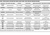 Table 2
Plasma proteins associated with cGVHD in human studies, unless otherwise indicated (continued)
We sought to identify plasma proteins that correlate with specific organ involvement in a large cohort of adult patients with well-characterized cGVHD. We hypothesized that plasma protein analysis might reflect tissue-specific pathogenesis and help explain heterogeneous organ involvement. Organ-specific treatment approaches could then be tailored if aberrant pathways were identified.

-
Results
Patient characteristics.
Patient characteristics are shown in Supplemental Table 1; supplemental material available online with this article; https://doi.org/10.1172/jci.insight.209042DS1 Sixteen institutions enrolled patients between 2007 and 2019, with over half accrued at Fred Hutchinson Cancer Center. The population was predominantly White (88%) and male (62%) who received peripheral blood grafts (90%). Acute myeloid leukemia, acute lymphoblastic leukemia, and myelodysplastic syndrome accounted for 63% of diagnoses. Approximately half the population received grafts from matched unrelated donors (50%), received calcineurin inhibitor and methotrexate acute GVHD (aGVHD) prophylaxis (53%), and had myeloablative conditioning (46%). Only 5 (1%) had haploidentical donors and 9 (1%) received posttransplant cyclophosphamide. Tables 3 and 4, and Supplemental Table 2 show GVHD characteristics at time of sample draw. cGVHD was mild in 18%, moderate in 47%, and severe in 35%; 63% were diagnosed within the previous 3 months. The most commonly involved organs were skin (67%), mouth (60%), and eye (49%). Prior grade II–IV aGVHD occurred in 48%. At the time of the blood sample, 60% were receiving prednisone and 48% were on a calcineurin inhibitor; 9.1% were not on any immunosuppression. Although most were continuing previous immunosuppressive agents, 73.1% had not started any new cGVHD-specific treatment right before the sample was collected. Previous treatments for cGVHD included calcineurin inhibitors (12.7%) and prednisone (10.2%). Less than 10% had previously received sirolimus, an antimetabolite (mycophenolate mofetil or methotrexate), extracorporeal photopheresis, or a B cell–targeting agent (BTK inhibitor or anti-CD20 monoclonal antibody) for cGVHD prior to sample acquisition. Three patients were on ruxolitinib, 1 on belumosudil, and none on axatilimab at the time of sample acquisition.
Plasma proteins.
Supplemental Figure 1 shows the correlations of the plasma proteins with each other. Strong correlations were seen between CXCL9 and CXCL10, as expected. Other correlations (Pearson’s correlation > 0.5) could reflect (a) Th1-Th17 and myeloid activation with IL-12, IL-21, and GM-CSF, and (b) antigen-presenting cell and Th1 and B cell activation with IL-12, CXCL9/CXCL10, and BAFF.
Association of individual plasma proteins with individual organ involvement, without adjusting for clinical variables.
Supplemental Table 3 shows the associations of the 19 plasma proteins with cGVHD organ involvement, adjusted for multiple testing but unadjusted for the 5 clinical variables. Five plasma proteins (GM-CSF, IL-12, IL-21, M-CSF, and IL-17A) were not associated with any organ involvement (at P < 0.05) and were not considered further. All organs except the eye had at least one plasma protein association (at P < 0.05). Despite the strong statistical associations, Figure 1 shows the overlapping distributions of plasma protein levels between different organ severity levels, presented as box-and-whisker plots and odds ratios (ORs).
 Figure 1
Plasma protein levels associated with various levels of organ involvement, not adjusting for clinical covariates.
Association of individual plasma proteins with individual organ involvement, adjusting for clinical variables.
Table 5 shows the marginal association of the 14 remaining plasma proteins with cGVHD organ manifestations, adjusting for the following 5 clinical variables: patient age, patient sex, time between HCT and blood draw, time since cGVHD diagnosis (more or less than 3 months), and steroid dose, as a continuous variable. P values were adjusted for multiple testing. Thirteen plasma proteins: IL-18, CXCL9, CXCL10, IL-6, IL-8, MCP1, BAFF, IL1RL1 (ST2), REG3a, MMP3, DKK3, and CD163 remained significantly associated with at least one organ manifestation, and there were 30 associations among the 7 organ systems (Supplemental Table 3). Only 2 of these associations were previously reported in the literature: IL-6 and joint involvement (6) and REG3a and gastrointestinal (GI) involvement (27), while the others have not been previously reported. MMP9 was not statistically associated with any organ manifestations once models were adjusted for the 5 clinical variables, although MMP9 was associated with joint and liver in our univariable analyses. Some previous publications have used an OR of greater than 1.3 or less than 0.7 to declare clinical importance as well as statistical significance; if this criterion is applied, MCP1 and MMP3 would no longer be associated with any organs. If a higher OR greater than 1.5 is required for clinical importance, only 4 plasma proteins associated with liver involvement (IL-18, IL-8, IL1RL1 [ST2], and CD163) and 2 associated with GI involvement (IL1RL1 [ST2] and REG3a) would be considered clinically important.
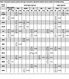 Table 5
Association of plasma proteins with organ involvement, adjusted for baseline clinical variables and multiple testing
Supplemental Figure 2A shows the area under the receiver operating characteristics curve (AUC) for joint involvement, as an example of high AUCs of 0.78 (0.71–0.85) for the 5 clinical variables alone without improvement after adding the plasma protein values. AUCs ranged from 0.77–0.81 using the different models for joint involvement. A similar pattern was seen for the other organs (data not shown). For the logistic regression model including only plasma proteins, the positive predictive value (PPV) was 0.76 (0.69–0.84) and the negative predictive value (NPV) was 0.46 (0.37–0.57) at the optimal cutoff. Thus, despite the high and usually acceptable AUCs for the correlative models, the PPV and NPV of the plasma proteins were modest. Supplemental Figure 2B shows the kernel-association testing procedure (KRV) coefficients demonstrating the very low explanatory value of plasma protein profiles for organ involvement.
Supplemental Table 4 displays the variables selected most often in our repeated application of the elastic net regression using the randomly chosen 80% training/20% sampling procedure for the 7 organs, as illustrated for joint in Supplemental Figure 2. In general, each of the 5 clinical variables was important in different models, with time since diagnosis important in 6 out of 7 models, years after transplant and patient age in 4 out of 7 models, and sex and prednisone dose in 3 out of 7 models.
Clustering of plasma proteins and organs.
Figure 2 shows the results of dimensionality reduction using t-distributed stochastic neighbor embedding (tSNE) clustering of plasma proteins for each organ. We were unable to identify any meaningful clusters defined by plasma proteins using this method. The results using uniform manifold approximation and projection (UMAP) were similarly unrevealing (not shown).
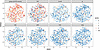 Figure 2
Dimensionality reduction using t-distributed stochastic neighbor embedding (tSNE) clustering of plasma proteins for each organ. The lack of identifiable clusters shows the lack of association between plasma protein profiles and organ involvement.


