
Go to The Journal of Clinical Investigation
  • About
  • Editors
  • Consulting Editors
  • For authors
  • Journal stats
  • Publication ethics
  • Publication alerts by email
  • Transfers
  • Advertising
  • Job board
  • Contact
  • Physician-Scientist Development
  • Current issue
  • Past issues
  • By specialty
    • COVID-19
    • Cardiology
    • Immunology
    • Metabolism
    • Nephrology
    • Oncology
    • Pulmonology
    • All ...
  • Videos
  • Collections
    • In-Press Preview
    • Resource and Technical Advances
    • Clinical Research and Public Health
    • Research Letters
    • Editorials
    • Perspectives
    • Physician-Scientist Development
    • Reviews
    • Top read articles

  • Current issue
  • Past issues
  • Specialties
  • In-Press Preview
  • Resource and Technical Advances
  • Clinical Research and Public Health
  • Research Letters
  • Editorials
  • Perspectives
  • Physician-Scientist Development
  • Reviews
  • Top read articles
  • About
  • Editors
  • Consulting Editors
  • For authors
  • Journal stats
  • Publication ethics
  • Publication alerts by email
  • Transfers
  • Advertising
  • Job board
  • Contact
Top
  • View PDF
  • Download citation information
  • Send a comment
  • Terms of use
  • Standard abbreviations
  • Need help? Email the journal
  • Top
  • Abstract
  • Introduction
  • Results
  • Discussion
  • Methods
  • Manuscript information
  • Supplemental material
  • Acknowledgments
  • Footnotes
  • References
  • Version history
  • Article usage
  • Citations to this article
Advertisement

Research ArticleInflammationPulmonology Open Access | 10.1172/jci.insight.208692

Sphingolipid metabolism contributes to sexual dimorphism in COPD pathogenesis

Elisabetta Granato,1,2,3 Xiaoyun Wang,2 Yun Zhang,2 Joselyn Rojas-Quintero,2 Ida Cerqua,1 Scott A. Ochsner,4 Jeff Thomas Kue,2 Luca Cecchetto,3 Maor Sauler,5 Farrah Kheradmand,2,6 Joshua Malo,7 Fiorentina Roviezzo,1 Irina Petrache,8 and Francesca Polverino2

1Department of Pharmacy, School of Medicine and Surgery, University of Naples Federico II, Naples, Italy.

2Section of Pulmonary, Critical Care and Sleep Medicine, Department of Medicine, Baylor College of Medicine, Houston, Texas, USA.

3Department of Genomic Medicine, The University of Texas MD Anderson Cancer Center, Houston, Texas, USA.

4Department of Molecular and Cellular Biology, Baylor College of Medicine, Houston, Texas, USA.

5Department of Internal Medicine, Division of Pulmonary and Critical Care Medicine, University of Michigan Medical School, Ann Arbor, Michigan, USA.

6Center for Translational Research on Inflammatory Diseases, Michael E. DeBakey Department of Veterans Affairs Medical Center, Houston, Texas, USA.

7Department of Medicine, University of Arizona, Tucson, Arizona, USA.

8Department of Medicine, National Jewish Health and University of Colorado, Denver, Colorado, USA.

Address for correspondence. Francesca Polverino, Department of Medicine, Baylor College of Medicine, 6501 Fannin Street, Houston, Texas 77030, USA. Email: fpolverino@copdnet.org.

Authorship note: EG and XW contributed equally to this work. IP and FP are co–senior authors.

Find articles by Granato, E. in: PubMed | Google Scholar

1Department of Pharmacy, School of Medicine and Surgery, University of Naples Federico II, Naples, Italy.

2Section of Pulmonary, Critical Care and Sleep Medicine, Department of Medicine, Baylor College of Medicine, Houston, Texas, USA.

3Department of Genomic Medicine, The University of Texas MD Anderson Cancer Center, Houston, Texas, USA.

4Department of Molecular and Cellular Biology, Baylor College of Medicine, Houston, Texas, USA.

5Department of Internal Medicine, Division of Pulmonary and Critical Care Medicine, University of Michigan Medical School, Ann Arbor, Michigan, USA.

6Center for Translational Research on Inflammatory Diseases, Michael E. DeBakey Department of Veterans Affairs Medical Center, Houston, Texas, USA.

7Department of Medicine, University of Arizona, Tucson, Arizona, USA.

8Department of Medicine, National Jewish Health and University of Colorado, Denver, Colorado, USA.

Address for correspondence. Francesca Polverino, Department of Medicine, Baylor College of Medicine, 6501 Fannin Street, Houston, Texas 77030, USA. Email: fpolverino@copdnet.org.

Authorship note: EG and XW contributed equally to this work. IP and FP are co–senior authors.

Find articles by Wang, X. in: PubMed | Google Scholar

1Department of Pharmacy, School of Medicine and Surgery, University of Naples Federico II, Naples, Italy.

2Section of Pulmonary, Critical Care and Sleep Medicine, Department of Medicine, Baylor College of Medicine, Houston, Texas, USA.

3Department of Genomic Medicine, The University of Texas MD Anderson Cancer Center, Houston, Texas, USA.

4Department of Molecular and Cellular Biology, Baylor College of Medicine, Houston, Texas, USA.

5Department of Internal Medicine, Division of Pulmonary and Critical Care Medicine, University of Michigan Medical School, Ann Arbor, Michigan, USA.

6Center for Translational Research on Inflammatory Diseases, Michael E. DeBakey Department of Veterans Affairs Medical Center, Houston, Texas, USA.

7Department of Medicine, University of Arizona, Tucson, Arizona, USA.

8Department of Medicine, National Jewish Health and University of Colorado, Denver, Colorado, USA.

Address for correspondence. Francesca Polverino, Department of Medicine, Baylor College of Medicine, 6501 Fannin Street, Houston, Texas 77030, USA. Email: fpolverino@copdnet.org.

Authorship note: EG and XW contributed equally to this work. IP and FP are co–senior authors.

Find articles by Zhang, Y. in: PubMed | Google Scholar

1Department of Pharmacy, School of Medicine and Surgery, University of Naples Federico II, Naples, Italy.

2Section of Pulmonary, Critical Care and Sleep Medicine, Department of Medicine, Baylor College of Medicine, Houston, Texas, USA.

3Department of Genomic Medicine, The University of Texas MD Anderson Cancer Center, Houston, Texas, USA.

4Department of Molecular and Cellular Biology, Baylor College of Medicine, Houston, Texas, USA.

5Department of Internal Medicine, Division of Pulmonary and Critical Care Medicine, University of Michigan Medical School, Ann Arbor, Michigan, USA.

6Center for Translational Research on Inflammatory Diseases, Michael E. DeBakey Department of Veterans Affairs Medical Center, Houston, Texas, USA.

7Department of Medicine, University of Arizona, Tucson, Arizona, USA.

8Department of Medicine, National Jewish Health and University of Colorado, Denver, Colorado, USA.

Address for correspondence. Francesca Polverino, Department of Medicine, Baylor College of Medicine, 6501 Fannin Street, Houston, Texas 77030, USA. Email: fpolverino@copdnet.org.

Authorship note: EG and XW contributed equally to this work. IP and FP are co–senior authors.

Find articles by Rojas-Quintero, J. in: PubMed | Google Scholar

1Department of Pharmacy, School of Medicine and Surgery, University of Naples Federico II, Naples, Italy.

2Section of Pulmonary, Critical Care and Sleep Medicine, Department of Medicine, Baylor College of Medicine, Houston, Texas, USA.

3Department of Genomic Medicine, The University of Texas MD Anderson Cancer Center, Houston, Texas, USA.

4Department of Molecular and Cellular Biology, Baylor College of Medicine, Houston, Texas, USA.

5Department of Internal Medicine, Division of Pulmonary and Critical Care Medicine, University of Michigan Medical School, Ann Arbor, Michigan, USA.

6Center for Translational Research on Inflammatory Diseases, Michael E. DeBakey Department of Veterans Affairs Medical Center, Houston, Texas, USA.

7Department of Medicine, University of Arizona, Tucson, Arizona, USA.

8Department of Medicine, National Jewish Health and University of Colorado, Denver, Colorado, USA.

Address for correspondence. Francesca Polverino, Department of Medicine, Baylor College of Medicine, 6501 Fannin Street, Houston, Texas 77030, USA. Email: fpolverino@copdnet.org.

Authorship note: EG and XW contributed equally to this work. IP and FP are co–senior authors.

Find articles by Cerqua, I. in: PubMed | Google Scholar

1Department of Pharmacy, School of Medicine and Surgery, University of Naples Federico II, Naples, Italy.

2Section of Pulmonary, Critical Care and Sleep Medicine, Department of Medicine, Baylor College of Medicine, Houston, Texas, USA.

3Department of Genomic Medicine, The University of Texas MD Anderson Cancer Center, Houston, Texas, USA.

4Department of Molecular and Cellular Biology, Baylor College of Medicine, Houston, Texas, USA.

5Department of Internal Medicine, Division of Pulmonary and Critical Care Medicine, University of Michigan Medical School, Ann Arbor, Michigan, USA.

6Center for Translational Research on Inflammatory Diseases, Michael E. DeBakey Department of Veterans Affairs Medical Center, Houston, Texas, USA.

7Department of Medicine, University of Arizona, Tucson, Arizona, USA.

8Department of Medicine, National Jewish Health and University of Colorado, Denver, Colorado, USA.

Address for correspondence. Francesca Polverino, Department of Medicine, Baylor College of Medicine, 6501 Fannin Street, Houston, Texas 77030, USA. Email: fpolverino@copdnet.org.

Authorship note: EG and XW contributed equally to this work. IP and FP are co–senior authors.

Find articles by Ochsner, S. in: PubMed | Google Scholar

1Department of Pharmacy, School of Medicine and Surgery, University of Naples Federico II, Naples, Italy.

2Section of Pulmonary, Critical Care and Sleep Medicine, Department of Medicine, Baylor College of Medicine, Houston, Texas, USA.

3Department of Genomic Medicine, The University of Texas MD Anderson Cancer Center, Houston, Texas, USA.

4Department of Molecular and Cellular Biology, Baylor College of Medicine, Houston, Texas, USA.

5Department of Internal Medicine, Division of Pulmonary and Critical Care Medicine, University of Michigan Medical School, Ann Arbor, Michigan, USA.

6Center for Translational Research on Inflammatory Diseases, Michael E. DeBakey Department of Veterans Affairs Medical Center, Houston, Texas, USA.

7Department of Medicine, University of Arizona, Tucson, Arizona, USA.

8Department of Medicine, National Jewish Health and University of Colorado, Denver, Colorado, USA.

Address for correspondence. Francesca Polverino, Department of Medicine, Baylor College of Medicine, 6501 Fannin Street, Houston, Texas 77030, USA. Email: fpolverino@copdnet.org.

Authorship note: EG and XW contributed equally to this work. IP and FP are co–senior authors.

Find articles by Kue, J. in: PubMed | Google Scholar

1Department of Pharmacy, School of Medicine and Surgery, University of Naples Federico II, Naples, Italy.

2Section of Pulmonary, Critical Care and Sleep Medicine, Department of Medicine, Baylor College of Medicine, Houston, Texas, USA.

3Department of Genomic Medicine, The University of Texas MD Anderson Cancer Center, Houston, Texas, USA.

4Department of Molecular and Cellular Biology, Baylor College of Medicine, Houston, Texas, USA.

5Department of Internal Medicine, Division of Pulmonary and Critical Care Medicine, University of Michigan Medical School, Ann Arbor, Michigan, USA.

6Center for Translational Research on Inflammatory Diseases, Michael E. DeBakey Department of Veterans Affairs Medical Center, Houston, Texas, USA.

7Department of Medicine, University of Arizona, Tucson, Arizona, USA.

8Department of Medicine, National Jewish Health and University of Colorado, Denver, Colorado, USA.

Address for correspondence. Francesca Polverino, Department of Medicine, Baylor College of Medicine, 6501 Fannin Street, Houston, Texas 77030, USA. Email: fpolverino@copdnet.org.

Authorship note: EG and XW contributed equally to this work. IP and FP are co–senior authors.

Find articles by Cecchetto, L. in: PubMed | Google Scholar

1Department of Pharmacy, School of Medicine and Surgery, University of Naples Federico II, Naples, Italy.

2Section of Pulmonary, Critical Care and Sleep Medicine, Department of Medicine, Baylor College of Medicine, Houston, Texas, USA.

3Department of Genomic Medicine, The University of Texas MD Anderson Cancer Center, Houston, Texas, USA.

4Department of Molecular and Cellular Biology, Baylor College of Medicine, Houston, Texas, USA.

5Department of Internal Medicine, Division of Pulmonary and Critical Care Medicine, University of Michigan Medical School, Ann Arbor, Michigan, USA.

6Center for Translational Research on Inflammatory Diseases, Michael E. DeBakey Department of Veterans Affairs Medical Center, Houston, Texas, USA.

7Department of Medicine, University of Arizona, Tucson, Arizona, USA.

8Department of Medicine, National Jewish Health and University of Colorado, Denver, Colorado, USA.

Address for correspondence. Francesca Polverino, Department of Medicine, Baylor College of Medicine, 6501 Fannin Street, Houston, Texas 77030, USA. Email: fpolverino@copdnet.org.

Authorship note: EG and XW contributed equally to this work. IP and FP are co–senior authors.

Find articles by Sauler, M. in: PubMed | Google Scholar

1Department of Pharmacy, School of Medicine and Surgery, University of Naples Federico II, Naples, Italy.

2Section of Pulmonary, Critical Care and Sleep Medicine, Department of Medicine, Baylor College of Medicine, Houston, Texas, USA.

3Department of Genomic Medicine, The University of Texas MD Anderson Cancer Center, Houston, Texas, USA.

4Department of Molecular and Cellular Biology, Baylor College of Medicine, Houston, Texas, USA.

5Department of Internal Medicine, Division of Pulmonary and Critical Care Medicine, University of Michigan Medical School, Ann Arbor, Michigan, USA.

6Center for Translational Research on Inflammatory Diseases, Michael E. DeBakey Department of Veterans Affairs Medical Center, Houston, Texas, USA.

7Department of Medicine, University of Arizona, Tucson, Arizona, USA.

8Department of Medicine, National Jewish Health and University of Colorado, Denver, Colorado, USA.

Address for correspondence. Francesca Polverino, Department of Medicine, Baylor College of Medicine, 6501 Fannin Street, Houston, Texas 77030, USA. Email: fpolverino@copdnet.org.

Authorship note: EG and XW contributed equally to this work. IP and FP are co–senior authors.

Find articles by Kheradmand, F. in: PubMed | Google Scholar |

1Department of Pharmacy, School of Medicine and Surgery, University of Naples Federico II, Naples, Italy.

2Section of Pulmonary, Critical Care and Sleep Medicine, Department of Medicine, Baylor College of Medicine, Houston, Texas, USA.

3Department of Genomic Medicine, The University of Texas MD Anderson Cancer Center, Houston, Texas, USA.

4Department of Molecular and Cellular Biology, Baylor College of Medicine, Houston, Texas, USA.

5Department of Internal Medicine, Division of Pulmonary and Critical Care Medicine, University of Michigan Medical School, Ann Arbor, Michigan, USA.

6Center for Translational Research on Inflammatory Diseases, Michael E. DeBakey Department of Veterans Affairs Medical Center, Houston, Texas, USA.

7Department of Medicine, University of Arizona, Tucson, Arizona, USA.

8Department of Medicine, National Jewish Health and University of Colorado, Denver, Colorado, USA.

Address for correspondence. Francesca Polverino, Department of Medicine, Baylor College of Medicine, 6501 Fannin Street, Houston, Texas 77030, USA. Email: fpolverino@copdnet.org.

Authorship note: EG and XW contributed equally to this work. IP and FP are co–senior authors.

Find articles by Malo, J. in: PubMed | Google Scholar |

1Department of Pharmacy, School of Medicine and Surgery, University of Naples Federico II, Naples, Italy.

2Section of Pulmonary, Critical Care and Sleep Medicine, Department of Medicine, Baylor College of Medicine, Houston, Texas, USA.

3Department of Genomic Medicine, The University of Texas MD Anderson Cancer Center, Houston, Texas, USA.

4Department of Molecular and Cellular Biology, Baylor College of Medicine, Houston, Texas, USA.

5Department of Internal Medicine, Division of Pulmonary and Critical Care Medicine, University of Michigan Medical School, Ann Arbor, Michigan, USA.

6Center for Translational Research on Inflammatory Diseases, Michael E. DeBakey Department of Veterans Affairs Medical Center, Houston, Texas, USA.

7Department of Medicine, University of Arizona, Tucson, Arizona, USA.

8Department of Medicine, National Jewish Health and University of Colorado, Denver, Colorado, USA.

Address for correspondence. Francesca Polverino, Department of Medicine, Baylor College of Medicine, 6501 Fannin Street, Houston, Texas 77030, USA. Email: fpolverino@copdnet.org.

Authorship note: EG and XW contributed equally to this work. IP and FP are co–senior authors.

Find articles by Roviezzo, F. in: PubMed | Google Scholar

1Department of Pharmacy, School of Medicine and Surgery, University of Naples Federico II, Naples, Italy.

2Section of Pulmonary, Critical Care and Sleep Medicine, Department of Medicine, Baylor College of Medicine, Houston, Texas, USA.

3Department of Genomic Medicine, The University of Texas MD Anderson Cancer Center, Houston, Texas, USA.

4Department of Molecular and Cellular Biology, Baylor College of Medicine, Houston, Texas, USA.

5Department of Internal Medicine, Division of Pulmonary and Critical Care Medicine, University of Michigan Medical School, Ann Arbor, Michigan, USA.

6Center for Translational Research on Inflammatory Diseases, Michael E. DeBakey Department of Veterans Affairs Medical Center, Houston, Texas, USA.

7Department of Medicine, University of Arizona, Tucson, Arizona, USA.

8Department of Medicine, National Jewish Health and University of Colorado, Denver, Colorado, USA.

Address for correspondence. Francesca Polverino, Department of Medicine, Baylor College of Medicine, 6501 Fannin Street, Houston, Texas 77030, USA. Email: fpolverino@copdnet.org.

Authorship note: EG and XW contributed equally to this work. IP and FP are co–senior authors.

Find articles by Petrache, I. in: PubMed | Google Scholar

1Department of Pharmacy, School of Medicine and Surgery, University of Naples Federico II, Naples, Italy.

2Section of Pulmonary, Critical Care and Sleep Medicine, Department of Medicine, Baylor College of Medicine, Houston, Texas, USA.

3Department of Genomic Medicine, The University of Texas MD Anderson Cancer Center, Houston, Texas, USA.

4Department of Molecular and Cellular Biology, Baylor College of Medicine, Houston, Texas, USA.

5Department of Internal Medicine, Division of Pulmonary and Critical Care Medicine, University of Michigan Medical School, Ann Arbor, Michigan, USA.

6Center for Translational Research on Inflammatory Diseases, Michael E. DeBakey Department of Veterans Affairs Medical Center, Houston, Texas, USA.

7Department of Medicine, University of Arizona, Tucson, Arizona, USA.

8Department of Medicine, National Jewish Health and University of Colorado, Denver, Colorado, USA.

Address for correspondence. Francesca Polverino, Department of Medicine, Baylor College of Medicine, 6501 Fannin Street, Houston, Texas 77030, USA. Email: fpolverino@copdnet.org.

Authorship note: EG and XW contributed equally to this work. IP and FP are co–senior authors.

Find articles by Polverino, F. in: PubMed | Google Scholar |

Authorship note: EG and XW contributed equally to this work. IP and FP are co–senior authors.

Published September 22, 2026 - More info

Published in Volume 11, Issue 21 on November 9, 2026
JCI Insight. 2026;11(21):e208692. https://doi.org/10.1172/jci.insight.208692.
© 2026 Granato et al. This work is licensed under the Creative Commons Attribution 4.0 International License. To view a copy of this license, visit http://creativecommons.org/licenses/by/4.0/.
Published September 22, 2026 - Version history
Received: May 1, 2026; Accepted: September 14, 2026
View PDF
Abstract

Sex-specific mechanisms in chronic obstructive pulmonary disease (COPD) remain poorly defined. Sphingolipids are bioactive mediators that regulate airway epithelial integrity, inflammation, and bronchial smooth muscle tone, and their dysregulation contributes to airway hyperresponsiveness. We tested whether sphingolipid metabolism is regulated by sex in COPD. Lung tissue and paired plasma from female and male never-smoking controls, ever-smoking controls, and individuals with COPD were analyzed for sphingolipid enzyme expression and metabolite levels. Bronchial rings from mice and human bronchial smooth muscle cells (HBSMCs) were exposed to estradiol or cigarette smoke extract (CSE) with and without ceramidase or sphingosine kinase inhibitors. Expression of sphingolipid regulators ASAH1 and ORMDL3 was increased in COPD airways, significantly higher in women, and correlated with airway smooth muscle remodeling. Plasma from women with COPD showed increased sphingosine-1-phosphate (S1P), whereas men exhibited higher ceramide levels. Female mice displayed greater CSE-induced bronchial reactivity, attenuated by sphingosine kinase inhibition. In male donor HBSMCs, estradiol increased ASAH1, ORMDL3, S1P, and smooth muscle protein expression and enhanced contractility. CSE further augmented molecular responses in estradiol-treated cells but reduced collagen gel contraction, whereas ceramidase inhibition attenuated estradiol-dependent effects. These findings suggest that estrogen-dependent alterations in sphingolipid metabolism contribute to airway hyperreactivity and remodeling in women with COPD.

Graphical Abstract
graphical abstract
Introduction

Chronic obstructive pulmonary disease (COPD) is characterized by chronic airflow obstruction and the presence of airway disease (bronchitis) and/or emphysema (1). Although exposure to cigarette smoke (CS) is the primary modifiable risk factor for COPD, the biological mechanisms that determine disease susceptibility and clinical phenotype predispositions are not fully understood. Sex-specific differences in the prevalence (2), age at onset (3), severity (4), and phenotypic manifestations of COPD (5) suggest that sex hormones play a key role in shaping lung biological responses to CS exposure.

In addition to chronic airway inflammation, airway hyperreactivity (AHR) and small airway remodeling (SAR) are frequently observed in COPD airways (6–8), which have partially overlapping pathophysiological features with asthma (9–11). Airway smooth muscle cells (ASMCs) contribute to COPD disease progression by amplifying inflammation and remodeling the airway lumen (12) through hyperplasia and metaplasia, particularly in the small airways, leading to airflow resistance and obstruction in COPD and asthma (9–11).

Sphingolipid metabolism functions as a rheostat that regulates the balance between sphingosine-1-phosphate (S1P) and ceramides, and involves key checkpoint enzymes such as ORM1-like-protein-3 (ORMDL3) and N-acylsphingosine-amidohydrolase-1, or acid ceramidase (ASAH1) (13) (Supplemental Figure 1; supplemental material available online with this article; https://doi.org/10.1172/jci.insight.208692DS1). We and others have previously demonstrated that increased S1P signaling in the lung (14, 15) drives airway inflammation, ASMC proliferation, AHR, and SAR in asthma (16–20). In contrast, ORMDL3 inhibits serine palmitoyltransferase, the rate-limiting enzyme in de novo sphingolipid synthesis. Experimental suppression of sphingolipid synthesis increases AHR in mice, and 17q21 asthma-risk genotypes associated with ORMDL3 have been linked to reduced circulating sphingolipid levels and AHR in children (21, 22). S1P exhibits sex-specific signaling properties (23–25), and its circulating levels correlate with estrogen concentrations (26). Additionally, CS impacts lipid and sphingolipid metabolism in COPD lungs (27, 28). For example, ORMDL3 (29, 30) and ASAH1 (31) accumulate in the airway epithelium after CS exposure in vitro. However, how CS exposure alters sphingolipid metabolism and contributes to sexual dimorphism in COPD remains unclear.

In this study, we hypothesized that sex-specific phenotypic differences in COPD, including the greater burden of airway-driven symptoms in women (5, 32), are driven by estrogen-dependent dysregulation of sphingolipid metabolism within the ASMC compartment. Using complementary human COPD cohorts and experimental CS exposure models, we identify a sex-specific sphingolipid mechanism that contributes to ASMC proliferation, AHR, and SAR in COPD.

Portions of these results were previously presented in abstract form (33).

Results

Human participant characterization. Formalin-fixed, paraffin-embedded (FFPE) lung tissue sections and plasma samples from 36 individuals, including never-smoker controls (NSCs), ever-smokers without COPD (SCs), and ever-smokers with COPD, were used in the analyses. Patient characteristics such as sex distribution, smoking habits, and computed tomography–defined (CT-defined) emphysema are summarized in Table 1. As expected, individuals with COPD had significantly lower lung function (predicted percentage forced expiratory volume in 1 second [FEV1%pred]) and higher emphysema scores (percentage of low-attenuation areas below the –950 Hounsfield units threshold [%LAA-950]) compared with either NSCs or SCs. No differences in age and sex were observed among the 3 groups.
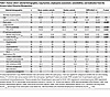
Table 1

Human cohort: selected demographics, lung function, emphysema assessment, comorbidities, and medication from the Arizona Cohort Polverino Biorepository

Sphingolipid metabolism is upregulated in airways of women with COPD. To assess small-airway sphingolipid transcriptomic signatures in women versus men with COPD, we performed GeoMx spatial transcriptomic analyses of the FFPE lung tissue sections. The airway regions of female COPD lungs exhibited a distinct sphingolipid signature compared with those of males, with significant upregulation of genes involved in sphingolipid metabolism. In particular, ASAH1 was significantly enriched in the airways of women (P = 0.004, Figure 1A). ASAH1 protects lung structural integrity by converting proapoptotic ceramide into prosurvival sphingosine/S1P and has been associated with COPD (34–36). This upregulation of ASAH1 in female COPD airways was also confirmed at the individual-participant level (Wilcoxon test, P = 0.00033; Figure 1B). Functionally, ASAH1 catalyzes the hydrolysis of proapoptotic ceramide to sphingosine, which is the immediate precursor of prosurvival S1P; however, the accumulation of S1P may drive increased tissue remodeling and inflammation (31).
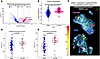
Sexual dimorphism in ASAH1 expression.Figure 1

Sexual dimorphism in ASAH1 expression. (A) A volcano plot derived from GeoMx spatial bulk–depth transcriptomics in airways from lungs of men and women with COPD. Significantly differentially expressed genes associated with sphingolipid metabolism are highlighted in blue or pink based on sex-specific enrichment. ASAH1 was significantly enriched in female COPD airways (P = 0.004). (B) ASAH1 expression stratified by sex using the Wilcoxon test. (C) Representative images from GeoMx lung slide scan and ROI sampling, showing ROI-specific expression of the ASAH1 gene. The top image is from a female participant with COPD, the lower image from a male with COPD. For GeoMx high-definition scanning, 3 morphological markers were used: DNA (blue), pan-CK (green), and CD45 (red). (D and E) Ceramide biosynthetic process and sphingolipid translocation GSVA scores, calculated using Gene Ontology Biological Process gene sets in airway regions across whole tissue sections. P values were determined using a nonparametric Wilcoxon test.

Figure 1C shows a representative overlay of immunofluorescence microscopic imaging and region-of-interest–specific (ROI-specific) expression of ASAH1 between men and women with COPD. Gene set enrichment analysis (GSEA) using the Hallmark pathway gene sets revealed significant positive enrichment in the airways of female COPD lungs for epithelial-mesenchymal transition (P = 1.3 × 10⁻⁴), inflammatory and immune activation signaling (IFN-α and IFN-γ responses, TNF-α signaling via NF-κB), and metabolic reprogramming pathways (glycolysis, oxidative phosphorylation, and mTORC1 signaling) (Supplemental Figure 2). Gene set variation analysis (GSVA) performed on the same transcriptomic dataset revealed significant upregulation of the ceramide biosynthetic process (Figure 1D) and sphingolipid translocation pathway (Figure 1E) in women represented by gene sets comprising 70 and 5 annotated genes, respectively (see Supplemental Table 3). This finding aligns with the increased expression of ASAH1.

Altogether, these transcriptomic data suggest increased ceramide synthesis and downstream metabolism to sphingosine via ASAH1 in women, consistent with a pattern of accelerated ceramide turnover. The concomitant upregulation of sphingolipid translocation pathway (reflecting inter-organelle trafficking) further supports the hypothesis of increased metabolic flux across cellular compartments, indicative of an overall activation of the sphingolipid biosynthetic pathway in women (Supplemental Figure 1). However, because direct metabolic flux was not measured, the functional consequences of these transcriptional changes require confirmation using isotope-tracing lipidomics.

ASAH1 and ORMDL3 are increased in small airways of women with COPD. Based on existing literature (20, 27–31, 34, 37–39) and the spatial transcriptomic results, we quantified the expression of 2 key enzymes of sphingolipid metabolism, ASAH1 and ORMDL3, in small airways from patients with COPD and controls. ORMDL3 is an estrogen-responsive ER protein that negatively regulates serine palmitoyltransferase (SPT) (29, 30), the rate-limiting enzyme of de novo ceramide synthesis, thereby modulating sphingolipid homeostasis in a sex-dependent manner (20). Furthermore, ORMDL3 promotes airway inflammation and AHR (40). Both ASAH1 and ORMDL3 enzymes have also been linked to sexual dimorphism in lung and airway-associated disease (14, 15, 40). Compared with controls and male COPD lungs, both ASAH1 and ORMDL3 were more abundant in female small airways, especially in ASMCs (Figure 2, A–D). Overall, these findings suggest an upregulation of sphingolipid metabolic enzymes in COPD ASMCs, with a more pronounced increase in women than in men.
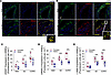
ASAH1 and ORMDL3 abundance in airway smooth muscle.Figure 2

ASAH1 and ORMDL3 abundance in airway smooth muscle. (A and B) Representative images of COPD lung sections co-immunostained for ASAH1 (red, A) or ORMDL3 (red, B), and α-SMA (green) and stained for nuclei with DAPI (blue). (C–E) Staining abundance was compared between male and female participants from the following groups: never-smoking controls (NSCs), smoking control (SCs), and COPD. Scale bars: 50 μm; 14 μm (inset). *P < 0.05; **P < 0.01; ***P < 0.001 by 1-way ANOVA followed by pair-wise testing (indicated by horizontal bars) with the Mann-Whitney U test.

Airway remodeling is increased in female COPD ASMCs and correlates with sphingolipid enzyme levels. Since an increase in sphingolipid metabolism induces ASMC proliferation and contraction (41), using immunostaining, we quantified α-smooth muscle actin (α-SMA) levels, a marker of ASMC proliferation (42). Both SC and COPD lungs showed higher α-SMA expression than NSCs; furthermore, within the SC and COPD groups, women had higher α-SMA expression compared with men (Figure 2E). A direct correlation was observed between the abundance of α-SMA and ORMDL3 (ρ = 0.692, P = 0.011; Figure 3A), with a similar trend observed between α-SMA and ASAH1 (ρ = 0.555, P = 0.070; Figure 3B). These results suggest the presence of active ASMC remodeling that is predominant in female patients and associated with sphingolipid enzyme activity. Importantly, α-SMA reflects smooth muscle proliferation and contractile activation, which primarily contributes to AHR and dynamic airflow limitation.
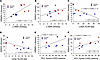
Associations of airway ORMDL3 and ASAH1 abundance with small-airway remodelFigure 3

Associations of airway ORMDL3 and ASAH1 abundance with small-airway remodeling, emphysema, and lung function in COPD. (A and B) Spearman correlation between ORMDL3 and α-SMA, and between ASAH1 and α-SMA, respectively. (C and D) Spearman correlation between ORMDL3 and lobar %LAA-950, and ASAH1 and lobar %LAA-950, respectively. (E and F) Spearman correlation between ORMDL3 and FEV1%pred, and ASAH1 and FEV1%pred.

ASAH1 and ORMDL3 expression is associated with emphysema and lung function. We next assessed the correlations between ASAH1 and ORMDL3 and COPD phenotypes, emphysema, and airflow obstruction, known to be differentially affected by sex. Lobar %LAA-950, a measure of emphysema on CT imaging, was strongly correlated with ORMDL3 (ρ = –0.804, P = 0.0003), in the female (ρ = –0.943, P = 0.016; Figure 3C), but not in the male group (ρ = –0.371, P = 0.497; Figure 3C). A similar significant correlation in women was noted between whole-lung %LAA-950, as well as regional emphysema burden and ORMDL3 (Supplemental Figure 3, A and B). ASAH1 did not correlate with lobar %LAA-950 (Figure 3D), although it correlated with whole-lung %LAA-950 across all participants (Supplemental Figure 3, A and B). FEV1%pred, a measure of airflow obstruction assessed by pulmonary function testing, was significantly associated with ORMDL3 in the entire cohort, with significance driven by the female subgroup (ρ = 0.650, P = 0.020; Figure 3E). FEV1%pred was also strongly correlated with ASAH1 in both sexes (ρ = 0.818, P = 0.0004; Figure 3F). Thus, the positive correlations between ORMDL3 and ASAH1 expression and FEV1%pred (Figure 3, E and F) suggest a predominantly airway-driven phenotype with relatively preserved lung parenchyma in individuals with higher sphingolipid enzyme expression. Overall, these data support a sex-dimorphic (with female predominance) association of airway remodeling and function with ASAH1 and ORMDL3.

Sexual dimorphism of circulating lipid metabolites in individuals with COPD. As both ASAH1 and ORMDL3 regulate the ceramide-S1P rheostat controlling airway inflammation, remodeling, and epithelial survival (43, 44), we quantified S1P and total ceramide in plasma from NSCs, SCs, and patients with COPD from the same human cohort used for lung studies shown above. Our goal was to identify a circulating lipid signature associated with the sexual dimorphism observed in the lung (see Figures 1 and 2).

Plasma S1P levels were significantly higher in women compared with men with COPD, while no sex differences were observed among control individuals (Figure 4A). There was a significant inverse correlation between plasma S1P levels and FEV1%pred in the entire cohort; when stratified by sex, this correlation reached statistical significance only in men (Figure 4B), suggesting that, in men, higher S1P levels are associated with worse lung function. Plasma total ceramide concentrations were significantly higher in men with COPD and SCs compared with women from the respective group (Figure 4C). There was a significant negative correlation between plasma ceramide levels and FEV1%pred in the entire cohort (Figure 4D). In women, ceramide levels tended to be negatively associated with FEV1%pred (P = 0.053), suggesting that, in women, higher ceramide levels are associated with worse lung function. These findings highlight a sexual dimorphism in the impact of sphingolipids on lung function in COPD, with higher S1P levels detrimental in men and higher ceramide levels detrimental in women.
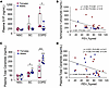
Circulating S1P and ceramide in women and men with COPD.Figure 4

Circulating S1P and ceramide in women and men with COPD. (A) Box-and-whisker plots of sphingosine-1-phosphate (S1P) concentrations in plasma from never-smoker controls (NSCs), ever-smoker controls (SCs), and participants with COPD. (B) Spearman (ρ) correlation between S1P and FEV1%pred, highlighting differences between sexes. (C) Box-and-whisker plots of total ceramide concentrations in plasma from NSCs, SCs, and participants with COPD. (D) Spearman’s ρ correlation between total ceramide and FEV1%pred, highlighting differences between sexes. One-way ANOVA (all P < 0.001) followed by pair-wise testing (indicated by horizontal bars) with Mann-Whitney U test between indicated groups: *P < 0.05; **P < 0.01; ***P < 0.001.

To validate these findings in plasma, we interrogated the sphingolipid data in samples from the Lung Tissue Research Consortium (LTRC) cohort (45). Plasma samples from 69 COPD GOLD 1–4 individuals were selected. Demographic information, smoking history, lung function parameters, and comorbidities are found in Supplemental Table 1. As expected, COPD GOLD 3–4 patients had significantly lower lung function and a greater extent of emphysema than GOLD 1–2 patients, while there were no differences in age, sex, or race among the 4 groups. We analyzed S1P/sphingosine and ceramide C16/C24 ratios because they have been associated with airway obstruction and clinical outcomes in COPD (46). Furthermore, using ratios helps normalize interindividual variability and more clearly capture shifts in sphingolipid balance (47). First, we evaluated the S1P/sphingosine and ceramide C16/C24 ratios across GOLD stages and observed that women tended to exhibit higher S1P/sphingosine ratios, while men showed higher ceramide C16/C24 ratios (Supplemental Figure 4, A–D). We then applied a 2-way ANOVA to assess the relative contribution of sex and disease severity to these differences. For both ratios, sex emerged as the only variable significantly associated with the observed variation (S1P/sphingosine: % variation = 16.33, P = 0.0187; C16/C24: % variation = 15.22, P = 0.0229), whereas GOLD stage did not significantly contribute. When examining individual sphingolipid species by sex, S1P levels were significantly higher in women at GOLD stage 2 (Supplemental Figure 4B); in contrast, sphingosine levels showed a trend toward being higher in men with COPD at GOLD stage 1, although this did not reach statistical significance (Supplemental Figure 4C). C16-ceramide levels did not differ significantly between men and women across GOLD stages (Supplemental Figure 4E), while C24-ceramide levels were significantly higher in women only at GOLD stage 2 (Supplemental Figure 4F).

Overall, these data support the peripheral blood findings, indicating that sex is a key determinant of sphingolipid profiles in individuals with COPD.

Bronchial hyperreactivity to CS predominates in female murine bronchi. We next assessed the physiological impact of sex-specific sphingolipid accumulation on airways, using isolated bronchi tested in an organ bath (48). Bronchi harvested from male and female C57BL/6J WT mice were incubated with 2% CS extract (CSE) for 24 hours at 37°C (Figure 5A). The airway reactivity measurement showed that 2% CSE exposure induced bronchial hyperactivity to carbachol in female mice, without any significant effect in bronchi from male mice (Figure 5B). Bronchial contraction was assessed with and without SKI-II, a sphingosine kinase inhibitor. SKI-II significantly reduced bronchial contraction in females (Figure 5B) but had no effect on the bronchi from male mice. These results suggest that S1P accumulation may be responsible for female susceptibility to AHR. Three-way ANOVA revealed that sex was the only variable with a significant effect (P = 0.0143). There were strong 2-way interactions between sex × CSE (P < 0.0001) and sex × SKI-II (P < 0.0001).
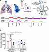
Sex-specific mouse bronchial constriction response to cigarette smoke exposFigure 5

Sex-specific mouse bronchial constriction response to cigarette smoke exposure. (A) Schematic representation of the functional study with representative contraction tracings (dyne/mg of bronchial tissue) following stimulation with carbachol at 1 × 10⁻⁶ M and 3 × 10⁻⁶ M (“Ca 10-6” and “Ca 3 × 10⁻⁶”). The bronchial rings were exposed to vehicle (V), 2% cigarette smoke extract (CSE), and 100 μM SKI-II, a sphingosine kinase inhibitor. (B) Bar graph with overlay scatter plot of contraction data (mean ± SEM; n = 3–4 per group). Three-way ANOVA was performed to assess sex, CSE, and inhibitor’s effect on bronchial contraction, showing a significant 3-way interaction (P = 0.0069). Pairwise comparison was performed using Tukey’s method (α = 0.05). *P < 0.05; **P < 0.01; ***P < 0.001.

Estradiol enhances CSE-induced ORMDL3 and ASAH1 expression in HBSMCs. To investigate whether sexual dimorphism in AHR is linked to sexual hormones, we quantified ASAH1 and ORMDL3 expression in primary human bronchial smooth muscle cells (HBSMCs) from male and female donors following CSE exposure, with and without estradiol supplementation.

HBSMCs harvested from a male donor were exposed to 0.25 μM estradiol (to simulate a female hormonal microenvironment) or vehicle every other day for 7 days; on the last day, vehicle- and estradiol-treated cells were exposed to 2% CSE for 24 hours. Quantification of immunofluorescence demonstrated that, compared with the vehicle, estradiol increased ORMDL3 and ASAH1 protein levels and that exposure to 2% CSE induced a significantly greater increase of both proteins only in cells exposed to estradiol (Figure 6, A–D). No significant difference in protein expression was detected between vehicle- and CSE-exposed male-derived cells in the absence of estradiol (Figure 6, A–D). Conversely, HBSMCs from a female donor exposed to 2% CSE for 24 hours showed a significant increase in ORMDL3 expression (Figure 6, C and D), indicating an intrinsic, sex-associated difference in baseline responsiveness to CS exposure, although this effect remained weaker than that observed in estradiol-treated cells, suggesting that hormonal status is the primary driver of ORMDL3 and ASAH1 induction in this model.
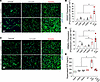
Effect of cigarette smoke and estradiol on ASAH1 and ORMDL3 expression in hFigure 6

Effect of cigarette smoke and estradiol on ASAH1 and ORMDL3 expression in human bronchial smooth muscle cells (HBSMCs). (A and B) ASAH1 and (C and D) ORMDL3 expression assessed by immunofluorescence in HBSMCs from a female donor (vehicle [“VehF”] or 2% CSE [“CSEF”]) and a male donor (vehicle [“VehM”, 2% CSE [“CSEM”], 0.25 μM estradiol [E2], or E2 + 2% CSE). Representative images and corresponding quantification are shown. Scale bars: 100 μm. Data are expressed as mean ± SEM with scatter plot overlaid. One-way ANOVA followed by 2-tailed Student’s t test. *P < 0.05; **P < 0.01 versus corresponding vehicle or the group indicated (n = 3/group). (E) Box-and-whisker plots of S1P levels in supernatants of male-derived HBSMCs exposed to vehicle (“VehM”), E2, 2% CSE (“CSEM”), 10 μM OEA (“OEAM”), or combinations. One-way ANOVA followed by pair-wise testing with the Mann-Whitney U test. *P < 0.05 versus corresponding vehicle or the group indicated (n = 3 per group).

Measurements of S1P levels in cell supernatants by ELISA showed that estradiol supplementation increased S1P production, and exposure to 2% CSE further elevated S1P levels, but only in estradiol-treated cells (Figure 6E). Under 2% CSE conditions, pretreatment with 10 μM N-oleoylethanolamine (OEA, an ASAH1 inhibitor) restored S1P levels to baseline in estradiol-treated cells (Figure 6E). Taken together, these data indicate that CS enhances ceramide to S1P conversion in the presence of estradiol, consistent with increased sphingolipid-mediated signaling.

Effect of estradiol and CS on the sphingolipid regulation of ASMC function. To evaluate sphingolipid-associated regulation of smooth muscle functionality, we assessed smooth muscle myosin heavy chain (SMMHC) expression by immunofluorescence microscopy in male-derived HBSMCs treated with estradiol and OEA. The expression of SMMHC, a marker of the contractile phenotype in ASMCs, is increased in their differentiated, force-generating state and is reduced during their synthetic, proliferative state (49). Estradiol supplementation increased SMMHC expression (Figure 7, A and B) and ASMC proliferation (Figure 7C). Incubation for 24 hours with 2% CSE further increased SMMHC levels and cell proliferation in estradiol-treated cells (Figure 7C). Preincubation with the ASAH1 inhibitor OEA (10 μM) attenuated the effects of estradiol and 2% CSE on SMMHC, restoring SMMHC expression and cellular proliferation to levels observed in vehicle-treated cells (Figure 7, A and B). Spearman’s analysis revealed significant correlations between SMMHC and ASAH1 (Figure 7D) and ORMDL3 (Figure 7E) levels, further supporting the notion that AHR is controlled, at least in part, by sphingolipids.
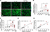
ASAH1 and ORMDL3 correlations with CSE-induced SMMHC expression in HBSMCs.Figure 7

ASAH1 and ORMDL3 correlations with CSE-induced SMMHC expression in HBSMCs. (A) Representative immunofluorescence images of SMMHC and (B) quantification of abundance of SMMHC in male-derived HBSMCs exposed to estradiol (E2), 2% CSE, and/or 10 μM N-oleoylethanolamine (OEA). Scale bar: 100 μm. Data are expressed as mean ± SEM with scatter plot overlaid. One-way ANOVA followed by 2-tailed Student’s t test. *P < 0.05 versus corresponding vehicle or the group indicated (n = 3–4 per group). (C) Cellular proliferation, expressed as the number of positive nuclei per high-power field at 24 hours after exposure to vehicle, E2, 2% CSE, and/or 10 μM OEA. One-way ANOVA followed by pair-wise testing with the Mann-Whitney U test. *P < 0.05; ***P < 0.001 versus corresponding vehicle or the group indicated (n = 7–12 per group). (D and E) Spearman correlations between SMMHC expression and ASAH1 (D) or ORMDL3 (E) expression in HBSMCs. The red dots represent E2 + 2% CSE treatment.

We complemented these studies with collagen contraction and scratch assays in male-derived HBSMCs treated with estradiol and OEA. Contraction in response to carbachol was measured at the 3-, 6-, 24-, and 48-hour time points (Figure 8A). Estradiol enhanced contraction in response to carbachol (Figure 8B), whereas CSE exposure reduced contraction (Figure 8A). This finding is consistent with previous reports demonstrating that CSE impairs the contractile capacity of normal HBSMCs through mechanisms involving, at least in part, reactive oxygen species (ROS) (50). Pretreatment with the ASAH1 inhibitor OEA (10 μM) reduced the contraction induced by estradiol but did not affect the hypocontractility induced by CSE (Figure 8, A–C). The speed of cellular migration was assessed at 6, 24, and 48 hours in scratch assays (Figure 8D). Overall, 2% CSE significantly reduced cell migration speed (Figure 8E). Estradiol abrogated the effects of 2% CSE, while OEA abrogated the estradiol effect on migration speed (Figure 8F). Overall, these results indicate that estradiol signaling increases contractility and cell migration in part via ASAH1.
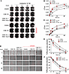
Effects of cigarette smoke and estradiol on contractility and scratch-woundFigure 8

Effects of cigarette smoke and estradiol on contractility and scratch-wound closure in male-donor HBSMCs. (A) Representative sequential images of cell plates with type I collagen mixed with human male–derived HBSMC “pellets” (2 × 10⁵) exposed to carbachol. (B and C) Tracings of kinetics of differential change in measurement of the cell pellets in cm (DTcm) following incubation with vehicle (carbachol only), 2% CSE, 0.25 μM estradiol (E), and/or 10 μM OEA. (D) Representative images with tracings of edges of male-derived HBSMCs (2 × 10⁵ cells) after generation of a linear scratch wound. (E and F) Kinetics of change in the wound area or scratch gap following incubation with vehicle (media only), 2% CSE, 0.25 μM estradiol (E), and/or 10 μM OEA. Panels B and C and panels E and F, respectively, display selected treatment groups from the same contraction and scratch experiments. The 2% CSE and estradiol + 2% CSE groups are included in both panels to facilitate comparisons with different treatment groups. Thus, these groups are intentionally duplicated across panels and do not represent independent experiments. ANOVA of repeated measures (P < 0.001) followed by pairwise multiple comparisons using Bonferroni’s t test. *P < 0.05; ***P < 0.001 versus indicated group (n = 3–6 per group per time point).
Discussion

In this study, we demonstrate that female COPD airways display a distinct sphingolipid transcriptional profile marked by upregulation of ceramide and sphingolipid metabolic pathways, along with enhanced sphingolipid biosynthesis. These alterations are associated with increased airway smooth muscle remodeling and reactivity. Consistent with this, women with COPD exhibit higher airway expression of ASAH1 and ORMDL3, as well as elevated circulating S1P levels, whereas men with COPD show higher circulating ceramide levels. Notably, ASAH1, ORMDL3, and ceramides are differentially associated with SAR, lung function, and emphysema severity. ASAH1 activity contributes to estradiol-associated changes in smooth muscle cell contractility and migration, potentially through increased downstream S1P signaling. These results provide insights into sex-specific differences in COPD and align with the clinical observation that women experience more pronounced airway remodeling (51) and rapid-onset loss of lung function (52), while men exhibit more severe emphysema (5).

We recently reported that ORMDL3 contributes to sex-associated differences in asthma and in atopic female mice (20), serving as an estrogen-responsive regulator of AHR (20). Our current findings in COPD are consistent with these previous results.

Among the sphingolipid enzymes (Supplemental Figure 1), we focused on ASAH1 and ORMDL3 due to their established relevance in chronic lung disease. ASAH1 encodes acid ceramidase, which hydrolyzes ceramides to sphingosine and has been genomically associated with COPD susceptibility (34). In contrast, ORMDL3 inhibits the de novo synthesis of ceramides (53) and has emerged as an important genetic determinant of asthma susceptibility and severity (40) (Supplemental Figure 1). Our data do not establish a direct causal link between ORMDL3 expression and S1P production. We interpret increased ORMDL3 expression as a potential compensatory response to CS-induced alterations in sphingolipid metabolism, particularly ceramide accumulation, which may suppress de novo sphingolipid synthesis. Ceramide availability may nevertheless be maintained through pathways operating independently of ORMDL3/SPT regulation. In parallel, increased ASAH1 expression may favor the conversion of ceramide to sphingosine, thereby increasing substrate availability for S1P production. ORMDL3 and ASAH1 may therefore participate in distinct but interconnected aspects of sphingolipid regulation.

S1P and its precursor, ceramide, function as opposing arms of a metabolic rheostat that governs structural cell survival within the lung parenchyma. The relative abundance of these sphingolipids influences airway disease pathobiology; a shift toward ceramide accumulation promotes apoptosis and contributes to emphysema development (35, 36), whereas S1P accumulation may enhance AHR and inflammation. This sphingolipid balance may therefore represent a key determinant of divergent disease trajectories across chronic obstructive and allergic respiratory disorders.

In our results, men with COPD have higher circulating levels of ceramides, which are associated with emphysema severity. Men generally develop more extensive emphysema than women (54) and show greater ceramide production in skeletal muscle, a feature linked to metabolic disorders such as type 2 diabetes (55). Elevated ceramide levels, particularly C16:0-ceramide, are associated with major adverse cardiac events (56) and emphysema severity (38, 57–59).

These data have direct relevance to airway remodeling and lung function. In HBSMCs, S1P upregulated α‑SMA and SMMHC, and estradiol enhanced contractility, supporting an estrogen‑linked pathway that may promote AHR. Notably, the estrogen-induced increase in S1P was not abolished by CSE exposure. Consistent with this, CSE exposure induced a trend toward greater increases in sphingolipid enzyme expression in female-derived than in male-derived HBSMCs. However, this effect remained weaker than that observed in estradiol-treated male-derived cells, suggesting that hormonal status is the primary driver of ORMDL3 and ASAH1 induction.

Although elevated S1P may counteract CS-induced ceramide accumulation (34–36) and reduce apoptosis, this potential benefit may be counterbalanced by increased airway inflammation and hyperreactivity (20, 40, 60, 61). Our murine data further support these sex-dependent differences. Female bronchi exposed to CSE exhibit S1P-dependent hyperreactivity, which was abrogated by sphingosine kinase inhibitors. These findings suggest that ceramide-driven pathology may preferentially contribute to parenchymal injury rather than airway remodeling in males, as evidenced by the lack of significant bronchial hyperreactivity in male mice.

This study has several limitations, including a small sample size, targeted rather than comprehensive lipidomic analyses, and cross-sectional measurements. Furthermore, common genetic variants at the 17q21 locus, known to regulate ORMDL3 expression and influence sphingolipid metabolism (40), were not assessed in our cohort due to the absence of genomic data. Although CS exposure was associated with increased ORMDL3 and ASAH1 expression and S1P accumulation, the upstream molecular mechanisms linking CS to the regulation of these enzymes were not directly investigated. A more complete understanding of the interaction between CS exposure and ORMDL3 and ASAH1 upregulation leading to the consequent alterations in sphingolipid metabolism and S1P production, and the cell‑type specificity of this pathway, will require isotope‑labeled lipid tracing (62) coupled with spatial and single‑cell–resolved metabolomic approaches (63).

Although S1P is a well‑established lymphocyte chemoattractant that could contribute to sex‑specific airway disease manifestations (64), inflammation was not assessed in the present study. Future work will be needed to define how lymphocytic infiltration influences airway remodeling and disease phenotype in women with COPD. In addition, given the complex interplay between steroid hormones and CS (65, 66), further mechanistic studies are warranted to delineate how these factors modulate sphingolipid signaling in the airways.

Lastly, beyond the ceramide/S1P axis investigated here, additional sphingolipid species may contribute to COPD pathobiology in a sex-dependent manner. These include glucosylceramide (GlcCer), whose deficiency promotes endothelial apoptosis (67); ceramide-1-phosphate (C1P), a proinflammatory mediator regulating eicosanoid biosynthesis and macrophage migration (68); sphingomyelin, decreased by CS exposure and associated with increased airway resistance and remodeling (69); and dihydroceramide, with emerging roles in ER stress and autophagy (70). Whether these species are differentially regulated by sex in COPD remains to be determined through future comprehensive sex-stratified lipidomic studies.

In conclusion, the sex-specific alterations in sphingolipid metabolism identified here have direct implications for COPD pathobiology and treatment. In men, strategies aimed at modulating ASAH1 activity may help reduce ceramide accumulation, a pathway linked to emphysema progression and lung function decline. In women, targeting S1P signaling may offer a means to limit airway smooth muscle remodeling and hyperreactivity, key features of airway disease in COPD. These concepts require validation in larger, well-phenotyped COPD cohorts and in experimental models capable of dissecting hormonal regulation of sphingolipid pathways. Ultimately, it will be critical to determine whether sex-specific modulation of sphingolipid metabolism can be translated into effective, disease-modifying therapies for COPD.

Methods

Sex as a biological variable

Both female and male human participants and mice were included in this study, and sex-associated differences were evaluated in the human and murine cohorts. Primary HBSMC experiments were conducted predominantly with cells from 1 male donor treated with estradiol to model hormonal effects; cells from 1 female donor were used for selected validation experiments. Thus, sex was considered as a biological variable, although the single-donor cell studies were not designed to establish donor-sex effects independently of interindividual variability.

Human studies

Human lung cohort. Lung tissue sections and plasma samples from 36 NSCs, SCs, and COPD GOLD 1–4 patients, both male and female. Lung tissue sections were obtained from explanted lungs at transplantation or from wedge resections/lobectomies performed for clinical indications (71). Normal lung tissue sections were identified by a clinical expert from surgical tissue obtained from individuals without COPD or other underlying primary lung disease. Blood samples from these individuals were collected in EDTA, centrifuged to obtain plasma, and stored at –80°C until further use (see below). CT-defined whole-lung and lobar emphysema were defined by the percentage of low-attenuation areas below the –950 Hounsfield units threshold (%LAA-950), as previously described (71).

Human blood LTRC cohort. Blood samples were collected in EDTA tubes to obtain plasma, and deidentified clinical data were provided by the LTRC. Plasma was stored at –80°C until processing and analysis of sphingolipid levels using liquid chromatography/mass spectrometry, as previously described (58). The results from 69 participants in this LTRC cohort were previously published (58) and were reanalyzed to determine sex differences in plasma ratios for S1P/sphingosine and ceramides C16 and C24 in COPD GOLD 1–4 participants.

GeoMX digital spatial profiling

FFPE lung sections from emphysematous men and women were used for digital spatial profiling of the Whole Transcriptomics Atlas using GeoMX technology, as previously published (71–73). Lung tissue samples were treated according to the manufacturer’s instructions. Airway ROIs were sampled for transcriptional analysis. RNA libraries were constructed and submitted for sequencing on an Illumina NextSeq 500. The resulting .fastq files were translated into .dcc files to be paired (“stitched”) with corresponding spatial selection information and analyzed by the spatial transcriptomics scripts for the R package (SpatialDecon: https://bioconductor.org/packages/release/bioc/html/SpatialDecon.html; GeomxTools: https://www.bioconductor.org/packages/release/bioc/html/GeomxTools.html).

Cell culture and treatments

HBSMCs from 1 male and 1 female donor (Lonza, CC-2576) were grown according to Lonza’s instructions. Cells from the male donor were treated with estradiol (0.25 μM; 50-28-2, MilliporeSigma) or vehicle every other day for 1 week, whereas cells from the female donor received vehicle treatment only. On day 6, the cells were treated with 2% CSE or vehicle for 24 hours. An additional group of male HBSMCs was pretreated with OEA (10 μM; O0383, MilliporeSigma) (74), an ASAH1 inhibitor, before 2% CSE incubation. CSE was prepared as previously described (75).

Each cell culture experiment was independently repeated 3 times, with each repetition representing a biological replicate.

Contraction assay

Male HBSMCs treated for 1 week with estradiol or vehicle were mixed in rat tail collagen type I (3440-100-01, R&D Systems) by mixing 6 parts of cells at a density of 2 × 10⁵ cells per dish, 2.5 parts of 3 mg/mL rat tail collagen, and 0.046 parts of 1 M NaOH. A quantity of 0.5 mL of the cell-collagen mixture was added per well in a 24-well plate, incubated for 1 hour at 37°C. After collagen polymerization, 0.5 mL of culture medium was added to each collagen gel, with the experimental treatments (± 10 μM OEA, ± 2% CSE, ± 0.25 μM estradiol, and combinations). Carbachol (212385, MilliporeSigma) was used as a contractile agonist, at a concentration of 10 μM in each well. Rat tail collagen only served as a control. Images were taken using Bio-Rad ChemiDoc chemiluminescent detection system after 3, 6, 24, and 48 hours. The collagen gel diameter change was measured using ImageJ/Fiji software (76).

Scratch assay

HBSMCs treated for 1 week with estradiol (or vehicle) were seeded into a 12-well tissue culture plate at a density that, after 24 hours of growth, reached 70%–80% confluence. Once at confluence, the cell layer was scraped in a straight line using a 1 mm pipette tip. After scratching, the cell monolayer was gently washed to remove detached cells, then replenished with fresh medium with the experimental treatments (± 10 μM OEA, ± 2% CSE, ± 0.25 μM estradiol, and combinations). Images were acquired using a phase-contrast microscope at ×4 magnification after 6, 24, and 48 hours at the same point coordinates (77).

Conventional immunofluorescence

FFPE lung sections from NSCs (n = 11; 5 females, 6 males), SCs (n = 11; 6 females, 5 males), and COPD GOLD 1–4 patients (n = 12; 6 females, 6 males) were deparaffinized and prepped for conventional immunofluorescence as previously described (71, 78, 79). Lung sections were immunostained for ASAH1, ORMDL3, and α-SMA; further details are in Supplemental Table 2. HBSMCs, after 1 week of estradiol/vehicle treatment, were plated at a density of 3 × 10⁴ cells on an 8-chamber polystyrene vessel tissue culture glass slide (Falcon, Corning) and treated with and without 10 μM OEA and with and without 2% CSE for 24 hours. Then, cells were fixed with cold glacial methanol 100% for 20 minutes. These glass slides were immunostained for ASAH1, ORMDL3, and SMMHC (smooth muscle myosin heavy chain, a marker of smooth muscle contraction); further details are in Supplemental Table 2. Pictures were acquired using an Olympus BX43 and a Vectra Polaris fluorescence microscope at ×20 magnification. Semiquantitative determination of protein expression in the bronchi was obtained using ImageJ/Fiji software by measuring fluorescence intensity.

Measurement of S1P and ceramide levels

S1P (MBS163661, MyBiosource) and ceramide (MBS3801246, MyBiosource) levels were quantified in human plasma from NSCs, SCs, and participants with COPD from the human lung cohort, as well as in HBSMC supernatants, using commercially available ELISA kits according the manufacturer’s instructions. Levels of sphingolipids were expressed as ng/mL.

Animal studies

All experiments were conducted during daylight hours in accordance with Italian regulations on the protection of animals used for experiments and other scientific purposes. Animal studies are reported in compliance with the ARRIVE guidelines (80). All animals were housed in a controlled environment with a 12-hour light/dark cycle under specific pathogen–free conditions and received water ad libitum.

Ex vivo airway responsiveness assessment

Female and male C57BL/6J WT mice, aged 8–9 weeks, were purchased from Charles River Laboratories. After humane euthanasia, bronchial rings were harvested from WT mice. The rings were 1–2 mm in length. They were placed in media with 2% CSE or vehicle for 24 hours. Afterward, the rings were transferred into organ baths (3 mL) filled with Krebs solution at 37°C and oxygenated with a 95% O2/5% CO2 mixture. The rings were then mounted to isometric force transducers (type 7006, Ugo Basile) and connected to a Powerlab 800 (ADInstruments). Bronchial rings were initially stretched to a resting tension of 0.5 g and allowed to equilibrate for more than 30 minutes. In each experiment, the bronchial rings were challenged with carbachol (1 × 10⁻⁶ M and 3 × 10⁻⁶ M) (81). In another set of experiments, the main bronchi were incubated with non-selective sphingosine kinase inhibitor SKI-II (2097, Tocris/R&D Systems) at 100 μM for 1 hour. After incubation, the bronchi were stimulated with carbachol (1 × 10⁻⁶ M) as previously described. Data were expressed as contraction capacity (dyne/mg of tissue).

Statistics

Nontranscriptomic data were analyzed using GraphPad InStat (Graphpad Software Inc) and SigmaPlot version 16 (Graffiti LLC). Statistical differences were assessed using 2-tailed Student’s t tests, Mann-Whitney U tests, or Wilcoxon tests, as appropriate. One-way, 2-way, or 3-way ANOVA was performed as applicable. Repeated-measures ANOVA was used to assess changes in contraction and scratch assays. Categorical variables were assessed using a z test for proportions. Correlations were evaluated using Spearman’s rank correlation coefficient (ρ). Statistical significance was defined as P < 0.05. Transcriptomic data were analyzed using R script packages for GeoMx (GeoMxTools, NanoStringNCTools, GeoMxWorkflows, lme4, and fgesea). Data were normalized and batch corrected. We chose a P value of less than 0.05 and log2(fold change) (log2FC) of greater than 0.5 for differential gene expression analyses. In box-and-whisker plots, the boxes represent the interquartile range (25th to 75th percentiles), the line within each box indicates the median, the whiskers extend to the minimum and maximum values.

Study approval

All human studies were approved by the Institutional Review Board of the University of Arizona (IRB 1811124026) or by the University of Illinois Chicago and Indiana University. All animal procedures were approved by the Italian regulations on the protection of animals used for experiments and other scientific purposes (D.Lgs. 26/2014) and European Union regulations (EU Directive 2010/63/EU).

Data availability

All data associated with this study are present in the paper or in the supplemental material. Values for all data points in graphs are reported in the Supporting Data Values file. Materials generated in this study are commercially available or will be made available upon request from the corresponding author.

Manuscript information

Author contributions. EG, FR, and FP conceived the project and designed the experiments. FP and IP designed experiments and contributed to data analyses and interpretation. EG, XW, JR, IC, YZ, JTK, SAO, JM, LC, MS, FK, and IP conducted experiments and/or contributed to data analysis and interpretation. EG, XW, JR, IP, and FP contributed to the writing and editing of the manuscript.

AI disclosure. The authors declare that no AI tools were used in the original research for or writing of this article.

Conflict of interest. FP has served as a consultant for Amgen, AstraZeneca, Chiesi Pharmaceuticals, Apogee Therapeutics, GlaxoSmithKline, Genentech, Aerogen, Pfizer, Windward Bio, and Sanofi-Regeneron. IP is a scientific co-founder of and served as a consultant for Allinaire Therapeutics.

Funding. This study was supported by the following:

  • Ministero dell’Università e della Ricerca grant PRIN 2022MMSBKL (to FR).

NIH grants R01 HL171622-01A1/04 (to FP), R01 HL077328 (to IP), and R03 HL095440 (to IP).

Supplemental material

View Supplemental data

Acknowledgments

None.

Address for correspondence. Francesca Polverino, Department of Medicine, Baylor College of Medicine, 6501 Fannin Street, Houston, Texas 77030, USA. Email: fpolverino@copdnet.org.

Footnotes

Copyright: © 2026, Granato et al. This is an open access article published under the terms of the Creative Commons Attribution 4.0 International License.

Reference information: JCI Insight. 2026;11(21):e208692.https://doi.org/10.1172/jci.insight.208692.

References
  1. WHO. Chronic obstructive pulmonary disease (COPD). https://www.who.int/news-room/fact-sheets/detail/chronic-obstructive-pulmonary-disease-(copd) Accessed September 21, 2026.
  2. Akinbami LJ, Liu X. Chronic obstructive pulmonary disease among adults aged 18 and over in the United States, 1998-2009. NCHS Data Brief. 2011;(63):1–8.
    View this article via: PubMed Google Scholar
  3. Diaz AA, et al. Prevalence and prognostic significance of COPD in adults younger than 50 years of age. NEJM Evid. 2025;4(8):EVIDoa2400424.
    View this article via: CrossRef PubMed Google Scholar
  4. Silverman EK, et al. Gender-related differences in severe, early-onset chronic obstructive pulmonary disease. Am J Respir Crit Care Med. 2000;162(6):2152–2158.
    View this article via: CrossRef PubMed Google Scholar
  5. Martinez FJ, et al. Sex differences in severe pulmonary emphysema. Am J Respir Crit Care Med. 2007;176(3):243–252.
    View this article via: CrossRef PubMed Google Scholar
  6. Singh D, et al. Small airway disease in chronic obstructive pulmonary disease: insights and implications for the clinician. Curr Opin Pulm Med. 2020;26(2):162–168.
    View this article via: CrossRef PubMed Google Scholar
  7. Hogg JC, et al. The contribution of small airway obstruction to the pathogenesis of chronic obstructive pulmonary disease. Physiol Rev. 2017;97(2):529–552.
    View this article via: CrossRef PubMed Google Scholar
  8. Maestrelli P, et al. Remodeling in response to infection and injury. Airway inflammation and hypersecretion of mucus in smoking subjects with chronic obstructive pulmonary disease. Am J Respir Crit Care Med. 2001;164(10 pt 2):S76–S80.
    View this article via: CrossRef PubMed Google Scholar
  9. Sutherland ER, Martin RJ. Airway inflammation in chronic obstructive pulmonary disease: comparisons with asthma. J Allergy Clin Immunol. 2003;112(5):819–827.
    View this article via: CrossRef PubMed Google Scholar
  10. Pare PD, et al. Airway hyperreactivity in COPD. Cause or effect, an in vivo in vitro comparison. Chest. 1987;91(5 suppl):40S–44S.
    View this article via: CrossRef PubMed Google Scholar
  11. Dey S, et al. Differential airway remodeling changes were observed in patients with asthma COPD overlap compared to patients with asthma and COPD alone. Am J Physiol Lung Cell Mol Physiol. 2022;323(4):L473–L483.
    View this article via: CrossRef PubMed Google Scholar
  12. Jia M, et al. Cigarette smoke particles-induced airway hyperreactivity in vivo and in vitro. Biol Pharm Bull. 2019;42(5):703–711.
    View this article via: CrossRef PubMed Google Scholar
  13. Rojas-Quintero J, Polverino F. Molecular maestro in the lung: the sphingolipid rheostat. Am J Respir Cell Mol Biol. 2025;73(2):170–172.
    View this article via: CrossRef PubMed Google Scholar
  14. Spiegel S, Milstien S. Sphingosine-1-phosphate: an enigmatic signalling lipid. Nat Rev Mol Cell Biol. 2003;4(5):397–407.
    View this article via: CrossRef PubMed Google Scholar
  15. Ghidoni R, et al. Role of sphingolipids in the pathobiology of lung inflammation. Mediators Inflamm. 2015;2015:487508.
    View this article via: CrossRef PubMed Google Scholar
  16. Kim SH, et al. Ceramide/sphingosine-1-phosphate imbalance is associated with distinct inflammatory phenotypes of uncontrolled asthma. Allergy. 2020;75(8):1991–2004.
    View this article via: CrossRef PubMed Google Scholar
  17. Park SJ, Im DS. Blockage of sphingosine-1-phosphate receptor 2 attenuates allergic asthma in mice. Br J Pharmacol. 2019;176(7):938–949.
    View this article via: CrossRef PubMed Google Scholar
  18. Price MM, et al. A specific sphingosine kinase 1 inhibitor attenuates airway hyperresponsiveness and inflammation in a mast cell-dependent murine model of allergic asthma. J Allergy Clin Immunol. 2013;131(2):501–511.
    View this article via: CrossRef PubMed Google Scholar
  19. Ammit AJ, et al. Sphingosine 1-phosphate modulates human airway smooth muscle cell functions that promote inflammation and airway remodeling in asthma. FASEB J. 2001;15(7):1212–1214.
    View this article via: CrossRef PubMed Google Scholar
  20. Granato E, et al. Sex hormones influence ORMDL3 expression: Implications for sex-associated asthma phenotype. J Allergy Clin Immunol. 2025;157(3):602–615.
    View this article via: CrossRef PubMed Google Scholar
  21. Worgall TS, et al. Impaired sphingolipid synthesis in the respiratory tract induces airway hyperreactivity. Sci Transl Med. 2013;5(186):186ra67.
    View this article via: CrossRef PubMed Google Scholar
  22. Ono JG, et al. Decreased sphingolipid synthesis in children with 17q21 asthma-risk genotypes. J Clin Invest. 2020;130(2):921–926.
    View this article via: JCI CrossRef PubMed Google Scholar
  23. Nava VE, et al. Sphingosine kinase type 1 promotes estrogen-dependent tumorigenesis of breast cancer MCF-7 cells. Exp Cell Res. 2002;281(1):115–127.
    View this article via: CrossRef PubMed Google Scholar
  24. Sukocheva O, et al. Estrogen transactivates EGFR via the sphingosine 1-phosphate receptor Edg-3: the role of sphingosine kinase-1. J Cell Biol. 2006;173(2):301–310.
    View this article via: CrossRef PubMed Google Scholar
  25. Montefusco D, et al. Sphingosine kinase 1 mediates sexual dimorphism in fibrosis in a mouse model of NASH. Mol Metab. 2022;62:101523.
    View this article via: CrossRef PubMed Google Scholar
  26. Guo S, et al. Higher level of plasma bioactive molecule sphingosine 1-phosphate in women is associated with estrogen. Biochim Biophys Acta. 2014;1841(6):836–846.
    View this article via: CrossRef PubMed Google Scholar
  27. Agarwal AR, et al. Short-term cigarette smoke exposure leads to metabolic alterations in lung alveolar cells. Am J Respir Cell Mol Biol. 2014;51(2):284–293.
    View this article via: CrossRef PubMed Google Scholar
  28. Gai X, et al. Serum glycerophospholipid profile in acute exacerbation of chronic obstructive pulmonary disease. Front Physiol. 2021;12:646010.
    View this article via: CrossRef PubMed Google Scholar
  29. Chen R, et al. Cigarette smoke induces endoplasmic reticulum stress-associated mucus hypersecretion via orosomucoid 1-like protein 3 in airway epithelia. Free Radic Res. 2025;59(4):342–355.
    View this article via: CrossRef PubMed Google Scholar
  30. Chen R, et al. ORMDL3 regulates cigarette smoke-induced endoplasmic reticulum stress in airway smooth muscle cells. J Allergy Clin Immunol. 2022;149(4):1445–1457.
    View this article via: CrossRef PubMed Google Scholar
  31. Bodas M, et al. Autophagy augmentation alleviates cigarette smoke-induced CFTR-dysfunction, ceramide-accumulation and COPD-emphysema pathogenesis. Free Radic Biol Med. 2019;131:81–97.
    View this article via: CrossRef PubMed Google Scholar
  32. Perez TA, et al. Sex differences between women and men with COPD: a new analysis of the 3CIA study. Respir Med. 2020;171:106105.
    View this article via: CrossRef PubMed Google Scholar
  33. Granato E, et al. A108 COPD pathogenesis: opening the pandora box. Am J Respir Crit Care Med. 2024;209:A2950.
  34. Bahr TM, et al. Peripheral blood mononuclear cell gene expression in chronic obstructive pulmonary disease. Am J Respir Cell Mol Biol. 2013;49(2):316–323.
    View this article via: CrossRef PubMed Google Scholar
  35. Goel K, et al. Pharmacological sphingosine-1 phosphate receptor 1 targeting in cigarette smoke-induced emphysema in mice. Am J Physiol Lung Cell Mol Physiol. 2022;322(6):L794–L803.
    View this article via: CrossRef PubMed Google Scholar
  36. Goel K, et al. Sphingosine 1 phosphate (S1P) receptor 1 is decreased in human lung microvascular endothelial cells of smokers and mediates S1P effect on autophagy. Cells. 2021;10(5):1200.
    View this article via: CrossRef PubMed Google Scholar
  37. Yu FPS, et al. Chronic lung injury and impaired pulmonary function in a mouse model of acid ceramidase deficiency. Am J Physiol Lung Cell Mol Physiol. 2018;314(3):L406–L420.
    View this article via: CrossRef PubMed Google Scholar
  38. Huang Q, et al. Extracellular vesicular delivery of ceramides from pulmonary macrophages to endothelial cells facilitates chronic obstructive pulmonary disease. Cell Commun Signal. 2025;23(1):124.
    View this article via: CrossRef PubMed Google Scholar
  39. Qi Q, et al. Decreased sphingosine due to down-regulation of acid ceramidase expression in airway of bronchiectasis patients: a potential contributor to Pseudomonas aeruginosa infection. Infect Drug Resist. 2023;16:2573–2588.
    View this article via: CrossRef PubMed Google Scholar
  40. Moffatt MF, et al. Genetic variants regulating ORMDL3 expression contribute to the risk of childhood asthma. Nature. 2007;448(7152):470–473.
    View this article via: CrossRef PubMed Google Scholar
  41. Liu L, et al. Sphingosine-1-phosphate induces airway smooth muscle cell proliferation, migration, and contraction by modulating Hippo signaling effector YAP. Am J Physiol Lung Cell Mol Physiol. 2018;315(4):L609–L621.
    View this article via: CrossRef PubMed Google Scholar
  42. Shinde AV, et al. The role of α-smooth muscle actin in fibroblast-mediated matrix contraction and remodeling. Biochim Biophys Acta Mol Basis Dis. 2017;1863(1):298–309.
    View this article via: CrossRef PubMed Google Scholar
  43. Rother N, et al. Acid ceramidase regulates innate immune memory. Cell Rep. 2023;42(12):113458.
    View this article via: CrossRef PubMed Google Scholar
  44. Debeuf N, et al. The ORMDL3 asthma susceptibility gene regulates systemic ceramide levels without altering key asthma features in mice. J Allergy Clin Immunol. 2019;144(6):1648–1659.
    View this article via: CrossRef PubMed Google Scholar
  45. Rojas-Quintero J, et al. Maternal smoking and CC-16: implications for lung development and COPD across the lifespan. Am J Respir Crit Care Med. 2025;212(3):591–604.
    View this article via: CrossRef PubMed Google Scholar
  46. Gharib AR, et al. Plasma sphingolipids, lung function and COPD: the cardiovascular health study. ERJ Open Res. 2023;9(2):–.:00346-2022.
    View this article via: CrossRef PubMed Google Scholar
  47. Mantovani A, et al. Ceramide-based risk score CERT-1 improves risk prediction for overall mortality and adverse cardiovascular outcomes in patients with and without cardiovascular disease: a prospective cohort study. Diabetes Obes Metab. 2025;27(3):1488–1497.
    View this article via: CrossRef PubMed Google Scholar
  48. Herlihy JT, et al. Assessment of airway hyperresponsiveness in murine tracheal rings. Methods Mol Biol. 2013;1032:257–269.
    View this article via: PubMed CrossRef Google Scholar
  49. Regan CP, et al. Molecular mechanisms of decreased smooth muscle differentiation marker expression after vascular injury. J Clin Invest. 2000;106(9):1139–1147.
    View this article via: JCI CrossRef PubMed Google Scholar
  50. Yoon CH, et al. Cigarette smoke extract-induced reduction in migration and contraction in normal human bronchial smooth muscle cells. Korean J Physiol Pharmacol. 2011;15(6):397–403.
    View this article via: CrossRef PubMed Google Scholar
  51. Polverino M, et al. Smoking pattern in men and women: a possible contributor to sex differences in smoke-related lung diseases. Am J Respir Crit Care Med. 2020;202(7):1048–1051.
    View this article via: CrossRef PubMed Google Scholar
  52. Sorheim IC, et al. Gender differences in COPD: are women more susceptible to smoking effects than men? Thorax. 2010;65(6):480–485.
    View this article via: CrossRef PubMed Google Scholar
  53. James B, et al. ORMDL3 and allergic asthma: from physiology to pathology. J Allergy Clin Immunol. 2019;144(3):634–640.
    View this article via: CrossRef PubMed Google Scholar
  54. Camp PG, et al. Sex differences in emphysema and airway disease in smokers. Chest. 2009;136(6):1480–1488.
    View this article via: CrossRef PubMed Google Scholar
  55. Straczkowski M, et al. Increased skeletal muscle ceramide level in men at risk of developing type 2 diabetes. Diabetologia. 2007;50(11):2366–2373.
    View this article via: CrossRef PubMed Google Scholar
  56. Meeusen JW, et al. Plasma ceramides. Arterioscler Thromb Vasc Biol. 2018;38(8):1933–1939.
    View this article via: CrossRef PubMed Google Scholar
  57. Petrache I, et al. Ceramide upregulation causes pulmonary cell apoptosis and emphysema-like disease in mice. Nat Med. 2005;11(5):491–498.
    View this article via: CrossRef PubMed Google Scholar
  58. Berdyshev EV, et al. Ceramide and sphingosine-1 phosphate in COPD lungs. [published online January 29, 2021]. Thorax. https://doi.org/10.1136/thoraxjnl-2020-215892.
    View this article via: PubMed Google Scholar
  59. Scarpa MC, et al. Ceramide expression and cell homeostasis in chronic obstructive pulmonary disease. Respiration. 2013;85(4):342–349.
    View this article via: CrossRef PubMed Google Scholar
  60. Rutherford C, et al. Regulation of cell survival by sphingosine-1-phosphate receptor S1P1 via reciprocal ERK-dependent suppression of Bim and PI-3-kinase/protein kinase C-mediated upregulation of Mcl-1. Cell Death Dis. 2013;4(11):e927.
    View this article via: CrossRef PubMed Google Scholar
  61. Marsolais D, et al. Modulation of chemokines and allergic airway inflammation by selective local sphingosine-1-phosphate receptor 1 agonism in lungs. Mol Pharmacol. 2011;79(1):61–68.
    View this article via: CrossRef PubMed Google Scholar
  62. Magkos F, Mittendorfer B. Stable isotope-labeled tracers for the investigation of fatty acid and triglyceride metabolism in humans in vivo. Clin Lipidol. 2009;4(2):215–230.
    View this article via: CrossRef PubMed Google Scholar
  63. Lane AN, et al. Challenges of spatially resolved metabolism in cancer research. Metabolites. 2024;14(7):383.
    View this article via: CrossRef PubMed Google Scholar
  64. Ryser MF, et al. S1P(1) overexpression stimulates S1P-dependent chemotaxis of human CD34+ hematopoietic progenitor cells but strongly inhibits SDF-1/CXCR4-dependent migration and in vivo homing. Mol Immunol. 2008;46(1):166–171.
    View this article via: CrossRef PubMed Google Scholar
  65. Peng J, et al. Estrogen metabolism within the lung and its modulation by tobacco smoke. Carcinogenesis. 2013;34(4):909–915.
    View this article via: CrossRef PubMed Google Scholar
  66. Gomez-Alonso MDC, et al. DNA methylation and lipid metabolism: an EWAS of 226 metabolic measures. Clin Epigenetics. 2021;13(1):7.
    View this article via: CrossRef PubMed Google Scholar
  67. Koike K, et al. Role of glucosylceramide in lung endothelial cell fate and emphysema. Am J Respir Crit Care Med. 2019;200(9):1113–1125.
    View this article via: CrossRef PubMed Google Scholar
  68. Presa N, et al. Regulation of cell migration and inflammation by ceramide 1-phosphate. Biochim Biophys Acta. 2016;1861(5):402–409.
    View this article via: CrossRef PubMed Google Scholar
  69. Gupta G, et al. Airway resistance caused by sphingomyelin synthase 2 insufficiency in response to cigarette smoke. Am J Respir Cell Mol Biol. 2020;62(3):342–353.
    View this article via: CrossRef PubMed Google Scholar
  70. Hernandez-Tiedra S, et al. Dihydroceramide accumulation mediates cytotoxic autophagy of cancer cells via autolysosome destabilization. Autophagy. 2016;12(11):2213–2229.
    View this article via: CrossRef PubMed Google Scholar
  71. Rojas-Quintero J, et al. Spatial transcriptomics resolve an emphysema-specific lymphoid follicle B cell signature in chronic obstructive pulmonary disease. Am J Respir Crit Care Med. 2024;209(1):48–58.
    View this article via: CrossRef PubMed Google Scholar
  72. Summers B, et al. Mice with lymphatic dysfunction develop pathogenic lung tertiary lymphoid organs that model an autoimmune emphysema phenotype of COPD. Am J Physiol Lung Cell Mol Physiol. 2025;328(1):L1–L14.
    View this article via: CrossRef PubMed Google Scholar
  73. Rojas-Quintero J, et al. Skewed adaptive immune responses are involved in alpha-1 antitrypsin deficiency emphysema. Eur Respir J. 2025;65(1):2400839.
    View this article via: CrossRef PubMed Google Scholar
  74. Riganti L, et al. Sphingosine-1-phosphate (S1P) impacts presynaptic functions by regulating synapsin I localization in the presynaptic compartment. J Neurosci. 2016;36(16):4624–4634.
    View this article via: CrossRef PubMed Google Scholar
  75. Igari K, et al. Cigarette smoke extract activates tartrate-resistant acid phosphatase-positive macrophage. J Vasc Res. 2019;56(3):139–151.
    View this article via: CrossRef PubMed Google Scholar
  76. Ngo P, et al. Collagen gel contraction assay. Methods Mol Biol. 2006;341:103–109.
    View this article via: PubMed CrossRef Google Scholar
  77. Martinotti S, Ranzato E. Scratch wound healing assay. Methods Mol Biol. 2020;2109:225–229.
    View this article via: PubMed CrossRef Google Scholar
  78. Polverino F, et al. A disintegrin and metalloproteinase domain-8: a novel protective proteinase in chronic obstructive pulmonary disease. Am J Respir Crit Care Med. 2018;198(10):1254–1267.
    View this article via: CrossRef PubMed Google Scholar
  79. Polverino F, et al. Metformin: experimental and clinical evidence for a potential role in emphysema treatment. Am J Respir Crit Care Med. 2021;204(6):651–666.
    View this article via: CrossRef PubMed Google Scholar
  80. Kilkenny C, et al. Improving bioscience research reporting: the ARRIVE guidelines for reporting animal research. PLoS Biol. 2010;8(6):e1000412.
    View this article via: CrossRef PubMed Google Scholar
  81. Cerqua I, et al. Enhanced efficacy of formoterol-montelukast salt in relieving asthma features and in preserving β2-agonists rescue therapy. Pharmacol Res. 2022;186:106536.
    View this article via: CrossRef PubMed Google Scholar
Version history
  • Version 1 (September 22, 2026): In-Press Preview
  • Version 2 (November 9, 2026): Electronic publication

Article tools

  • View PDF
  • Download citation information
  • Send a comment
  • Terms of use
  • Standard abbreviations
  • Need help? Email the journal

Metrics

  • Article usage
  • Citations to this article

Go to

  • Top
  • Abstract
  • Introduction
  • Results
  • Discussion
  • Methods
  • Manuscript information
  • Supplemental material
  • Acknowledgments
  • Footnotes
  • References
  • Version history
Advertisement
Advertisement

Copyright © 2026 American Society for Clinical Investigation
ISSN 2379-3708

Sign up for email alerts