
Go to The Journal of Clinical Investigation
  • About
  • Editors
  • Consulting Editors
  • For authors
  • Journal stats
  • Publication ethics
  • Publication alerts by email
  • Transfers
  • Advertising
  • Job board
  • Contact
  • Physician-Scientist Development
  • Current issue
  • Past issues
  • By specialty
    • COVID-19
    • Cardiology
    • Immunology
    • Metabolism
    • Nephrology
    • Oncology
    • Pulmonology
    • All ...
  • Videos
  • Collections
    • In-Press Preview
    • Resource and Technical Advances
    • Clinical Research and Public Health
    • Research Letters
    • Editorials
    • Perspectives
    • Physician-Scientist Development
    • Reviews
    • Top read articles

  • Current issue
  • Past issues
  • Specialties
  • In-Press Preview
  • Resource and Technical Advances
  • Clinical Research and Public Health
  • Research Letters
  • Editorials
  • Perspectives
  • Physician-Scientist Development
  • Reviews
  • Top read articles
  • About
  • Editors
  • Consulting Editors
  • For authors
  • Journal stats
  • Publication ethics
  • Publication alerts by email
  • Transfers
  • Advertising
  • Job board
  • Contact
Top
  • View PDF
  • Download citation information
  • Send a comment
  • Terms of use
  • Standard abbreviations
  • Need help? Email the journal
  • Top
  • Abstract
  • Introduction
  • A multidimensional framework for cardiac aging: the A×G×E×D model
  • Modeling cardiac aging for clinical impact: a NAM-based framework
  • Ability to simulate the passage of time: aging heart in a dish
  • Long-term monitoring platforms that capture dynamic age-related changes
  • Limitations, challenges, and future directions
  • Author contributions
  • Conflict of interest
  • Funding support
  • Acknowledgments
  • Footnotes
  • References
  • Version history
  • Article usage
  • Citations to this article
Advertisement

Review Series Open Access | 10.1172/jci.insight.203447

From aging biology to cardiac biotechnology: emerging platforms for modeling cardiac aging

Kritika Chaddha,1 Mabel Bartlett,2 Tzahi Cohen-Karni,2,3 and Aditi Gurkar1,4

1Aging Institute, Department of Medicine, University of Pittsburgh, Pittsburgh, Pennsylvania, USA.

2Biomedical Engineering and

3Materials Science and Engineering, Carnegie Mellon University, Pittsburgh, Pennsylvania, USA.

4Division of Geriatric Medicine, Department of Medicine, University of Pittsburgh, Pittsburgh, Pennsylvania, USA.

Address correspondence to: Aditi U. Gurkar, Aging Institute, Suite 563, 100 Technology Drive, University of Pittsburgh, Pittsburgh, PA 15219, USA. Phone: 412.624.7494; Email: agurkar1@pitt.edu. Or to: Tzahi Cohen-Karni, Biomedical Engineering and Materials Science & Engineering Carnegie Mellon University, Pittsburgh, Pennsylvania, USA. Email: tzahi@andrew.cmu.edu.

Authorship note: KC and MB contributed equally to this work.

Find articles by Chaddha, K. in: PubMed | Google Scholar

1Aging Institute, Department of Medicine, University of Pittsburgh, Pittsburgh, Pennsylvania, USA.

2Biomedical Engineering and

3Materials Science and Engineering, Carnegie Mellon University, Pittsburgh, Pennsylvania, USA.

4Division of Geriatric Medicine, Department of Medicine, University of Pittsburgh, Pittsburgh, Pennsylvania, USA.

Address correspondence to: Aditi U. Gurkar, Aging Institute, Suite 563, 100 Technology Drive, University of Pittsburgh, Pittsburgh, PA 15219, USA. Phone: 412.624.7494; Email: agurkar1@pitt.edu. Or to: Tzahi Cohen-Karni, Biomedical Engineering and Materials Science & Engineering Carnegie Mellon University, Pittsburgh, Pennsylvania, USA. Email: tzahi@andrew.cmu.edu.

Authorship note: KC and MB contributed equally to this work.

Find articles by Bartlett, M. in: PubMed | Google Scholar

1Aging Institute, Department of Medicine, University of Pittsburgh, Pittsburgh, Pennsylvania, USA.

2Biomedical Engineering and

3Materials Science and Engineering, Carnegie Mellon University, Pittsburgh, Pennsylvania, USA.

4Division of Geriatric Medicine, Department of Medicine, University of Pittsburgh, Pittsburgh, Pennsylvania, USA.

Address correspondence to: Aditi U. Gurkar, Aging Institute, Suite 563, 100 Technology Drive, University of Pittsburgh, Pittsburgh, PA 15219, USA. Phone: 412.624.7494; Email: agurkar1@pitt.edu. Or to: Tzahi Cohen-Karni, Biomedical Engineering and Materials Science & Engineering Carnegie Mellon University, Pittsburgh, Pennsylvania, USA. Email: tzahi@andrew.cmu.edu.

Authorship note: KC and MB contributed equally to this work.

Find articles by Cohen-Karni, T. in: PubMed | Google Scholar

1Aging Institute, Department of Medicine, University of Pittsburgh, Pittsburgh, Pennsylvania, USA.

2Biomedical Engineering and

3Materials Science and Engineering, Carnegie Mellon University, Pittsburgh, Pennsylvania, USA.

4Division of Geriatric Medicine, Department of Medicine, University of Pittsburgh, Pittsburgh, Pennsylvania, USA.

Address correspondence to: Aditi U. Gurkar, Aging Institute, Suite 563, 100 Technology Drive, University of Pittsburgh, Pittsburgh, PA 15219, USA. Phone: 412.624.7494; Email: agurkar1@pitt.edu. Or to: Tzahi Cohen-Karni, Biomedical Engineering and Materials Science & Engineering Carnegie Mellon University, Pittsburgh, Pennsylvania, USA. Email: tzahi@andrew.cmu.edu.

Authorship note: KC and MB contributed equally to this work.

Find articles by Gurkar, A. in: PubMed | Google Scholar |

Authorship note: KC and MB contributed equally to this work.

Published August 10, 2026 - More info

Published in Volume 11, Issue 15 on August 10, 2026
JCI Insight. 2026;11(15):e203447. https://doi.org/10.1172/jci.insight.203447.
© 2026 Chaddha et al. This work is licensed under the Creative Commons Attribution 4.0 International License. To view a copy of this license, visit http://creativecommons.org/licenses/by/4.0/.
Published August 10, 2026 - Version history
View PDF
Abstract

Aging is a major contributor to cardiovascular disease and mortality in older adults. Yet most preclinical and experimental cardiac studies fail to account for age as a primary biological variable, leaving a critical gap in our understanding of how aging contributes to disease progression. Bridging this gap requires integrating aging biology, cardiac pathophysiology, and cutting-edge biotechnology to uncover the mechanisms underlying age-related cardiac dysfunction. We offer a new approach methodologies (NAMs) perspective on how emerging bioengineering strategies may reshape the study of cardiac aging by enabling multidimensional monitoring of cardiac function, aging trajectories, and therapeutic responses. To capture this complexity, we propose the A×G×E×D framework, where A stands for age, G for genetics, E for environment, and D for drug exposure, as a multidimensional lens for understanding how these factors converge to determine cardiac vulnerability during aging. We highlight the integration of long-term cardiac microtissues with advancements in biotechnology to model age. This Perspective opens new frontiers for understanding how A×G×E×D interactions manifest at the molecular, cellular, and electrophysiological levels and for designing responsive, personalized interventions that align with each individual’s evolving physiology. By developing robust bioengineered platforms that recapitulate human cardiac aging, we can advance toward precision geromedicine for cardiovascular health.

Introduction

Cardiac aging represents a fundamental challenge in modern medicine, as age-related changes in cardiac structure and function underlie the majority of cardiovascular diseases in older adults (1). The aging heart exhibits progressive deterioration, including cardiomyocyte (CM) hypertrophy, increased fibrosis, impaired diastolic function, reduced contractile reserve, and electrical remodeling, all of which increase vulnerability to heart failure, arrhythmias, and ischemic disease (2, 3). Aging interacts with genetic predisposition (4), environmental exposures (5), and pharmacological interventions to determine cardiovascular outcomes, requiring models that recapitulate human cardiac aging with high fidelity (6).

Small animal models, particularly mice, have provided valuable insights into cardiac biology; however, fundamental differences in heart rate (500–600 bpm vs. 60–100 bpm in humans), action potential duration, ion channel expression, and metabolic characteristics limit translational relevance (7–12). Inbred mouse strains also fail to capture the genetic diversity and environmental heterogeneity of human populations (13). The FDA Modernization Act 3.0 and recent NIH initiatives promote new approach methodologies (NAMs) (14): alternative methods to traditional animal testing that incorporate human-relevant models and advanced technologies, representing a paradigm shift toward human-centric, mechanistically informed functional platforms (15). These platforms capture complex cardiac behaviors, including electrophysiological activity, action potential dynamics, conduction velocity, contractile force, and emergent rhythmic properties, such as heart rate variability, moving beyond endpoint toxicity toward continuous, multiparametric readouts of living cardiac tissue (16, 17).

Human induced pluripotent stem cell–derived (hiPSC-derived) cardiac models offer unprecedented opportunities to study cardiac aging in human genetic backgrounds (18). hiPSCs are amenable to genome editing (19), which offers a powerful approach to investigate the effect of specific genetic polymorphisms on cardiac aging in isogenic backgrounds by eliminating confounding effects of genetic variation and enabling precise dissection of genotype-phenotype relationships (20). Combined with 3D organoids, organ-on-chip systems, and integrated bioelectronics, NAMs enable long-term culture, functional monitoring, and mechanistic interrogation of aging processes (21). This Perspective explores how NAMs can model cardiac aging, emphasizing (a) the A×G×E×D framework for understanding multifactorial axes, (b) strategies to simulate biological and chronological aging, and (c) advanced platforms for longitudinal assessment of aging trajectories.

A multidimensional framework for cardiac aging: the A×G×E×D model

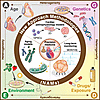
Cardiac aging arises through the interplay of multiple biological axes that determine cardiac vulnerability and resilience. We define this as the A×G×E×D framework, reflecting the convergence of four axes: age (A), genetics (G), environment (E), and drug (D) exposure (Figure 1).
Integrating age, genetics, environment, and drug exposure through new approFigure 1

Integrating age, genetics, environment, and drug exposure through new approach methodologies to model cardiac pathophysiology and heterogeneity. New approach methodologies (NAMs) have the potential to allow a comprehensive understanding of cardiac aging and disease by integrating multiple axes — age (A), genetics (G), environment (E), and drug exposure (D). By combining hiPSC-derived cardiac constructs and advanced monitoring technologies, the NAMs framework provides a multidimensional view of how A×G×E×D interactions govern cardiac health and therapeutic response, driving innovation toward targeted, patient-specific interventions (precision medicine).

Age encompasses progressive deterioration of cardiac resilience throughout the lifespan, impairing mitochondrial competency, proteostasis, DNA repair, and inflammatory regulation, diminishing the myocardium’s ability to withstand metabolic, oxidative, and mechanical stress (22). The rate of functional decline varies substantially between individuals, reflecting interactions with other axes.

Genetics, including SNPs, gene expression profiles, and epigenetic modifications, defines intrinsic susceptibility by governing mitochondrial robustness, drug metabolism, redox homeostasis, and sarcomeric stability (23). For example, variants in the genes encoding myosin binding protein C3 (MYBPC3) (24) and titin (TTN) usually remain clinically silent in youth but manifest as cardiomyopathy or hemodynamic stress with age (25). Polygenic risk scores for coronary artery disease show stronger predictive power in older cohorts, suggesting that age amplifies genetic vulnerability (A×G) (26).

Environment encompasses diet, physical activity, toxin exposure, and comorbidities such as diabetes, obesity, and hypertension. Sedentary behavior, for example, compounds age-related arterial stiffness more dramatically in older adults (A×E), disrupting metabolic flux, cardiac homeostasis, and inflammatory balance (27).

Drug/exposure reflects the dose, duration, and mechanisms of pharmacological agents, including therapeutics, pollutants, and occupational exposures, that introduce cardiac stress. Common drugs, such as NSAIDs and fluoroquinolones, carry disproportionately higher cardiac risk in aging populations (A×D). NSAIDs, widely prescribed for arthritis and musculoskeletal pain in older adults, inhibit COX-2–derived prostacyclin (PGI2) synthesis, shifting the hemostatic balance toward a prothrombotic state that increases the risk of myocardial infarction and stroke, particularly with chronic use (28, 29). Fluoroquinolones, commonly used broad-spectrum antibiotics for urinary and respiratory tract infections, block the cardiac hERG (KCNH2) potassium channel, prolonging the QT interval and predisposing to torsades de pointes while more than doubling the risk of aortic aneurysm or dissection in older patients (30, 31).

A defining strength of NAM platforms is the capacity to operationalize this framework, recognizing that these axes interact synergistically rather than independently. Adverse convergence, even minor perturbations, can trigger cascading electrical abnormalities, structural remodeling, and functional impairment. hiPSC-derived cardiac tissues and 3D organoid platforms are uniquely positioned to simulate these interactions in controlled, human-relevant systems, enabling mechanistic dissection of individual susceptibility, longitudinal disease tracking, and personalized risk stratification strategies beyond the reach of conventional animal models.
Modeling cardiac aging for clinical impact: a NAM-based framework

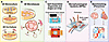
Modeling human-relevant cardiac aging demands platforms that capture the integrated A×G×E×D framework. hiPSCs provide the cellular foundation, enabling differentiation into CMs, fibroblasts, endothelial cells, and smooth muscle cells (32), while direct reprogramming (transdifferentiation) via lineage-specific transcription factors offers a complementary route bypassing the pluripotent state (33, 34). 2D monocultures support high-throughput screens, including genome-wide CRISPR/Cas9 screens (35) and high-content imaging studies tracking mitochondrial morphology, calcium dynamics, and contractile function at single-cell resolution (36), though they lack the structural complexity of native myocardium that 3D platforms are now addressing (37) (Figure 2).
Advancing human cardiac modeling platforms from 2D cultures to complex 3D sFigure 2

Advancing human cardiac modeling platforms from 2D cultures to complex 3D systems. Schematic overview of model platform progression. (A) 2D monoculture and coculture systems. (B) 3D microtissues enable multicellular organization. (C) Self-assembled organoids/organ-on-chip platforms. (D) Microphysiological systems (MPS) enhance physiological relevance with perfusion and multiorgan integration. (E) Panomic benchmarking (genomics, transcriptomics, proteomics, metabolomics) ensures model fidelity to human heart capturing A×G×E×D.

3D cardiac organoids and microtissues represent a significant advance in physiological relevance. Self-assembled multicellular human cardiac organoids partially recapitulate human cardiac function and enable tissue maturation and aging beyond what 2D cultures achieve (38–40). Organ-on-chip devices extend this further, culturing tissues within 3D scaffolds under controlled mechanical and electrical stimulation (40–42). Platforms such as Biowire II, which aligns cardiac tissues with field stimulation protocols (43), and PREDICT-96, a high-throughput microtissue array (44), now enable scalable functional readouts of aging phenotypes. Advanced microphysiological systems integrate multiple tissue types with perfusion (45, 46) (e.g., liver and kidney on the same chip), modeling how circulating metabolites, inflammatory factors, and damage signals from distal organs influence tissue function, recapitulating the multiorgan dimension of aging that isolated cardiac models cannot capture. Critically, perfusion architecture, whether unidirectional or recirculating, governs delivery of oxygen, nutrients, hormones, and paracrine signals between compartments, enabling modeling of how systemic metabolic dysfunction, organ crosstalk, and circulating damage-associated signals amplify cardiac aging phenotypes (45, 46). These systems raise a critical question: how do we confirm these platforms truly reflect age-related pathophysiology?

Rigorous benchmarking against primary human cardiac tissue is essential to confirm that these platforms reflect true age-related pathophysiology. Multi-omic datasets from donor hearts spanning transcriptomics, proteomics, metabolomics, and epigenomics provide molecular references for validation, while functional criteria, including contractile force, conduction velocity, calcium handling kinetics, action potential morphology, and stress response capacity, establish physiological benchmarks and define biological clocks that could track in vitro age progression (Figure 2). Once validated, these platforms can become powerful predictive tools to stratify patient-specific drug responses, identify senolytic and cardioprotective candidates before clinical trials, and model age-drug interactions that animal studies systematically miss. Thus, NAMs are positioned as indispensable bridges between cardiac aging research and therapeutic decision-making. Operationally, this translational impact unfolds across three interconnected stages: drug screening, where iPSC-based cardiac microphysiological systems enable high-throughput cardiotoxicity profiling with pharmacological fidelity (47); biomarker discovery, where longitudinal electrophysiological readouts such as action potential duration and arrhythmia incidence serve as validated translational biomarkers bridging preclinical and clinical risk assessment (48); and clinical stratification, where patient-specific iPSC-CM profiling preserves interindividual variation in drug response pathways, enabling genotype-informed risk stratification for precision cardiovascular medicine (49).
Ability to simulate the passage of time: aging heart in a dish

Understanding cardiac aging requires integrating molecular, cellular, and tissue-level changes that accumulate over time. The 12 interconnected hallmarks — genomic instability, telomere attrition, epigenetic alterations, loss of proteostasis, disabled macroautophagy, deregulated nutrient sensing, mitochondrial dysfunction, cellular senescence, stem cell exhaustion, altered intercellular communication, chronic inflammation, and dysbiosis — provide a useful organizational framework for cataloging biological processes that contribute to cardiac aging. Though how these hallmarks interact (22), generate an effect which predominates in cardiac tissue, and map onto clinical outcomes remains an active area of investigation.

The cascade begins at the genome. DNA damage accumulates in CMs with age (50, 51), activating ATM/ATR-p53 signaling that drives senescence (52) and metabolic rewiring (53), while telomere dysfunction (54) from replicative and oxidative stress compounds this decline. Genomic damage, in turn, reshapes the epigenome through histone modifications, chromatin accessibility changes, and altered chromatin remodeling factors, locking CMs into maladaptive gene-expression states that compromise regenerative and stress response capacity (55). Importantly, whole-genome sequencing (WGS) of single CMs from aged human hearts revealed age-dependent accumulation of somatic mutations, which were predominantly oxidative in origin (54). Future NAM platforms should incorporate strategies to capture this mutational burden, through single-cell WGS of long-term cultured iPSC-CMs or spatially resolved sequencing, to recapitulate the clonal mosaic landscape of the aged myocardium and its functional consequences. Beyond DNA methylation clocks, aging is associated with progressive shifts in histone modification landscapes, including loss of H3K27me3 at developmentally regulated loci, global histone deacetylation, and altered chromatin accessibility, that lock CMs into maladaptive transcriptional states. In NAMs, these changes can be profiled using ATAC-seq to assess chromatin accessibility dynamics, ChIP-seq targeting key histone marks (H3K27me3, H3K4me3), and live epigenetic reporters such as SIRT1-GFP to track deacetylase activity in real time (55). That failure manifests most acutely in mitochondria, where mtDNA damage, impaired electron transport chain (ETC) activity, and elevated ROS lead to adenosine triphosphate (ATP) exhaustion and arrhythmogenesis (56, 57). As they converge with impaired proteostasis and declining autophagy, these changes overwhelm cellular quality control, driving senescence-associated secretory phenotype (SASP) factor release that disrupts paracrine crosstalk among CMs, fibroblasts, and endothelial cells, ultimately attracting immune infiltration and promoting fibrosis (58). Together, these hallmarks form a mechanistic backbone that NAMs should recapitulate to faithfully model age-related cardiac dysfunction.

Establishing such NAMs requires careful consideration of both chronological age (passage of time) and biological age (deterioration of functional ability). Cardiac aging has been modeled through long-term culture or exposure to biological stressors, including aged ECM, telomere shortening, nuclear lamina disruption, mechanical and metabolic stress, and aged serum. Aged ECM recapitulates changes in stiffness, composition, and biochemical signaling characteristic of aged hearts (59, 60). Telomere damage can be induced via targeted oxidative stress (61), replicative exhaustion (62), inhibition of telomere shelterin proteins (e.g., TRF2) (54, 63), or CRISPR-based editing (64), while nuclear lamina disruption — as seen through progerin accumulation in Hutchinson-Gilford progeria syndrome (HGPS) — can also accelerate aging phenotypes (65). Patients with HGPS, telomere dysfunction, and other progeroid syndromes, including Werner, Cockayne, and Fanconi anemia, provide iPSC sources for studying biological cardiac aging, with DNA polymerase subunit γ (POLG) mutations highlighting the role of mitochondrial genome stability (66–70). Interestingly, iPSC-derived CMs from individuals with end-stage cardiomyopathy are well-established disease models that exhibit accelerated aging phenotypes (71), suggesting that NAM platforms established with these hiPSCs could be a rich resource for simulating age-related cardiac dysfunction and interrogating the molecular mechanisms that couple disease progression to biological aging. Recent literature also suggests that exposure to aged donor serum further recapitulates aging, inducing senescence markers and metabolic shifts in cultured cells (72, 73) (Figure 3, Table 1), suggesting that this strategy is effective to drive biological aging (Table 1).

Modeling chronological and biological aging in NAMs.Figure 3

Modeling chronological and biological aging in NAMs. Chronological aging is modeled through long-term culture, allowing time-dependent accumulation of cellular and molecular changes, such as metabolic slowdown, proteostatic decline, and oxidative damage. In contrast, biological aging captures functional decline, induced through stressors, such as telomere shortening, oxidative or mechanical stress, and nuclear lamina alterations. Moreover, biological aging can also be induced by culturing hiPSCs from aged donors, on aged ECM or serum/plasma exposure from older or diseased individuals. These approaches mimic intrinsic and extrinsic damage seen in aging tissue. Together, these complementary strategies reproduce the molecular, metabolic, and structural hallmarks of cardiac aging, enabling investigation of mechanisms driving age-associated cardiac dysfunction and disease susceptibility.
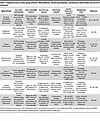
Table 1

Integrative map of cardiac aging hallmarks, NAM platforms, monitoring modalities, and biosensor toolkit within the A×G×E×D framework

However, challenges remain in rigorously modeling cardiac aging in vitro. Uncertainty persists about which hallmarks are most appropriate for in situ experiments, how many are needed to reliably track age-related changes, and which endpoint combinations best detect aging across cardiac cell types. Furthermore, heterogeneous aging within the same cardiac unit underscores the need for single-cell resolution approaches. Critically, capturing the progressive and slow-moving nature of cardiac aging demands longitudinal experimental designs, necessitating NAM platforms capable of stable, long-term recording and repeated functional assessment across extended timescales to distinguish transient stress responses from bona fide age-related decline.

Long-term monitoring platforms that capture dynamic age-related changes

Cardiac tissue functions as a complex bioelectrical system in which electrical signals are intricately regulated by metabolic processes and mitochondrial activity that determine heart rhythm and force of contraction (74). Disruptions from aging, genetic variants, environmental stressors, or drug exposures impair excitation-contraction coupling and drive progressive electrical instability. Conventional in vitro models that rely on brief electrophysiological snapshots are insufficient for capturing long-term aging effects; therefore, technologies capable of probing dynamic changes over extended timeframes are essential within the A×G×E×D framework. Advances in multielectrode arrays (MEAs) (75), voltage-sensitive dyes (76), optogenetic sensors (77), and genetically encoded calcium biosensors (78, 79), are enhancing our ability to study cumulative cardiac aging effects, revealing information about ion channel dynamics, intercellular communication, and single-cell activity across time. Below we discuss the biology underlying specific platforms and the biotechnology needed.

High spatial resolution. Cardiac tissue is inherently heterogeneous, with variable gene expression and metabolic states producing nonuniform responses to aging (80). Spatially resolved electrophysiological recordings detect distinct electrical behaviors linking single-cell activity to broader signal propagation, generating isochronal maps that reveal conduction dynamics, including silent zones, directional shifts, and regional amplitude changes, critical for identifying disruptions in cell-cell communication and structural remodeling within aged tissue (81).

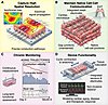
Electrode sizes of 0.1–1 mm and spacing of 0.1–3 mm are generally sufficient to capture localized aging phenotypes and tissue-wide activity (82, 83). Surface-mounted devices support surface-level reconstructions, while embedded devices enable internal recordings of extracellular field potentials (84). For engineered cardiac tissues, embedding 128+ electrodes allows comprehensive 3D mapping (85–88), and future high-density platforms should support electrode placement at 250 to 500 μm intervals (89, 90), while minimizing device footprint to preserve physiological relevance (91) (Figure 4).
Functional features of cardiac NAMs for high-resolution and long-term functFigure 4

Functional features of cardiac NAMs for high-resolution and long-term functional assessment. (A) Capture high spatial resolution. Bioengineered devices should be designed to enable high-spatial-resolution mapping of electrical activity, allowing precise visualization of conduction pathways, signal propagation, and local heterogeneity within cardiac tissues. (B) Maintain native cell-cell communication. Integrated mesh electrodes can be embedded within the cardiac microtissues to maintain native cell-cell junctions and tissue architecture, ensuring physiological signal transmission and mechanical integrity. (C) Chronic monitoring. Systems designed for chronic monitoring can enable continuous measurement of conduction velocity, contractile force, and beat rate over extended culture periods spanning several months. (D) Device functionality. Platforms that allow dynamic mechanical stimulation, including cyclic contraction and stretching, which closely mimics the mechanical environment of the native myocardium and promotes tissue maturation would enable a highly rigorous and reproducible NAM. Combined with integrated sensors and electrodes, these devices can provide a noninvasive, high-fidelity readout of electrical and mechanical performance, offering a powerful tool for modeling cardiac aging, disease, and therapeutic response in vitro.

Minimal interference with cell-cell junctions. Physiological heart function depends on precise electrical and mechanical coupling between CMs (92), mediated by desmosomes, integrins, and cadherins and synchronized via gap junctions at intercalated discs (93, 94). Aging compounds this challenge through structural remodeling, fibrosis, myocyte hypertrophy, and reduced cellular density. Bioelectronic devices must therefore be biocompatible not just in materials but in their capacity to preserve these cellular interactions (95).

Electrodes of 10–20 μm diameter and polymer-encapsulated traces under 10 μm in width and thickness minimize device footprint between adjacent cells (96, 97). Soft, flexible materials enable closer mimicry of the native cellular environment (98), allowing bioelectronic systems to function as biomimetic scaffolds rather than foreign implants, promoting long-term biocompatibility and stable chronic interfacing with the aging heart (Figure 4).

Preserving device functionality during cardiac tissue contraction. Reliable long-term recordings require electrode materials and insulating layers to retain structural integrity across tens of thousands of contraction cycles. This challenge is amplified in aging models, where altered tissue stiffness and irregular contraction patterns impose nonuniform mechanical loads that accelerate device fatigue.

Kirigami-inspired structures allow devices to stretch biaxially while keeping material stress below elastic limits (90, 99, 100). Intrinsically stretchable materials and serpentine metal traces offer an alternative, though often at the cost of resolution (101, 102). Kirigami-based devices fabricated via high-resolution photolithography accommodate microscale features needed for precise tissue interfacing (103), offering a promising combination of mechanical resilience and fabrication precision for chronic monitoring over weeks to months (104).

Tissue monitoring over time (with age). As next-generation bioelectronics advance, continuous monitoring capabilities within electrophysiological recording platforms emerge as a critical need. Rather than relying on isolated snapshots, continuous monitoring provides a temporal map of aging progression, capturing subtle early-stage changes that might otherwise go undetected (105), which are particularly important when the timing of pathological changes is unknown. In aging cardiac tissue, electrical alterations may emerge gradually or sporadically, and without ongoing monitoring, critical onset of dysfunction may be missed entirely. This is illustrated by doxorubicin-induced cardiotoxicity, where progressive mitochondrial dysfunction and contractile decline are only detectable days after initial exposure (106) and by age-related calcium handling. An important emerging consideration is that acute drug responses frequently diverge from long-term outcomes: studies have demonstrated that 24- to 48-hour drug exposures fail to capture clinically relevant cardiotoxic phenotypes that only emerge over weeks of treatment, underscoring the critical need for extended monitoring windows within the A×G×E×D framework. Once the timescale of interest is identified, periodic sampling protocols, such as recording several minutes every hour or day, can balance data capture with system longevity. A complement to electrophysiology, intermittent fluorescent imaging uses genetically encoded reporters, including calcium indicators such as GCaMP, metabolic sensors such as SoNar (107), and senescence markers (108–110), enables longitudinal molecular readouts that can be correlated with functional electrophysiological outputs without disrupting tissue culture.

Despite progress in long-term electrophysiological recordings (111), most existing studies have been confined to 2D models (112) and short timeframes. A critical gap remains: no existing framework has demonstrated sustained electrical monitoring of 3D cardiac tissue within a fully controlled incubator environment, where maintaining both tissue and electronics in a single stable system reduces risks of thermal stress, physical disruption, or device degradation. Practical implementation would involve integrating perfusion systems alongside electronics capable of routing signals outside the incubator without compromising signal quality. Chronic recordings from 3D cardiac models offer detailed views of how electrophysiological changes unfold in response to intrinsic aging and extrinsic stressors (113, 114), enabling identification of early deviations in electrical patterns that may precede overt dysfunction, ultimately guiding preventative interventions and, in the context of personalized medicine, tailoring treatment to an individual’s unique biological aging profile. Notably, incorporating vascular-like perfusion dynamics, including physiological shear stress, oxygen and nutrient delivery, and endothelial cell–CM coupling is critical for long-term engineered cardiac tissues, as the absence of perfusion induces metabolic stress, inflammatory signaling, and electrophysiological instability that confound aging phenotypes, whereas dynamic flow restores near-adult contractile force, conduction velocity, and metabolic maturation (115, 116) (Figure 4).

Integrate hallmarks of aging with tissue function. Traditional aging models have largely focused on single stressors, such as DNA damage or mitochondrial dysfunction. However, aging in vivo is driven by simultaneous and nonlinear activation of multiple hallmarks shaped by intrinsic and environmental factors. Combinatorial stressor models that combine genotoxic stress, mitochondrial uncouplers, and inflammatory cytokines more accurately simulate age-related cardiac dysfunction than single-pathway approaches. Applying these models in iPSC-derived tissues or organoids generates rich, high-dimensional datasets that, when paired with artificial intelligence (AI) and machine learning (ML) (117), can reveal hallmark interactions and support so-called “intervention stacking” strategies that target multiple hallmarks in sequence or tandem. Dynamic aging scores integrating hallmark biology with molecular, cellular, and functional metrics are needed to predict disease risk and functional decline across tissues (118).

Multimodal biosensors are central to this shift toward integration. Expanding beyond electrophysiology to fluorescence-based and electrochemical sensors enables real-time monitoring of ATP, ROS, NADH, glucose, and lactate (119–121), while genetically encoded biosensors report on autophagy, redox state, proteasome activity, ER stress, and cellular senescence (122–126) in live cells. A key limitation is that most biosensors are deployed individually rather than multiplexed, preventing simultaneous monitoring of interacting pathways. Future NAM platforms should integrate multiplexed biosensors to capture dynamic hallmark interactions at a systems level. AI and ML are critical enablers: supervised algorithms classify samples by age or disease state, unsupervised models uncover hidden aging subtypes, and time-series models predict transitions between health and disease by detecting early shifts in mitochondrial dynamics or immune activity (127, 128). Coupled with interpretable AI frameworks and trained on interventional datasets, these models identify therapeutic targets and support causal inferencing. Building this technological and analytical infrastructure for real-time, systems-level aging assessment will lay the groundwork for precision geroscience-enabling tailored interventions, extended health span, and informed aging policy (129, 130) (Figure 4).
Limitations, challenges, and future directions

Despite their transformative potential, current NAMs exhibit critical limitations. Immune cells cannot be overlooked, as chronic inflammation and immune-cardiac crosstalk are central drivers of age-related cardiac dysfunction. Macrophages can be introduced through coculture systems, including iPSC-derived macrophages that preserve donor-specific genetic context, enabling modeling of SASP-mediated recruitment, inflammatory infiltration, and profibrotic signaling. However, most immune cells have finite lifespans in culture, necessitating periodic replenishment or microfluidic perfusion to sustain physiologically relevant immune-cardiac interactions over the timescales required to study aging meaningfully.

Long-term viability beyond 90–120 days remains technically challenging, limiting the ability to model chronic damage accumulation over timeframes that better approximate human aging. Environmental variables, such as oxygen gradients, nutrient availability, and ECM stiffness, are difficult to control in 3D systems, introducing variability that complicates interpretation. These limitations underscore the need for continued technical refinement, multi-omics benchmarking against primary human tissues, and standardized age-scoring systems. Validation across multiple laboratories and integration of computational models will be essential for translating NAM-based discoveries into clinical applications.

Aging is not merely a background variable in cardiac disease, it is a central mechanistic driver shaping how biological stress is sensed, processed, and amplified within the heart. Realizing the full potential of this platform will require integrating vascularized organoids, immunocompetent microtissues, and embedded multimodal bioelectronics capable of continuous metabolic and electrophysiological monitoring, alongside AI-driven pipelines to interpret high-dimensional time-series data and predict inflection points in tissue aging. By deploying the A×G×E×D framework and modeling biological age directly in vitro, NAMs must ultimately evolve toward closed-loop systems that unite sensors, analytics, and interventions, advancing a new era of precision geromedicine where prediction replaces surveillance and early intervention replaces reactive treatment.

Author contributions

KC and MB reviewed the literature. KC wrote the first draft. All authors revised and contributed to the article. KC and AUG approved the submitted version.

Conflict of interest

The authors have declared that no conflict of interest exists.

Funding support

This work is the result of NIH funding, in whole or in part, and is subject to the NIH Public Access Policy. Through acceptance of this federal funding, the NIH has been given a right to make the work publicly available in PubMed Central.

  • NIH grants R01HL161106 to AG, KC, MB, and TCK and R01ES035733 and R21 AG086744 to AG.
  • American Federation for Aging Research/Hevolution to AG.
  • Richard King Mellon Foundation grant.
Acknowledgments

We thank Sri Ravali Pulipaka and Syed Afroz Ali for active discussions and feedback on the manuscript.

Address correspondence to: Aditi U. Gurkar, Aging Institute, Suite 563, 100 Technology Drive, University of Pittsburgh, Pittsburgh, PA 15219, USA. Phone: 412.624.7494; Email: agurkar1@pitt.edu. Or to: Tzahi Cohen-Karni, Biomedical Engineering and Materials Science & Engineering Carnegie Mellon University, Pittsburgh, Pennsylvania, USA. Email: tzahi@andrew.cmu.edu.

Footnotes

Copyright: © 2026, Chaddha et al. This is an open access article published under the terms of the Creative Commons Attribution 4.0 International License.

Reference information: JCI Insight. 2026;11(15):e203447.https://doi.org/10.1172/jci.insight.203447.

References
  1. Dai DF, et al. Cardiac aging: from molecular mechanisms to significance in human health and disease. Antioxid Redox Signal. 2012;16(12):1492–1526.
    View this article via: CrossRef PubMed Google Scholar
  2. Hastings MH, et al. Cardiac ageing: from hallmarks to therapeutic opportunities. Cardiovasc Res. 2025;121(10):1474–1488.
    View this article via: CrossRef PubMed Google Scholar
  3. Lazzeroni D, et al. The aging heart: a molecular and clinical challenge. Int J Mol Sci. 2022;23(24):16033.
    View this article via: CrossRef PubMed Google Scholar
  4. Rodriguez-Rodero S, et al. Aging genetics and aging. Aging Dis. 2011;2(3):186–195.
    View this article via: PubMed Google Scholar
  5. Belsky DW, Baccarelli AA. To promote healthy aging, focus on the environment. Nat Aging. 2023;3(11):1334–1344.
    View this article via: CrossRef PubMed Google Scholar
  6. Zheng P, et al. Cardiovascular ageing: hallmarks, signaling pathways, diseases and therapeutic targets. Signal Transduct Target Ther. 2026;11(1):142.
    View this article via: CrossRef PubMed Google Scholar
  7. Joukar S. A comparative review on heart ion channels, action potentials and electrocardiogram in rodents and human: extrapolation of experimental insights to clinic. Lab Anim Res. 2021;37(1):25.
    View this article via: CrossRef PubMed Google Scholar
  8. Kaese S, Verheule S. Cardiac electrophysiology in mice: a matter of size. Front Physiol. 2012;3:345.
    View this article via: CrossRef PubMed Google Scholar
  9. Varro A, et al. Cardiac transmembrane ion channels and action potentials: cellular physiology and arrhythmogenic behavior. Physiol Rev. 2021;101(3):1083–1176.
    View this article via: CrossRef PubMed Google Scholar
  10. Blackwell DJ, et al. Animal models to study cardiac arrhythmias. Circ Res. 2022;130(12):1926–1964.
    View this article via: CrossRef PubMed Google Scholar
  11. van den Dolder FW, et al. Experimental models of hypertrophic cardiomyopathy: a systematic review. JACC Basic Transl Sci. 2025;10(4):511–546.
    View this article via: CrossRef PubMed Google Scholar
  12. Withaar C, et al. Heart failure with preserved ejection fraction in humans and mice: embracing clinical complexity in mouse models. Eur Heart J. 2021;42(43):4420–4430.
    View this article via: CrossRef PubMed Google Scholar
  13. Dumont BL, et al. Into the wild: a novel wild-derived inbred strain resource expands the genomic and phenotypic diversity of laboratory mouse models. PLoS Genet. 2024;20(4):e1011228.
    View this article via: CrossRef PubMed Google Scholar
  14. Gerke S, et al. The FDA’s plan to phase out animal testing. Trends Biotechnol. 2026;
    View this article via: CrossRef Google Scholar
  15. Sewell F, et al. New approach methodologies (NAMs): identifying and overcoming hurdles to accelerated adoption. Toxicol Res (Camb). 2024;13(2):tfae044.
    View this article via: CrossRef PubMed Google Scholar
  16. Wang W, et al. Microfluidic platforms for monitoring cardiomyocyte electromechanical activity. Microsyst Nanoeng. 2025;11(1):4.
    View this article via: CrossRef PubMed Google Scholar
  17. Dou W, et al. Microengineered platforms for characterizing the contractile function of in vitro cardiac models. Microsyst Nanoeng. 2022;8:26.
    View this article via: CrossRef PubMed Google Scholar
  18. Ergir E, et al. Generation and maturation of human iPSC-derived 3D organotypic cardiac microtissues in long-term culture. Sci Rep. 2022;12(1):17409.
    View this article via: CrossRef PubMed Google Scholar
  19. Higo S, et al. Genome editing in human induced pluripotent stem cells (hiPSCs). Methods Mol Biol. 2021;2320:235–245.
    View this article via: PubMed CrossRef Google Scholar
  20. Sharma A, et al. Induced pluripotent stem cell-derived cardiomyocytes for cardiovascular disease modeling and drug screening. Stem Cell Res Ther. 2013;4(6):150.
    View this article via: CrossRef PubMed Google Scholar
  21. Zhu L, et al. Cardiac organoids: a 3D Technology for modeling heart development and disease. Stem Cell Rev Rep. 2022;18(8):2593–2605.
    View this article via: CrossRef PubMed Google Scholar
  22. Lopez-Otin C, et al. Hallmarks of aging: an expanding universe. Cell. 2023;186(2):243–278.
    View this article via: CrossRef PubMed Google Scholar
  23. Ferro A, et al. Exploring the role of genetic and genomic factors in therapeutic response to heart failure: a comprehensive analytical review. Genes (Basel). 2025;16(7):801.
    View this article via: CrossRef PubMed Google Scholar
  24. Ananthamohan K, et al. Hypertrophic cardiomyopathy in MYBPC3 carriers in aging. J Cardiovasc Aging. 2024;4(1):9.
    View this article via: CrossRef PubMed Google Scholar
  25. Fatkin D, Huttner IG. Titin-truncating mutations in dilated cardiomyopathy: the long and short of it. Curr Opin Cardiol. 2017;32(3):232–238.
    View this article via: CrossRef PubMed Google Scholar
  26. Sherafati A, et al. Polygenic risk and coronary artery disease severity. Circ Genom Precis Med. 2024;17(5):e004470.
    View this article via: CrossRef PubMed Google Scholar
  27. Herzog MJ, et al. Arterial stiffness and vascular aging: mechanisms, prevention, and therapy. Signal Transduct Target Ther. 2025;10(1):282.
    View this article via: CrossRef PubMed Google Scholar
  28. Antman EM, et al. Cyclooxygenase inhibition and cardiovascular risk. Circulation. 2005;112(5):759–770.
    View this article via: CrossRef PubMed Google Scholar
  29. Gunaydin C, Bilge SS. Effects of nonsteroidal anti-inflammatory drugs at the molecular level. Eurasian J Med. 2018;50(2):116–121.
    View this article via: CrossRef PubMed Google Scholar
  30. Chan T, Bunce PE. Fluoroquinolone antimicrobial drugs. CMAJ. 2017;189(17):E638.
    View this article via: CrossRef PubMed Google Scholar
  31. Gorelik E, et al. Fluoroquinolones and cardiovascular risk: a systematic review, meta-analysis and network meta-analysis. Drug Saf. 2019;42(4):529–538.
    View this article via: CrossRef PubMed Google Scholar
  32. Lyra-Leite DM, et al. A review of protocols for human iPSC culture, cardiac differentiation, subtype-specification, maturation, and direct reprogramming. STAR Protoc. 2022;3(3):101560.
    View this article via: CrossRef PubMed Google Scholar
  33. Jahng JWS, et al. The role of metabolism in directed differentiation versus trans-differentiation of cardiomyocytes. Semin Cell Dev Biol. 2022;122:56–65.
    View this article via: CrossRef PubMed Google Scholar
  34. Takashima S, et al. Myocyte-specific enhancer factor 2c triggers transdifferentiation of adipose tissue-derived stromal cells into spontaneously beating cardiomyocyte-like cells. Sci Rep. 2021;11(1):1520.
    View this article via: CrossRef PubMed Google Scholar
  35. Nishiga M, et al. CRISPRi/a screening with human iPSCs. Methods Mol Biol. 2021;2320:261–281.
    View this article via: PubMed CrossRef Google Scholar
  36. Menduti G, Boido M. Recent advances in high-content imaging and analysis in iPSC-based modelling of neurodegenerative diseases. Int J Mol Sci. 2023;24(19):14689.
    View this article via: CrossRef PubMed Google Scholar
  37. Roland TJ, Song K. Advances in the generation of constructed cardiac tissue derived from induced pluripotent stem cells for disease modeling and therapeutic discovery. Cells. 2024;13(3):250.
    View this article via: CrossRef PubMed Google Scholar
  38. Arslan U, et al. Perspectives for future use of cardiac microtissues from human pluripotent stem cells. ACS Biomater Sci Eng. 2022;8(11):4605–4609.
    View this article via: CrossRef PubMed Google Scholar
  39. Cho J, et al. From engineered heart tissue to cardiac organoid. Theranostics. 2022;12(6):2758–2772.
    View this article via: CrossRef PubMed Google Scholar
  40. Kim H, et al. Progress in multicellular human cardiac organoids for clinical applications. Cell Stem Cell. 2022;29(4):503–514.
    View this article via: CrossRef PubMed Google Scholar
  41. Lewis-Israeli YR, et al. Self-assembling human heart organoids for the modeling of cardiac development and congenital heart disease. Nat Commun. 2021;12(1):5142.
    View this article via: CrossRef PubMed Google Scholar
  42. Juguilon C, et al. In vitro modeling of interorgan crosstalk: multi-organ-on-a-chip for studying cardiovascular-kidney-metabolic syndrome. Circ Res. 2025;136(11):1476–1493.
    View this article via: CrossRef PubMed Google Scholar
  43. Feric NT, et al. Engineered cardiac tissues generated in the Biowire II: a platform for human-based drug discovery. Toxicol Sci. 2019;172(1):89–97.
    View this article via: CrossRef PubMed Google Scholar
  44. Tan K, et al. Correction: a high-throughput microfluidic microphysiological system (PREDICT-96) to recapitulate hepatocyte function in dynamic, re-circulating flow conditions. Lab Chip. 2020;20(19):3653.
    View this article via: CrossRef PubMed Google Scholar
  45. Renggli K, et al. Integrated microphysiological systems: transferable organ models and recirculating flow. Adv Biosyst. 2019;3(5):e1900018.
    View this article via: CrossRef PubMed Google Scholar
  46. Ronaldson-Bouchard K, et al. A multi-organ chip with matured tissue niches linked by vascular flow. Nat Biomed Eng. 2022;6(4):351–371.
    View this article via: CrossRef PubMed Google Scholar
  47. Mathur A, et al. Human iPSC-based cardiac microphysiological system for drug screening applications. Sci Rep. 2015;5:8883.
    View this article via: CrossRef PubMed Google Scholar
  48. Blinova K, et al. Comprehensive translational assessment of human-induced pluripotent stem cell derived cardiomyocytes for evaluating drug-induced arrhythmias. Toxicol Sci. 2017;155(1):234–247.
    View this article via: CrossRef PubMed Google Scholar
  49. Matsa E, et al. Transcriptome profiling of patient-specific human ipsc-cardiomyocytes predicts individual drug safety and efficacy responses in vitro. Cell Stem Cell. 2016;19(3):311–325.
    View this article via: CrossRef PubMed Google Scholar
  50. Henpita C, et al. Loss of DNA repair mechanisms in cardiac myocytes induce dilated cardiomyopathy. Aging Cell. 2023;22(4):e13782.
    View this article via: CrossRef PubMed Google Scholar
  51. Choudhury S, et al. Somatic mutations in single human cardiomyocytes reveal age-associated DNA damage and widespread oxidative genotoxicity. Nat Aging. 2022;2(8):714–725.
    View this article via: CrossRef PubMed Google Scholar
  52. Luan Y, et al. Cardiac cell senescence: molecular mechanisms, key proteins and therapeutic targets. Cell Death Discov. 2024;10(1):78.
    View this article via: CrossRef PubMed Google Scholar
  53. Xie S, et al. Metabolic landscape in cardiac aging: insights into molecular biology and therapeutic implications. Signal Transduct Target Ther. 2023;8(1):114.
    View this article via: CrossRef PubMed Google Scholar
  54. Rossiello F, et al. Telomere dysfunction in ageing and age-related diseases. Nat Cell Biol. 2022;24(2):135–147.
    View this article via: CrossRef PubMed Google Scholar
  55. Chen Y, et al. Chromatin accessibility: biological functions, molecular mechanisms and therapeutic application. Signal Transduct Target Ther. 2024;9(1):340.
    View this article via: CrossRef PubMed Google Scholar
  56. Xu X, et al. Mitochondria in oxidative stress, inflammation and aging: from mechanisms to therapeutic advances. Signal Transduct Target Ther. 2025;10(1):190.
    View this article via: CrossRef PubMed Google Scholar
  57. Gambardella J, et al. Functional role of mitochondria in arrhythmogenesis. Adv Exp Med Biol. 2017;982:191–202.
    View this article via: PubMed CrossRef Google Scholar
  58. Hamazaki J, Murata S. Relationships between protein degradation, cellular senescence, and organismal aging. J Biochem. 2024;175(5):473–480.
    View this article via: CrossRef PubMed Google Scholar
  59. Angelini A, et al. Mechanosensing dysregulation in the fibroblast: A hallmark of the aging heart. Ageing Res Rev. 2020;63:101150.
    View this article via: CrossRef PubMed Google Scholar
  60. Meschiari CA, et al. The impact of aging on cardiac extracellular matrix. Geroscience. 2017;39(1):7–18.
    View this article via: CrossRef PubMed Google Scholar
  61. Barnes RP, et al. Telomeric 8-oxo-guanine drives rapid premature senescence in the absence of telomere shortening. Nat Struct Mol Biol. 2022;29(7):639–652.
    View this article via: CrossRef PubMed Google Scholar
  62. Liu J, et al. Roles of telomere biology in cell senescence, replicative and chronological ageing. Cells. 2019;8(1):54.
    View this article via: CrossRef PubMed Google Scholar
  63. Sfeir A, de Lange T. Removal of shelterin reveals the telomere end-protection problem. Science. 2012;336(6081):593–597.
    View this article via: CrossRef PubMed Google Scholar
  64. Brane AC, Tollefsbol TO. Targeting telomeres and telomerase: studies in aging and disease utilizing CRISPR/Cas9 technology. Cells. 2019;8(2):186.
    View this article via: CrossRef PubMed Google Scholar
  65. Monnerat G, et al. Modelling premature cardiac aging with induced pluripotent stem cells from a hutchinson-gilford Progeria syndrome patient. Front Physiol. 2022;13:1007418.
    View this article via: CrossRef PubMed Google Scholar
  66. Capell BC, et al. Mechanisms of cardiovascular disease in accelerated aging syndromes. Circ Res. 2007;101(1):13–26.
    View this article via: CrossRef PubMed Google Scholar
  67. Ikeda Y, et al. Understanding vascular diseases: lessons from premature aging syndromes. Can J Cardiol. 2016;32(5):650–658.
    View this article via: PubMed CrossRef Google Scholar
  68. Dai DF, et al. Age-dependent cardiomyopathy in mitochondrial mutator mice is attenuated by overexpression of catalase targeted to mitochondria. Aging Cell. 2010;9(4):536–544.
    View this article via: CrossRef PubMed Google Scholar
  69. Lewis W, et al. Decreased mtDNA, oxidative stress, cardiomyopathy, and death from transgenic cardiac targeted human mutant polymerase gamma. Lab Invest. 2007;87(4):326–335.
    View this article via: CrossRef PubMed Google Scholar
  70. Jothi D, Kulka LAM. Strategies for modeling aging and age-related diseases. NPJ Aging. 2024;10(1):32.
    View this article via: CrossRef PubMed Google Scholar
  71. Vo QD, et al. Induced pluripotent stem cells in cardiomyopathy: advancing disease modeling, therapeutic development, and regenerative therapy. Int J Mol Sci. 2025;26(11):4984.
    View this article via: CrossRef PubMed Google Scholar
  72. Jeon OH, et al. Systemic induction of senescence in young mice after single heterochronic blood exchange. Nat Metab. 2022;4(8):995–1006.
    View this article via: CrossRef PubMed Google Scholar
  73. Chung TD, et al. Aged human serum induces vascular changes in an isogenic co-culture venule model. Stem Cell Reports. 2025;20(7):102544.
    View this article via: CrossRef PubMed Google Scholar
  74. Kleber AG, Jin Q. Coupling between cardiac cells-an important determinant of electrical impulse propagation and arrhythmogenesis. Biophys Rev (Melville). 2021;2(3):031301.
    View this article via: CrossRef PubMed Google Scholar
  75. Clements M. Multielectrode array (MEA) assay for profiling electrophysiological drug effects in human stem cell-derived cardiomyocytes. Curr Protoc Toxicol. 2016;68:22.4.1–22.4.32.
    View this article via: CrossRef PubMed Google Scholar
  76. Girouard SD, et al. Unique properties of cardiac action potentials recorded with voltage-sensitive dyes. J Cardiovasc Electrophysiol. 1996;7(11):1024–1038.
    View this article via: CrossRef PubMed Google Scholar
  77. Koopman CD, et al. Cardiac optogenetics: using light to monitor cardiac physiology. Basic Res Cardiol. 2017;112(5):56.
    View this article via: CrossRef PubMed Google Scholar
  78. Tallini YN, et al. Imaging cellular signals in the heart in vivo: Cardiac expression of the high-signal Ca2+ indicator GCaMP2. Proc Natl Acad Sci U S A. 2006;103(12):4753–4758.
    View this article via: CrossRef PubMed Google Scholar
  79. Kaestner L, et al. Genetically encoded Ca2+ indicators in cardiac myocytes. Circ Res. 2014;114(10):1623–1639.
    View this article via: CrossRef PubMed Google Scholar
  80. Anto Michel N, et al. Cellular heterogeneity of the heart. Front Cardiovasc Med. 2022;9:868466.
    View this article via: CrossRef PubMed Google Scholar
  81. Kalmykov A, et al. Organ-on-e-chip: three-dimensional self-rolled biosensor array for electrical interrogations of human electrogenic spheroids. Sci Adv. 2019;5(8):eaax0729.
    View this article via: CrossRef PubMed Google Scholar
  82. Takigawa M, et al. Effect of electrode size and spacing on electrograms: optimized electrode configuration for near-field electrogram characterization. Heart Rhythm. 2022;19(1):102–112.
    View this article via: CrossRef PubMed Google Scholar
  83. Takigawa M, et al. Impact of spacing and orientation on the scar threshold with a high-density grid catheter. Circ Arrhythm Electrophysiol. 2019;12(9):e007158.
    View this article via: CrossRef PubMed Google Scholar
  84. Cheng L, et al. Recent advances in flexible noninvasive electrodes for surface electromyography acquisition. npj Flex Electron. 2023;7:39).
    View this article via: CrossRef Google Scholar
  85. Schroter M, et al. Advances in large-scale electrophysiology with high-density microelectrode arrays. Lab Chip. 2025;25(19):4844–4885.
    View this article via: CrossRef PubMed Google Scholar
  86. Chen Z, et al. Soft, bioresorbable, transparent microelectrode arrays for multimodal spatiotemporal mapping and modulation of cardiac physiology. Sci Adv. 2023;9(27):eadi0757.
    View this article via: CrossRef PubMed Google Scholar
  87. Xu F, et al. Architecture design and advanced manufacturing of heart-on-a-chip: scaffolds, stimulation and sensors. Microsyst Nanoeng. 2024;10:96.
    View this article via: CrossRef PubMed Google Scholar
  88. Fullenkamp DE, et al. Simultaneous electromechanical monitoring in engineered heart tissues using a mesoscale framework. Sci Adv. 2024;10(37):eado7089.
    View this article via: CrossRef PubMed Google Scholar
  89. Lee JY, et al. Foldable three dimensional neural electrode arrays for simultaneous brain interfacing of cortical surface and intracortical multilayers. npj Flex Electron. 2022;6:86.
    View this article via: CrossRef Google Scholar
  90. Yang X, et al. Kirigami electronics for long-term electrophysiological recording of human neural organoids and assembloids. Nat Biotechnol. 2024;42(12):1836–1843.
    View this article via: CrossRef PubMed Google Scholar
  91. Dai X, et al. Three-dimensional mapping and regulation of action potential propagation in nanoelectronics-innervated tissues. Nat Nanotechnol. 2016;11(9):776–782.
    View this article via: CrossRef PubMed Google Scholar
  92. Bartlett M, et al. A snapshot review on materials enabled multimodal bioelectronics for neurological and cardiac research. MRS Adv. 2023;8(19):1047–1060.
    View this article via: CrossRef PubMed Google Scholar
  93. Nielsen MS, et al. The intercalated disc: a unique organelle for electromechanical synchrony in cardiomyocytes. Physiol Rev. 2023;103(3):2271–2319.
    View this article via: CrossRef PubMed Google Scholar
  94. Noorman M, et al. Cardiac cell-cell junctions in health and disease: electrical versus mechanical coupling. J Mol Cell Cardiol. 2009;47(1):23–31.
    View this article via: CrossRef PubMed Google Scholar
  95. Cox-Pridmore DM, et al. Emerging bioelectronic strategies for cardiovascular tissue engineering and implantation. Small. 2022;18(17):e2105281.
    View this article via: CrossRef PubMed Google Scholar
  96. Feiner R, et al. Engineered hybrid cardiac patches with multifunctional electronics for online monitoring and regulation of tissue function. Nat Mater. 2016;15(6):679–685.
    View this article via: CrossRef PubMed Google Scholar
  97. Qiang Y, et al. Transparent arrays of bilayer-nanomesh microelectrodes for simultaneous electrophysiology and two-photon imaging in the brain. Sci Adv. 2018;4(9):eaat0626.
    View this article via: CrossRef PubMed Google Scholar
  98. Sang M, et al. Ultra-thin flexible encapsulating materials for soft bio-integrated electronics. Adv Sci (Weinh). 2022;9(30):e2202980.
    View this article via: PubMed CrossRef Google Scholar
  99. Tan S, et al. Cell-cell interactions in the heart: advanced cardiac models and omics technologies. Stem Cell Res Ther. 2024;15(1):362.
    View this article via: CrossRef PubMed Google Scholar
  100. Lin Z, et al. Cyborg organoids integrated with stretchable nanoelectronics can be functionally mapped during development. Nat Protoc. 2025;20(9):2528–2559.
    View this article via: CrossRef PubMed Google Scholar
  101. Zhang Y, et al. Buckling in serpentine microstructures and applications in elastomer-supported ultra-stretchable electronics with high areal coverage. Soft Matter. 2013;9(33):8062–8070.
    View this article via: CrossRef PubMed Google Scholar
  102. Li TL, et al. Stretchable mesh microelectronics for the biointegration and stimulation of human neural organoids. Biomaterials. 2022;290:121825.
    View this article via: CrossRef PubMed Google Scholar
  103. Morikawa Y, et al. Ultrastretchable Kirigami bioprobes. Adv Healthc Mater. 2018;7(3)
    View this article via: PubMed CrossRef Google Scholar
  104. Brooks AK, et al. Kirigami-inspired biodesign for applications in healthcare. Adv Mater. 2022;34(18):e2109550.
    View this article via: CrossRef PubMed Google Scholar
  105. Pyrkov TV, et al. Longitudinal analysis of blood markers reveals progressive loss of resilience and predicts human lifespan limit. Nat Commun. 2021;12(1):2765.
    View this article via: CrossRef PubMed Google Scholar
  106. Wallace KB, et al. Mitochondrial determinants of doxorubicin-induced cardiomyopathy. Circ Res. 2020;126(7):926–941.
    View this article via: CrossRef PubMed Google Scholar
  107. Zhao Y, et al. SoNar, a highly responsive NAD+/NADH sensor, allows high-throughput metabolic screening of anti-tumor agents. Cell Metab. 2015;21(5):777–789.
    View this article via: CrossRef PubMed Google Scholar
  108. Yi Z, et al. Generation of a p21 reporter mouse and its use to identify and eliminate p21high cells in vivo. Int J Mol Sci. 2023;24(6):5565.
    View this article via: CrossRef PubMed Google Scholar
  109. Wang B, et al. Intermittent clearance of p21-highly-expressing cells extends lifespan and confers sustained benefits to health and physical function. Cell Metab. 2024;36(8):1795–1805.e6.
    View this article via: CrossRef PubMed Google Scholar
  110. Burd CE, et al. Monitoring tumorigenesis and senescence in vivo with a p16(INK4a)-luciferase model. Cell. 2013;152(1-2):340–351.
    View this article via: CrossRef PubMed Google Scholar
  111. Bloch Thomsen PE, et al. Long-term recording of cardiac arrhythmias with an implantable cardiac monitor in patients with reduced ejection fraction after acute myocardial infarction: the Cardiac Arrhythmias and Risk Stratification After Acute Myocardial Infarction (CARISMA) study. Circulation. 2010;122(13):1258–1264.
    View this article via: CrossRef PubMed Google Scholar
  112. Liu H, et al. Heart-on-a-chip model with integrated extra- and intracellular bioelectronics for monitoring cardiac electrophysiology under acute hypoxia. Nano Lett. 2020;20(4):2585–2593.
    View this article via: CrossRef PubMed Google Scholar
  113. Shi K, et al. Advanced passive 3D bioelectronics: powerful tool for the cardiac electrophysiology investigation. Microsyst Nanoeng. 2025;11(1):50.
    View this article via: CrossRef PubMed Google Scholar
  114. Jackman CP, et al. Dynamic culture yields engineered myocardium with near-adult functional output. Biomaterials. 2016;111:66–79.
    View this article via: CrossRef PubMed Google Scholar
  115. Min S, et al. Versatile human cardiac tissues engineered with perfusable heart extracellular microenvironment for biomedical applications. Nat Commun. 2024;15(1):2564.
    View this article via: CrossRef PubMed Google Scholar
  116. Besser RR, et al. Engineered microenvironments for maturation of stem cell derived cardiac myocytes. Theranostics. 2018;8(1):124–140.
    View this article via: CrossRef PubMed Google Scholar
  117. Maramraju S, et al. AI-organoid integrated systems for biomedical studies and applications. Bioeng Transl Med. 2024;9(2):e10641.
    View this article via: CrossRef PubMed Google Scholar
  118. Ashiqur Rahman S, et al. Deep learning for biological age estimation. Brief Bioinform. 2021;22(2):1767–1781.
    View this article via: CrossRef PubMed Google Scholar
  119. Erard M, et al. Biosensors for spatiotemporal detection of reactive oxygen species in cells and tissues. Am J Physiol Regul Integr Comp Physiol. 2018;314(5):R667–R683.
    View this article via: CrossRef PubMed Google Scholar
  120. Li X, et al. Elucidating the spatiotemporal dynamics of glucose metabolism with genetically encoded fluorescent biosensors. Cell Rep Methods. 2024;4(11):100904.
    View this article via: CrossRef PubMed Google Scholar
  121. Nasu Y, et al. A genetically encoded fluorescent biosensor for extracellular L-lactate. Nat Commun. 2021;12(1):7058.
    View this article via: CrossRef PubMed Google Scholar
  122. Nasrollahpour H, et al. Biosensors; a novel concept in real-time detection of autophagy. Biosens Bioelectron. 2024;254:116204.
    View this article via: CrossRef PubMed Google Scholar
  123. Pedre B. A guide to genetically-encoded redox biosensors: State of the art and opportunities. Arch Biochem Biophys. 2024;758:110067.
    View this article via: CrossRef PubMed Google Scholar
  124. Floro EJ, Rocheleau JV. Apollo-IRE1: a genetically encoded sensor for live cell and multiplexed imaging of ER stress. Biophys J. 2023;122(3_suppl 1):436a.
    View this article via: CrossRef Google Scholar
  125. Mo GC, et al. Genetically encoded biosensors for visualizing live-cell biochemical activity at super-resolution. Nat Methods. 2017;14(4):427–434.
    View this article via: CrossRef PubMed Google Scholar
  126. Oldach L, Zhang J. Genetically encoded fluorescent biosensors for live-cell visualization of protein phosphorylation. Chem Biol. 2014;21(2):186–197.
    View this article via: CrossRef PubMed Google Scholar
  127. Marino N, et al. Towards AI-driven longevity research: an overview. Front Aging. 2023;4:1057204.
    View this article via: CrossRef PubMed Google Scholar
  128. Li J, et al. Machine learning and data-driven inverse modeling of metabolomics unveil key processes of active aging. NPJ Syst Biol Appl. 2025;11(1):103.
    View this article via: CrossRef PubMed Google Scholar
  129. Theodorakis N, et al. Integrating machine learning with multi-omics technologies in geroscience: towards personalized medicine. J Pers Med. 2024;14(9):931.
    View this article via: CrossRef PubMed Google Scholar
  130. Kroemer G, et al. From geroscience to precision geromedicine: Understanding and managing aging. Cell. 2025;188(8):2043–2062.
    View this article via: CrossRef PubMed Google Scholar
  131. Anderson R, et al. Length-independent telomere damage drives post-mitotic cardiomyocyte senescence. EMBO J. 2019;38(5):e100492.
    View this article via: CrossRef PubMed Google Scholar
  132. Dai DF, Rabinovitch PS. Cardiac aging in mice and humans: the role of mitochondrial oxidative stress. Trends Cardiovasc Med. 2009;19(7):213–220.
    View this article via: CrossRef PubMed Google Scholar
  133. Trifunovic A, et al. Premature ageing in mice expressing defective mitochondrial DNA polymerase. Nature. 2004;429(6990):417–423.
    View this article via: CrossRef PubMed Google Scholar
  134. Gilsbach R, et al. Distinct epigenetic programs regulate cardiac myocyte development and disease in the human heart in vivo. Nat Commun. 2018;9(1):391.
    View this article via: CrossRef PubMed Google Scholar
  135. Herman AB, et al. Epigenetic dysregulation in cardiovascular aging and disease. J Cardiovasc Aging. 2021;1:10.
    View this article via: PubMed CrossRef Google Scholar
  136. Horvath S. DNA methylation age of human tissues and cell types. Genome Biol. 2013;14(10):R115.
    View this article via: CrossRef PubMed Google Scholar
  137. Mongelli A, et al. Distinguishable DNA methylation defines a cardiac-specific epigenetic clock. Clin Epigenetics. 2023;15(1):53.
    View this article via: CrossRef PubMed Google Scholar
  138. Zhang W, et al. Epigenetic modifications in cardiovascular aging and diseases. Circ Res. 2018;123(7):773–786.
    View this article via: CrossRef PubMed Google Scholar
  139. Dookun E, et al. Therapeutic potential of senolytics in cardiovascular disease. Cardiovasc Drugs Ther. 2022;36(1):187–196.
    View this article via: CrossRef PubMed Google Scholar
  140. Gurkar AU, et al. Large-scale clustered transcriptional silencing associated with cellular senescence. Aging Cell. 2025;24(4):e70015.
    View this article via: CrossRef PubMed Google Scholar
  141. Mehdizadeh M, et al. The role of cellular senescence in cardiac disease: basic biology and clinical relevance. Nat Rev Cardiol. 2022;19(4):250–264.
    View this article via: CrossRef PubMed Google Scholar
  142. Walaszczyk A, et al. Pharmacological clearance of senescent cells improves survival and recovery in aged mice following acute myocardial infarction. Aging Cell. 2019;18(3):e12945.
    View this article via: CrossRef PubMed Google Scholar
  143. Zhai P, Sadoshima J. Cardiomyocyte senescence and the potential therapeutic role of senolytics in the heart. J Cardiovasc Aging. 2024;4(2):18.
    View this article via: CrossRef PubMed Google Scholar
  144. Boengler K, et al. Mitochondria and ageing: role in heart, skeletal muscle and adipose tissue. J Cachexia Sarcopenia Muscle. 2017;8(3):349–369.
    View this article via: CrossRef PubMed Google Scholar
  145. Dai DF, et al. Mitochondrial oxidative stress in aging and healthspan. Longev Healthspan. 2014;3:6.
    View this article via: CrossRef PubMed Google Scholar
  146. Lesnefsky EJ, et al. Mitochondrial metabolism in aging heart. Circ Res. 2016;118(10):1593–1611.
    View this article via: CrossRef PubMed Google Scholar
  147. Tokuyama T, et al. Disease modeling of mitochondrial cardiomyopathy using patient-specific induced pluripotent stem cells. Biology (Basel). 2021;10(10):981.
    View this article via: PubMed CrossRef Google Scholar
  148. Zhao Y, et al. A platform for generation of chamber-specific cardiac tissues and disease modeling. Cell. 2019;176(4):913–927.e18.
    View this article via: CrossRef PubMed Google Scholar
  149. Abdellatif M, et al. Autophagy in cardiovascular aging. Circ Res. 2018;123(7):803–824.
    View this article via: CrossRef PubMed Google Scholar
  150. Bravo-San Pedro JM, et al. Autophagy and mitophagy in cardiovascular disease. Circ Res. 2017;120(11):1812–1824.
    View this article via: CrossRef PubMed Google Scholar
  151. Packer M. Longevity genes, cardiac ageing, and the pathogenesis of cardiomyopathy: implications for understanding the effects of current and future treatments for heart failure. Eur Heart J. 2020;41(39):3856–3861.
    View this article via: CrossRef PubMed Google Scholar
  152. Sciarretta S, et al. The role of autophagy in the heart. Annu Rev Physiol. 2018;80:1–26.
    View this article via: CrossRef PubMed Google Scholar
  153. Conboy IM, et al. Rejuvenation of aged progenitor cells by exposure to a young systemic environment. Nature. 2005;433(7027):760–764.
    View this article via: CrossRef PubMed Google Scholar
  154. Kuro-o M. Klotho and aging. Biochim Biophys Acta. 2009;1790(10):1049–1058.
    View this article via: CrossRef PubMed Google Scholar
  155. Kussauer S, et al. hiPSCs derived cardiac cells for drug and toxicity screening and disease modeling: what micro- electrode-array analyses can tell us. Cells. 2019;8(11):1331.
    View this article via: CrossRef PubMed Google Scholar
  156. Lee P, et al. Simultaneous voltage and calcium mapping of genetically purified human induced pluripotent stem cell-derived cardiac myocyte monolayers. Circ Res. 2012;110(12):1556–1563.
    View this article via: CrossRef PubMed Google Scholar
  157. Loffredo FS, et al. Growth differentiation factor 11 is a circulating factor that reverses age-related cardiac hypertrophy. Cell. 2013;153(4):828–839.
    View this article via: CrossRef PubMed Google Scholar
  158. Ozcebe SG, et al. Effect of cellular and ECM aging on human iPSC-derived cardiomyocyte performance, maturity and senescence. Biomaterials. 2021;268:120554.
    View this article via: CrossRef PubMed Google Scholar
  159. Ferrucci L, Fabbri E. Inflammageing: chronic inflammation in ageing, cardiovascular disease, and frailty. Nat Rev Cardiol. 2018;15(9):505–522.
    View this article via: CrossRef PubMed Google Scholar
  160. Mauro AG, et al. NLRP3 inflammasome in acute myocardial infarction. J Cardiovasc Pharmacol. 2019;74(3):175–187.
    View this article via: CrossRef PubMed Google Scholar
  161. Mo B, et al. NLRP3 inflammasome in cardiovascular diseases: an update. Front Immunol. 2025;16:1550226.
    View this article via: CrossRef PubMed Google Scholar
  162. Sano S, et al. Tet2-mediated clonal hematopoiesis accelerates heart failure through a mechanism involving the IL-1β/NLRP3 inflammasome. J Am Coll Cardiol. 2018;71(8):875–886.
    View this article via: CrossRef PubMed Google Scholar
  163. Swirski FK, Nahrendorf M. Cardioimmunology: the immune system in cardiac homeostasis and disease. Nat Rev Immunol. 2018;18(12):733–744.
    View this article via: CrossRef PubMed Google Scholar
  164. Tardif JC, et al. Efficacy and safety of low-dose colchicine after myocardial infarction. N Engl J Med. 2019;381(26):2497–2505.
    View this article via: CrossRef PubMed Google Scholar
Version history
  • Version 1 (August 10, 2026): Electronic publication

Article tools

  • View PDF
  • Download citation information
  • Send a comment
  • Terms of use
  • Standard abbreviations
  • Need help? Email the journal

Metrics

  • Article usage
  • Citations to this article

Go to

  • Top
  • Abstract
  • Introduction
  • A multidimensional framework for cardiac aging: the A×G×E×D model
  • Modeling cardiac aging for clinical impact: a NAM-based framework
  • Ability to simulate the passage of time: aging heart in a dish
  • Long-term monitoring platforms that capture dynamic age-related changes
  • Limitations, challenges, and future directions
  • Author contributions
  • Conflict of interest
  • Funding support
  • Acknowledgments
  • Footnotes
  • References
  • Version history
Advertisement
Advertisement

Copyright © 2026 American Society for Clinical Investigation
ISSN 2379-3708

Sign up for email alerts