
Research ArticleCardiologyInflammation
Open Access |  10.1172/jci.insight.202383
Human pericardial macrophages suppress cardiac fibrosis through cystatin C signaling after myocardial infarction
Ali Fatehi Hassanabad,1 Sarthak Sinha,2 Arzina Jaffer,2 Darrell Belke,3 Nicole L. Rosin,2 Elodie Labit,2 Daniel Young,4,5,6 Friederike I. Schoettler,3 Keerthana Chockalingam,2 Benjamin Haeyul Lee,3 Jameson A. Dundas,3,4 Emilie de Chantal,3,4 Carmina A. Isidoro,3,4 Alexander Tam,3 Hanjoo B. Shim,4,7 Anna N. Zarzycki,3 Afshin Derakhshani,4,5,6,7 Elisabeth Gorgiogianni,3 Jeannine D. Turnbull,3 Antoine Dufour,4,5,6 Shalina S. Ousman,8 Jeff A. Biernaskie,2,9,10 Paul W.M. Fedak,1 and Justin F. Deniset3,4
1Section of Cardiac Surgery, Department of Cardiac Sciences, Libin Cardiovascular Institute, Cumming School of Medicine;
2Department of Comparative Biology and Experimental Medicine;
3Libin Cardiovascular Institute;
4Department of Physiology and Pharmacology, Cumming School of Medicine;
5Department of Biochemistry and Molecular Biology, Cumming School of Medicine;
6McCaig Institute for Bone and Joint Health, Cumming School of Medicine;
7Snyder Institute for Chronic Diseases, Cumming School of Medicine;
8Department of Clinical Neurosciences and Department of Cell Biology and Anatomy, Cumming School of Medicine;
9Hotchkiss Brain Institute; and
10Alberta Children’s Hospital Research Institute; University of Calgary, Calgary, Alberta, Canada.
Address correspondence to: Justin F. Deniset, Health Research Innovation Centre, 3330 Hospital Drive NW, Room GAC56, Calgary, Alberta T2N 4N1, Canada. Phone: 204.995.5110; Email: jdeniset@ucalgary.ca.
Authorship note: PWMF and JFD are co–senior authors.
Find articles by Fatehi Hassanabad, A. in: PubMed | Google Scholar
1Section of Cardiac Surgery, Department of Cardiac Sciences, Libin Cardiovascular Institute, Cumming School of Medicine;
2Department of Comparative Biology and Experimental Medicine;
3Libin Cardiovascular Institute;
4Department of Physiology and Pharmacology, Cumming School of Medicine;
5Department of Biochemistry and Molecular Biology, Cumming School of Medicine;
6McCaig Institute for Bone and Joint Health, Cumming School of Medicine;
7Snyder Institute for Chronic Diseases, Cumming School of Medicine;
8Department of Clinical Neurosciences and Department of Cell Biology and Anatomy, Cumming School of Medicine;
9Hotchkiss Brain Institute; and
10Alberta Children’s Hospital Research Institute; University of Calgary, Calgary, Alberta, Canada.
Address correspondence to: Justin F. Deniset, Health Research Innovation Centre, 3330 Hospital Drive NW, Room GAC56, Calgary, Alberta T2N 4N1, Canada. Phone: 204.995.5110; Email: jdeniset@ucalgary.ca.
Authorship note: PWMF and JFD are co–senior authors.
Find articles by
Sinha, S.
in:
PubMed
|
Google Scholar
|

1Section of Cardiac Surgery, Department of Cardiac Sciences, Libin Cardiovascular Institute, Cumming School of Medicine;
2Department of Comparative Biology and Experimental Medicine;
3Libin Cardiovascular Institute;
4Department of Physiology and Pharmacology, Cumming School of Medicine;
5Department of Biochemistry and Molecular Biology, Cumming School of Medicine;
6McCaig Institute for Bone and Joint Health, Cumming School of Medicine;
7Snyder Institute for Chronic Diseases, Cumming School of Medicine;
8Department of Clinical Neurosciences and Department of Cell Biology and Anatomy, Cumming School of Medicine;
9Hotchkiss Brain Institute; and
10Alberta Children’s Hospital Research Institute; University of Calgary, Calgary, Alberta, Canada.
Address correspondence to: Justin F. Deniset, Health Research Innovation Centre, 3330 Hospital Drive NW, Room GAC56, Calgary, Alberta T2N 4N1, Canada. Phone: 204.995.5110; Email: jdeniset@ucalgary.ca.
Authorship note: PWMF and JFD are co–senior authors.
Find articles by Jaffer, A. in: PubMed | Google Scholar
1Section of Cardiac Surgery, Department of Cardiac Sciences, Libin Cardiovascular Institute, Cumming School of Medicine;
2Department of Comparative Biology and Experimental Medicine;
3Libin Cardiovascular Institute;
4Department of Physiology and Pharmacology, Cumming School of Medicine;
5Department of Biochemistry and Molecular Biology, Cumming School of Medicine;
6McCaig Institute for Bone and Joint Health, Cumming School of Medicine;
7Snyder Institute for Chronic Diseases, Cumming School of Medicine;
8Department of Clinical Neurosciences and Department of Cell Biology and Anatomy, Cumming School of Medicine;
9Hotchkiss Brain Institute; and
10Alberta Children’s Hospital Research Institute; University of Calgary, Calgary, Alberta, Canada.
Address correspondence to: Justin F. Deniset, Health Research Innovation Centre, 3330 Hospital Drive NW, Room GAC56, Calgary, Alberta T2N 4N1, Canada. Phone: 204.995.5110; Email: jdeniset@ucalgary.ca.
Authorship note: PWMF and JFD are co–senior authors.
Find articles by Belke, D. in: PubMed | Google Scholar
1Section of Cardiac Surgery, Department of Cardiac Sciences, Libin Cardiovascular Institute, Cumming School of Medicine;
2Department of Comparative Biology and Experimental Medicine;
3Libin Cardiovascular Institute;
4Department of Physiology and Pharmacology, Cumming School of Medicine;
5Department of Biochemistry and Molecular Biology, Cumming School of Medicine;
6McCaig Institute for Bone and Joint Health, Cumming School of Medicine;
7Snyder Institute for Chronic Diseases, Cumming School of Medicine;
8Department of Clinical Neurosciences and Department of Cell Biology and Anatomy, Cumming School of Medicine;
9Hotchkiss Brain Institute; and
10Alberta Children’s Hospital Research Institute; University of Calgary, Calgary, Alberta, Canada.
Address correspondence to: Justin F. Deniset, Health Research Innovation Centre, 3330 Hospital Drive NW, Room GAC56, Calgary, Alberta T2N 4N1, Canada. Phone: 204.995.5110; Email: jdeniset@ucalgary.ca.
Authorship note: PWMF and JFD are co–senior authors.
Find articles by Rosin, N. in: PubMed | Google Scholar
1Section of Cardiac Surgery, Department of Cardiac Sciences, Libin Cardiovascular Institute, Cumming School of Medicine;
2Department of Comparative Biology and Experimental Medicine;
3Libin Cardiovascular Institute;
4Department of Physiology and Pharmacology, Cumming School of Medicine;
5Department of Biochemistry and Molecular Biology, Cumming School of Medicine;
6McCaig Institute for Bone and Joint Health, Cumming School of Medicine;
7Snyder Institute for Chronic Diseases, Cumming School of Medicine;
8Department of Clinical Neurosciences and Department of Cell Biology and Anatomy, Cumming School of Medicine;
9Hotchkiss Brain Institute; and
10Alberta Children’s Hospital Research Institute; University of Calgary, Calgary, Alberta, Canada.
Address correspondence to: Justin F. Deniset, Health Research Innovation Centre, 3330 Hospital Drive NW, Room GAC56, Calgary, Alberta T2N 4N1, Canada. Phone: 204.995.5110; Email: jdeniset@ucalgary.ca.
Authorship note: PWMF and JFD are co–senior authors.
Find articles by Labit, E. in: PubMed | Google Scholar
1Section of Cardiac Surgery, Department of Cardiac Sciences, Libin Cardiovascular Institute, Cumming School of Medicine;
2Department of Comparative Biology and Experimental Medicine;
3Libin Cardiovascular Institute;
4Department of Physiology and Pharmacology, Cumming School of Medicine;
5Department of Biochemistry and Molecular Biology, Cumming School of Medicine;
6McCaig Institute for Bone and Joint Health, Cumming School of Medicine;
7Snyder Institute for Chronic Diseases, Cumming School of Medicine;
8Department of Clinical Neurosciences and Department of Cell Biology and Anatomy, Cumming School of Medicine;
9Hotchkiss Brain Institute; and
10Alberta Children’s Hospital Research Institute; University of Calgary, Calgary, Alberta, Canada.
Address correspondence to: Justin F. Deniset, Health Research Innovation Centre, 3330 Hospital Drive NW, Room GAC56, Calgary, Alberta T2N 4N1, Canada. Phone: 204.995.5110; Email: jdeniset@ucalgary.ca.
Authorship note: PWMF and JFD are co–senior authors.
Find articles by Young, D. in: PubMed | Google Scholar
1Section of Cardiac Surgery, Department of Cardiac Sciences, Libin Cardiovascular Institute, Cumming School of Medicine;
2Department of Comparative Biology and Experimental Medicine;
3Libin Cardiovascular Institute;
4Department of Physiology and Pharmacology, Cumming School of Medicine;
5Department of Biochemistry and Molecular Biology, Cumming School of Medicine;
6McCaig Institute for Bone and Joint Health, Cumming School of Medicine;
7Snyder Institute for Chronic Diseases, Cumming School of Medicine;
8Department of Clinical Neurosciences and Department of Cell Biology and Anatomy, Cumming School of Medicine;
9Hotchkiss Brain Institute; and
10Alberta Children’s Hospital Research Institute; University of Calgary, Calgary, Alberta, Canada.
Address correspondence to: Justin F. Deniset, Health Research Innovation Centre, 3330 Hospital Drive NW, Room GAC56, Calgary, Alberta T2N 4N1, Canada. Phone: 204.995.5110; Email: jdeniset@ucalgary.ca.
Authorship note: PWMF and JFD are co–senior authors.
Find articles by Schoettler, F. in: PubMed | Google Scholar
1Section of Cardiac Surgery, Department of Cardiac Sciences, Libin Cardiovascular Institute, Cumming School of Medicine;
2Department of Comparative Biology and Experimental Medicine;
3Libin Cardiovascular Institute;
4Department of Physiology and Pharmacology, Cumming School of Medicine;
5Department of Biochemistry and Molecular Biology, Cumming School of Medicine;
6McCaig Institute for Bone and Joint Health, Cumming School of Medicine;
7Snyder Institute for Chronic Diseases, Cumming School of Medicine;
8Department of Clinical Neurosciences and Department of Cell Biology and Anatomy, Cumming School of Medicine;
9Hotchkiss Brain Institute; and
10Alberta Children’s Hospital Research Institute; University of Calgary, Calgary, Alberta, Canada.
Address correspondence to: Justin F. Deniset, Health Research Innovation Centre, 3330 Hospital Drive NW, Room GAC56, Calgary, Alberta T2N 4N1, Canada. Phone: 204.995.5110; Email: jdeniset@ucalgary.ca.
Authorship note: PWMF and JFD are co–senior authors.
Find articles by Chockalingam, K. in: PubMed | Google Scholar
1Section of Cardiac Surgery, Department of Cardiac Sciences, Libin Cardiovascular Institute, Cumming School of Medicine;
2Department of Comparative Biology and Experimental Medicine;
3Libin Cardiovascular Institute;
4Department of Physiology and Pharmacology, Cumming School of Medicine;
5Department of Biochemistry and Molecular Biology, Cumming School of Medicine;
6McCaig Institute for Bone and Joint Health, Cumming School of Medicine;
7Snyder Institute for Chronic Diseases, Cumming School of Medicine;
8Department of Clinical Neurosciences and Department of Cell Biology and Anatomy, Cumming School of Medicine;
9Hotchkiss Brain Institute; and
10Alberta Children’s Hospital Research Institute; University of Calgary, Calgary, Alberta, Canada.
Address correspondence to: Justin F. Deniset, Health Research Innovation Centre, 3330 Hospital Drive NW, Room GAC56, Calgary, Alberta T2N 4N1, Canada. Phone: 204.995.5110; Email: jdeniset@ucalgary.ca.
Authorship note: PWMF and JFD are co–senior authors.
Find articles by Lee, B. in: PubMed | Google Scholar
1Section of Cardiac Surgery, Department of Cardiac Sciences, Libin Cardiovascular Institute, Cumming School of Medicine;
2Department of Comparative Biology and Experimental Medicine;
3Libin Cardiovascular Institute;
4Department of Physiology and Pharmacology, Cumming School of Medicine;
5Department of Biochemistry and Molecular Biology, Cumming School of Medicine;
6McCaig Institute for Bone and Joint Health, Cumming School of Medicine;
7Snyder Institute for Chronic Diseases, Cumming School of Medicine;
8Department of Clinical Neurosciences and Department of Cell Biology and Anatomy, Cumming School of Medicine;
9Hotchkiss Brain Institute; and
10Alberta Children’s Hospital Research Institute; University of Calgary, Calgary, Alberta, Canada.
Address correspondence to: Justin F. Deniset, Health Research Innovation Centre, 3330 Hospital Drive NW, Room GAC56, Calgary, Alberta T2N 4N1, Canada. Phone: 204.995.5110; Email: jdeniset@ucalgary.ca.
Authorship note: PWMF and JFD are co–senior authors.
Find articles by Dundas, J. in: PubMed | Google Scholar
1Section of Cardiac Surgery, Department of Cardiac Sciences, Libin Cardiovascular Institute, Cumming School of Medicine;
2Department of Comparative Biology and Experimental Medicine;
3Libin Cardiovascular Institute;
4Department of Physiology and Pharmacology, Cumming School of Medicine;
5Department of Biochemistry and Molecular Biology, Cumming School of Medicine;
6McCaig Institute for Bone and Joint Health, Cumming School of Medicine;
7Snyder Institute for Chronic Diseases, Cumming School of Medicine;
8Department of Clinical Neurosciences and Department of Cell Biology and Anatomy, Cumming School of Medicine;
9Hotchkiss Brain Institute; and
10Alberta Children’s Hospital Research Institute; University of Calgary, Calgary, Alberta, Canada.
Address correspondence to: Justin F. Deniset, Health Research Innovation Centre, 3330 Hospital Drive NW, Room GAC56, Calgary, Alberta T2N 4N1, Canada. Phone: 204.995.5110; Email: jdeniset@ucalgary.ca.
Authorship note: PWMF and JFD are co–senior authors.
Find articles by de Chantal, E. in: PubMed | Google Scholar
1Section of Cardiac Surgery, Department of Cardiac Sciences, Libin Cardiovascular Institute, Cumming School of Medicine;
2Department of Comparative Biology and Experimental Medicine;
3Libin Cardiovascular Institute;
4Department of Physiology and Pharmacology, Cumming School of Medicine;
5Department of Biochemistry and Molecular Biology, Cumming School of Medicine;
6McCaig Institute for Bone and Joint Health, Cumming School of Medicine;
7Snyder Institute for Chronic Diseases, Cumming School of Medicine;
8Department of Clinical Neurosciences and Department of Cell Biology and Anatomy, Cumming School of Medicine;
9Hotchkiss Brain Institute; and
10Alberta Children’s Hospital Research Institute; University of Calgary, Calgary, Alberta, Canada.
Address correspondence to: Justin F. Deniset, Health Research Innovation Centre, 3330 Hospital Drive NW, Room GAC56, Calgary, Alberta T2N 4N1, Canada. Phone: 204.995.5110; Email: jdeniset@ucalgary.ca.
Authorship note: PWMF and JFD are co–senior authors.
Find articles by Isidoro, C. in: PubMed | Google Scholar
1Section of Cardiac Surgery, Department of Cardiac Sciences, Libin Cardiovascular Institute, Cumming School of Medicine;
2Department of Comparative Biology and Experimental Medicine;
3Libin Cardiovascular Institute;
4Department of Physiology and Pharmacology, Cumming School of Medicine;
5Department of Biochemistry and Molecular Biology, Cumming School of Medicine;
6McCaig Institute for Bone and Joint Health, Cumming School of Medicine;
7Snyder Institute for Chronic Diseases, Cumming School of Medicine;
8Department of Clinical Neurosciences and Department of Cell Biology and Anatomy, Cumming School of Medicine;
9Hotchkiss Brain Institute; and
10Alberta Children’s Hospital Research Institute; University of Calgary, Calgary, Alberta, Canada.
Address correspondence to: Justin F. Deniset, Health Research Innovation Centre, 3330 Hospital Drive NW, Room GAC56, Calgary, Alberta T2N 4N1, Canada. Phone: 204.995.5110; Email: jdeniset@ucalgary.ca.
Authorship note: PWMF and JFD are co–senior authors.
Find articles by Tam, A. in: PubMed | Google Scholar
1Section of Cardiac Surgery, Department of Cardiac Sciences, Libin Cardiovascular Institute, Cumming School of Medicine;
2Department of Comparative Biology and Experimental Medicine;
3Libin Cardiovascular Institute;
4Department of Physiology and Pharmacology, Cumming School of Medicine;
5Department of Biochemistry and Molecular Biology, Cumming School of Medicine;
6McCaig Institute for Bone and Joint Health, Cumming School of Medicine;
7Snyder Institute for Chronic Diseases, Cumming School of Medicine;
8Department of Clinical Neurosciences and Department of Cell Biology and Anatomy, Cumming School of Medicine;
9Hotchkiss Brain Institute; and
10Alberta Children’s Hospital Research Institute; University of Calgary, Calgary, Alberta, Canada.
Address correspondence to: Justin F. Deniset, Health Research Innovation Centre, 3330 Hospital Drive NW, Room GAC56, Calgary, Alberta T2N 4N1, Canada. Phone: 204.995.5110; Email: jdeniset@ucalgary.ca.
Authorship note: PWMF and JFD are co–senior authors.
Find articles by Shim, H. in: PubMed | Google Scholar
1Section of Cardiac Surgery, Department of Cardiac Sciences, Libin Cardiovascular Institute, Cumming School of Medicine;
2Department of Comparative Biology and Experimental Medicine;
3Libin Cardiovascular Institute;
4Department of Physiology and Pharmacology, Cumming School of Medicine;
5Department of Biochemistry and Molecular Biology, Cumming School of Medicine;
6McCaig Institute for Bone and Joint Health, Cumming School of Medicine;
7Snyder Institute for Chronic Diseases, Cumming School of Medicine;
8Department of Clinical Neurosciences and Department of Cell Biology and Anatomy, Cumming School of Medicine;
9Hotchkiss Brain Institute; and
10Alberta Children’s Hospital Research Institute; University of Calgary, Calgary, Alberta, Canada.
Address correspondence to: Justin F. Deniset, Health Research Innovation Centre, 3330 Hospital Drive NW, Room GAC56, Calgary, Alberta T2N 4N1, Canada. Phone: 204.995.5110; Email: jdeniset@ucalgary.ca.
Authorship note: PWMF and JFD are co–senior authors.
Find articles by Zarzycki, A. in: PubMed | Google Scholar
1Section of Cardiac Surgery, Department of Cardiac Sciences, Libin Cardiovascular Institute, Cumming School of Medicine;
2Department of Comparative Biology and Experimental Medicine;
3Libin Cardiovascular Institute;
4Department of Physiology and Pharmacology, Cumming School of Medicine;
5Department of Biochemistry and Molecular Biology, Cumming School of Medicine;
6McCaig Institute for Bone and Joint Health, Cumming School of Medicine;
7Snyder Institute for Chronic Diseases, Cumming School of Medicine;
8Department of Clinical Neurosciences and Department of Cell Biology and Anatomy, Cumming School of Medicine;
9Hotchkiss Brain Institute; and
10Alberta Children’s Hospital Research Institute; University of Calgary, Calgary, Alberta, Canada.
Address correspondence to: Justin F. Deniset, Health Research Innovation Centre, 3330 Hospital Drive NW, Room GAC56, Calgary, Alberta T2N 4N1, Canada. Phone: 204.995.5110; Email: jdeniset@ucalgary.ca.
Authorship note: PWMF and JFD are co–senior authors.
Find articles by Derakhshani, A. in: PubMed | Google Scholar
1Section of Cardiac Surgery, Department of Cardiac Sciences, Libin Cardiovascular Institute, Cumming School of Medicine;
2Department of Comparative Biology and Experimental Medicine;
3Libin Cardiovascular Institute;
4Department of Physiology and Pharmacology, Cumming School of Medicine;
5Department of Biochemistry and Molecular Biology, Cumming School of Medicine;
6McCaig Institute for Bone and Joint Health, Cumming School of Medicine;
7Snyder Institute for Chronic Diseases, Cumming School of Medicine;
8Department of Clinical Neurosciences and Department of Cell Biology and Anatomy, Cumming School of Medicine;
9Hotchkiss Brain Institute; and
10Alberta Children’s Hospital Research Institute; University of Calgary, Calgary, Alberta, Canada.
Address correspondence to: Justin F. Deniset, Health Research Innovation Centre, 3330 Hospital Drive NW, Room GAC56, Calgary, Alberta T2N 4N1, Canada. Phone: 204.995.5110; Email: jdeniset@ucalgary.ca.
Authorship note: PWMF and JFD are co–senior authors.
Find articles by Gorgiogianni, E. in: PubMed | Google Scholar
1Section of Cardiac Surgery, Department of Cardiac Sciences, Libin Cardiovascular Institute, Cumming School of Medicine;
2Department of Comparative Biology and Experimental Medicine;
3Libin Cardiovascular Institute;
4Department of Physiology and Pharmacology, Cumming School of Medicine;
5Department of Biochemistry and Molecular Biology, Cumming School of Medicine;
6McCaig Institute for Bone and Joint Health, Cumming School of Medicine;
7Snyder Institute for Chronic Diseases, Cumming School of Medicine;
8Department of Clinical Neurosciences and Department of Cell Biology and Anatomy, Cumming School of Medicine;
9Hotchkiss Brain Institute; and
10Alberta Children’s Hospital Research Institute; University of Calgary, Calgary, Alberta, Canada.
Address correspondence to: Justin F. Deniset, Health Research Innovation Centre, 3330 Hospital Drive NW, Room GAC56, Calgary, Alberta T2N 4N1, Canada. Phone: 204.995.5110; Email: jdeniset@ucalgary.ca.
Authorship note: PWMF and JFD are co–senior authors.
Find articles by Turnbull, J. in: PubMed | Google Scholar
1Section of Cardiac Surgery, Department of Cardiac Sciences, Libin Cardiovascular Institute, Cumming School of Medicine;
2Department of Comparative Biology and Experimental Medicine;
3Libin Cardiovascular Institute;
4Department of Physiology and Pharmacology, Cumming School of Medicine;
5Department of Biochemistry and Molecular Biology, Cumming School of Medicine;
6McCaig Institute for Bone and Joint Health, Cumming School of Medicine;
7Snyder Institute for Chronic Diseases, Cumming School of Medicine;
8Department of Clinical Neurosciences and Department of Cell Biology and Anatomy, Cumming School of Medicine;
9Hotchkiss Brain Institute; and
10Alberta Children’s Hospital Research Institute; University of Calgary, Calgary, Alberta, Canada.
Address correspondence to: Justin F. Deniset, Health Research Innovation Centre, 3330 Hospital Drive NW, Room GAC56, Calgary, Alberta T2N 4N1, Canada. Phone: 204.995.5110; Email: jdeniset@ucalgary.ca.
Authorship note: PWMF and JFD are co–senior authors.
Find articles by
Dufour, A.
in:
PubMed
|
Google Scholar
|
1Section of Cardiac Surgery, Department of Cardiac Sciences, Libin Cardiovascular Institute, Cumming School of Medicine;
2Department of Comparative Biology and Experimental Medicine;
3Libin Cardiovascular Institute;
4Department of Physiology and Pharmacology, Cumming School of Medicine;
5Department of Biochemistry and Molecular Biology, Cumming School of Medicine;
6McCaig Institute for Bone and Joint Health, Cumming School of Medicine;
7Snyder Institute for Chronic Diseases, Cumming School of Medicine;
8Department of Clinical Neurosciences and Department of Cell Biology and Anatomy, Cumming School of Medicine;
9Hotchkiss Brain Institute; and
10Alberta Children’s Hospital Research Institute; University of Calgary, Calgary, Alberta, Canada.
Address correspondence to: Justin F. Deniset, Health Research Innovation Centre, 3330 Hospital Drive NW, Room GAC56, Calgary, Alberta T2N 4N1, Canada. Phone: 204.995.5110; Email: jdeniset@ucalgary.ca.
Authorship note: PWMF and JFD are co–senior authors.
Find articles by Ousman, S. in: PubMed | Google Scholar
1Section of Cardiac Surgery, Department of Cardiac Sciences, Libin Cardiovascular Institute, Cumming School of Medicine;
2Department of Comparative Biology and Experimental Medicine;
3Libin Cardiovascular Institute;
4Department of Physiology and Pharmacology, Cumming School of Medicine;
5Department of Biochemistry and Molecular Biology, Cumming School of Medicine;
6McCaig Institute for Bone and Joint Health, Cumming School of Medicine;
7Snyder Institute for Chronic Diseases, Cumming School of Medicine;
8Department of Clinical Neurosciences and Department of Cell Biology and Anatomy, Cumming School of Medicine;
9Hotchkiss Brain Institute; and
10Alberta Children’s Hospital Research Institute; University of Calgary, Calgary, Alberta, Canada.
Address correspondence to: Justin F. Deniset, Health Research Innovation Centre, 3330 Hospital Drive NW, Room GAC56, Calgary, Alberta T2N 4N1, Canada. Phone: 204.995.5110; Email: jdeniset@ucalgary.ca.
Authorship note: PWMF and JFD are co–senior authors.
Find articles by Biernaskie, J. in: PubMed | Google Scholar
1Section of Cardiac Surgery, Department of Cardiac Sciences, Libin Cardiovascular Institute, Cumming School of Medicine;
2Department of Comparative Biology and Experimental Medicine;
3Libin Cardiovascular Institute;
4Department of Physiology and Pharmacology, Cumming School of Medicine;
5Department of Biochemistry and Molecular Biology, Cumming School of Medicine;
6McCaig Institute for Bone and Joint Health, Cumming School of Medicine;
7Snyder Institute for Chronic Diseases, Cumming School of Medicine;
8Department of Clinical Neurosciences and Department of Cell Biology and Anatomy, Cumming School of Medicine;
9Hotchkiss Brain Institute; and
10Alberta Children’s Hospital Research Institute; University of Calgary, Calgary, Alberta, Canada.
Address correspondence to: Justin F. Deniset, Health Research Innovation Centre, 3330 Hospital Drive NW, Room GAC56, Calgary, Alberta T2N 4N1, Canada. Phone: 204.995.5110; Email: jdeniset@ucalgary.ca.
Authorship note: PWMF and JFD are co–senior authors.
Find articles by
Fedak, P.
in:
PubMed
|
Google Scholar
|
1Section of Cardiac Surgery, Department of Cardiac Sciences, Libin Cardiovascular Institute, Cumming School of Medicine;
2Department of Comparative Biology and Experimental Medicine;
3Libin Cardiovascular Institute;
4Department of Physiology and Pharmacology, Cumming School of Medicine;
5Department of Biochemistry and Molecular Biology, Cumming School of Medicine;
6McCaig Institute for Bone and Joint Health, Cumming School of Medicine;
7Snyder Institute for Chronic Diseases, Cumming School of Medicine;
8Department of Clinical Neurosciences and Department of Cell Biology and Anatomy, Cumming School of Medicine;
9Hotchkiss Brain Institute; and
10Alberta Children’s Hospital Research Institute; University of Calgary, Calgary, Alberta, Canada.
Address correspondence to: Justin F. Deniset, Health Research Innovation Centre, 3330 Hospital Drive NW, Room GAC56, Calgary, Alberta T2N 4N1, Canada. Phone: 204.995.5110; Email: jdeniset@ucalgary.ca.
Authorship note: PWMF and JFD are co–senior authors.
Find articles by Deniset, J. in: PubMed | Google Scholar
Published July 16, 2026 - More info
JCI Insight. 2026;11(17):e202383. https://doi.org/10.1172/jci.insight.202383.
© 2026 Hassanabad et al. This work is licensed under the Creative Commons Attribution 4.0 International License. To view a copy of this license, visit http://creativecommons.org/licenses/by/4.0/.
Received: November 14, 2025; Accepted: July 14, 2026


-
Results
Human pericardial immune cells inhibit fibroblast activity. We previously showed that the acellular fraction of PF supernatant (PFsup) can promote cardiac fibroblast activation via TGF-β signaling (10). Since pericardial immune cells can also contribute to the local milieu through secretion of local factors (10), we hypothesized that these immune cells could directly contribute to this fibrotic signaling. To explore this, an established 3D human cardiac fibroblast gel contraction assay was adapted to a coculture system with patient-derived pericardial immune cells (PFcs) (Figure 1A) (18). Human cardiac fibroblasts seeded alone are responsive to TGF-β1 stimulation, resulting in increased gel contraction, with the degree of contraction serving as a surrogate for extent of fibroblast fibrotic activity (Figure 1B). Surprisingly, coculturing with patient-derived PFcs resulted in reduced baseline fibroblast activity and blunted TGF-β1–induced fibroblast activation (Figure 1B). To further confirm this action of PFcs in their natural context, we added an equivalent number of PFcs back to their respective acellular pericardial fraction (PFsup). PFc numbers in human PF vary between 3 × 10⁵ and 50 × 105 cells/mL; thus a standardized 8 × 10⁵ cells/mL was used to mimic the median value noted in native PF. The re-addition of PFcs blunted the fluid’s fibroblast activation capacity (Figure 1C). Next, we compared the antifibrotic capacity of PFcs from different patient groups and found that PFcs from patients with no flow-limiting coronary artery disease (NOCAD) exhibited a more substantial antifibrotic capacity than patients who had a recent myocardial infarction (MI group) (Figure 1D). These results show that PFcs have an antifibrotic activity that is inhibited in response to myocardial injury.
 Figure 1
MI elicits a distinct inflammatory response in the pericardial space. (A and B) Schematic of in vitro coculture setup (A) and quantification of cardiac fibroblast–induced (CFb-induced) collagen gel contraction in response to TGF-β1 stimulation in the absence (CFb) or presence of human PF cells (PFc) (CFb:PFc) (B). n = 31 patient pericardial samples and 27 independent experiments. **P < 0.01, ***P < 0.001, ****P < 0.0001, 1-way ANOVA with Tukey’s multiple-comparison test. (C) Quantification of CFb-induced collagen gel contraction in response to acellular PF without PFcs (PFsup) or addition of PFcs (PFsup + PFc) from the matching patient. Normalized to CFb contraction in culture medium. n = 23 patient pericardial samples and independent experiments. *P < 0.05, paired 2-tailed t test. (D) Quantification of CFb-induced collagen gel contraction in response to TGF-β1 stimulation in the presence of PFcs from NOCAD or MI patients. Normalized to CFb-alone (without PFc) contraction in response to TGF-β1 stimulation. n = 6 NOCAD patient pericardial samples; n = 8 MI patient pericardial samples, and 14 independent experiments. *P < 0.05, unpaired 2-tailed t test.
MI promotes phenotypic shifts in subsets of human pericardial macrophages. To explore the immune cell populations within the PF that contribute to this antifibrotic response and are altered following MI, we generated a queryable atlas of scRNA-seq data for PFcs collected from patients undergoing coronary artery bypass surgery (MI group, n = 4) and mitral valve surgery (NOCAD group, n = 4) (Figure 2A and Supplemental Table 1; supplemental material available online with this article; https://doi.org/10.1172/jci.insight.202383DS1). Using uniform manifold approximation and projection (UMAP), we analyzed these scRNA-seq data from all 8 patient samples (a total of 54,765 cells). We identified 19 distinct immune and non-immune cell populations/states (Figure 2B). We next confirmed cluster identity by plotting known lineage markers in addition to unique markers (Figure 2C and Supplemental Table 2). We identified 5 distinct macrophage clusters, each exhibiting the canonical macrophage markers CSF1R, CD68, and CD163 (Figure 2C). Macrophage subtypes were distinguished by specific expression profiles: cluster 0 and 10 macrophages (FN1), cluster 1 and 16 macrophages (CCL2), cluster 14 macrophages (TREM2, APOE, and SPP1), and cluster 6 proliferating macrophages (TOP2A and MKI67). Cluster 10 macrophages were further distinguished from cluster 0 by the expression of MT2A and SOD2, while cluster 16 macrophages were differentiated from cluster 1 macrophages by the expression of MSR1, CD93, and FCGBP. Among dendritic cells (DCs), cDC2 (cluster 9, FCER1A, CLEC10A) and myeloid DCs (cluster 4, CD68, CD163, CLEC10A) were the most abundant, with smaller populations of cDC1 (cluster 17, CLEC9A, XCR1, BATF3) and plasmacytoid DCs (pDCs)/AXL+SIGLEC6+ DCs (ASDCs) (cluster 18, CLEC4C, IL3RA, GZMB). The lymphoid compartment consisted primarily of CD4+ T cells (cluster 2, CD3E), memory-like CD4+ T cells (cluster 7, CD3E, CD40LG), and CD8+ T cells (cluster 3, NKG7, GZMA). Additional immune populations included innate lymphoid cells and γδ T cells (cluster 5, KLRD1, NKG7, KLRC1), B cells (cluster 11, IGKC, MS4A1, CD79A), and proliferating T/NK cells. Mesothelial cells (cluster 15, MSLN, KRT19, PRG4) were the only non-immune cell type in the PF. The absence of FCGR3B expression across all clusters confirms the low abundance of neutrophils.
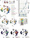 Figure 2
MI promotes phenotypic changes in specific subpopulations of pericardial macrophages. (A) Schematic of PF collection from the 2 patient groups, NOCAD (n = 4) and MI (n = 4), and subsequent analysis using scRNA-seq. (B) UMAP projection of 54,765 PFcs from the 8 patients, identifying 19 distinct states/populations of immune and non-immune cells. (C) Bubble plot with scRNA-seq expression of canonical lineage and subset markers. (D) UMAP projections of PFcs from NOCAD and MI patient samples and stacked plot quantification of cell clusters from NOCAD and MI patient samples. *P < 0.05, generalized linear mixed-effects model. (E) Kernel density estimates depicting magnitude of molecular response elicited by pericardial immune cell subsets in both patient groups (left) and those upregulated (right) compared with downregulated (middle) in the MI group relative to the NOCAD group, calculated by summing of differentially expressed gene fold changes for each cell cluster.
Comparison of these populations under different disease states revealed that macrophage cluster 0 was proportionally reduced in MI patient samples compared with NOCAD patient samples (Figure 2D and Supplemental Figure 1). Further, the global magnitude of molecular responses between the NOCAD and MI cohorts was largely concentrated within macrophage clusters 0 and 1 with 139 and 133 differentially expressed genes, respectively (Figure 2E and Supplemental Table 3). This is in contrast to the other macrophage clusters 10, 14, and 16, which displayed 9, 3, and 0 differentially expressed genes, respectively (Supplemental Table 3). Pathway analysis for differentially expressed genes in cluster 0 macrophages identified pathways related to interleukin signaling, hemostasis, and angiogenesis, enriched in the MI condition. In contrast, protein/peptide processing and phagocytosis were favored in the NOCAD setting (Supplemental Figure 2). Concerning cluster 1 macrophages, pathways related to allograft rejection and to complement were highlighted under NOCAD conditions (Supplemental Figure 2). In response to MI, cell stress responses and leukocyte differentiation–associated pathways were increased. In the MI setting, both macrophage clusters showed alterations in pathways related to antigen processing and to MHC class II antigen presentation (Supplemental Figure 2).
Human pericardial macrophage trajectories are responsive to cardiac injury. Since cluster 0 and 1 macrophages were the prominent immune cell type that responded to MI, we aimed to define their maturation dynamics by performing velocity analysis. Louvain clustering, disease group, individual patient, and velocity length were overlaid with velocity vector fields to generate a latent time plot (Figure 3A). Analyzing the velocity vectors in the 2 patient cohorts revealed 5 macrophage states (Figure 3B). In the NOCAD patients, macrophage state 0 predominated (Figure 3C). In contrast, MI patients demonstrated a shift to macrophage state 1 (Figure 3C). Modeling the differentiation paths revealed 2 trajectories defined by FN1 and CCL2 expression (Figure 3, D and E). The vector lengths in the NOCAD condition were longer and more directional toward the FN1hi state in comparison with MI, supporting more significant differentiation of this macrophage state (Figure 3B). In addition, the connection lineage between the CCL2hi state and the FN1hi state was lost in the MI setting (Figure 3D). To characterize these macrophage states/populations further, the gene expression trends of additional markers were queried using kernel density plots. FN1hi macrophages demonstrated higher gene expression of CD14, CD163, HLA-DRA, TIMD4, and LYVE1 regardless of disease context (Figure 3F and Supplemental Figure 3). The expression of the transcription factor GATA6, which is commonly used to define resident pericardial macrophages in mice, was also enhanced in the FN1hi macrophages (Figure 3F and Supplemental Figure 3). It should be noted, however, that the overall GATA6 expression was lower compared with these other markers and GATA6 was only expressed by approximately 5% of FN1hi macrophages. The CCL2 macrophage population demonstrated higher gene expression of LGMN and STAB1 (Figure 3F and Supplemental Figure 3).
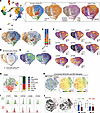 Figure 3
MI dictates maturation trajectories for pericardial macrophages. (A) Louvain clustering of pericardial macrophages represented by state, disease group, and individual patient and overlaid with velocity vector fields to generate a latent time plot. (B) UMAP plotting RNA velocity analysis of subclustered pericardial macrophages undergoing state transitions, colored by cluster ID. (C) Stacked bar plot depicting cluster composition of clinical cohorts examined. (D) UMAP colored by pericardial macrophage clusters and overlaid with summary path curves based on vector fields and pericardial macrophage state composition to determine macrophage states. (E and F) RNA gene expression of selected genes driving divergent maturation trajectories (E) and associated defining markers (F). (G) Flow cytometry analysis and quantification of pericardial immune populations in combined NOCAD and MI samples. (H and I) Flow cytometry–based representation of pericardial macrophage populations depicted by t-SNE plots (H) and expression of protein markers by histogram plots in FN1hi and FN1lo pericardial macrophage populations (I). (J) Quantification of FN1hi and FN1lo pericardial macrophage populations in both NOCAD and MI patient samples. n = 5 for NOCAD, n = 11 for MI. *P < 0.05, unpaired 2-tailed t test.
Pericardial macrophage populations were further explored by flow cytometry. Macrophages represented roughly 50% of the local pericardial CD45 compartment, and 2 subsets were identified by the presence and absence of combined CD163 and FN1 expression (Figure 3, G and H). Consistent with transcriptional data, CD163hiFN1+ macrophages were also defined by TIM4 and LYVE1 expression (Figure 3I). In contrast, CD163loFN1– macrophages did not display CCL2 protein expression (data not shown) nor unique expression of LGMN and STAB1 (Figure 3I). CD163hiFN1+ pericardial macrophages were enriched in NOCAD patient samples relative to MI patients, whereas CD163loFN1– macrophages remained unchanged, mimicking changes in clusters 0 and 1 observed by scRNA-seq (Figure 2D and Figure 3J).
Human pericardial macrophage subsets have unique gene regulatory and niche networks. To determine the factors potentially driving these macrophage trajectories, gene regulatory network development using single-cell regulatory network inference and clustering (SCENIC) was performed (Figure 4A) (19). The regulon density was also assessed for the 2 macrophage clusters (Figure 4A). This analysis revealed that the transcription factors IRF2 and HOXB6 are predicted as key drivers of FN1hi cluster 0 macrophage–related genes (Figure 4B and Supplemental Figure 4). Meanwhile, predicted genes downstream of PPARG, MXD1, and XBP1 activity primarily localized to CCL2hi cluster 1 macrophages (Figure 4C and Supplemental Figure 4). These findings suggest that the divergently activated macrophage states are controlled by distinct regulatory programs, uncovering key upstream regulators that dictate pericardial macrophage populations under homeostatic and pathological conditions.
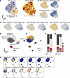 Figure 4
Gene regulatory and signaling networks for pericardial macrophages. (A) Gene regulatory network (GRN) clustering and regulon density in cluster 0 (c0) and cluster 1 (c1) pericardial macrophages. (B and C) Gene regulatory networks preferentially driving cluster 0 (IRF2, HOXB6; B) and cluster 1 (PPARG, MXD1, XBP1; C) pericardial macrophage profiles. (D and E) UMAP plotting of signaling pathways associated with pericardial macrophages by NICHES analysis (D) and representation of cluster 0 and 1 pericardial macrophages within these signaling clusters (E). (F and G) Stacked bar plots depicting signaling cluster representation between disease states (F) or pericardial macrophage clusters (G). (H) UMAP plotting of expression distribution for PDGFB and FN1; CCL2, CCL13, CALM3, CALM1, and CALR; C3; WNT5A; VEGFA; and PLAU.
Given the divergent trajectories and regulatory networks of cluster 0 and 1 pericardial macrophages, we next explored their cell-to-cell signaling capacity locally. Performing niche interactions and communication heterogeneity in extracellular signaling (NICHES) analysis on these macrophage subsets revealed their involvement in several signaling pathways (Figure 4, D and E). FN1-, PDGFB-, C3-, and WNT5A-related signaling pathways were enriched under NOCAD conditions and in cluster 0 macrophages (Figure 4, D and F–H). In response to MI, CCL2- and CALM-related signaling was enhanced largely by cluster 1 macrophages (Figure 4, D and F–H). Both PLAU signaling and VEGFA signaling were relatively equally represented across disease states and macrophage clusters (Figure 4, D and F–H). This supports largely differing functions for these pericardial macrophage subsets within their local milieu.
FN1+ pericardial macrophages inhibit fibroblast activity via cystatin C secretion. Since the FN1+CD163hi pericardial macrophage subset is the most abundant cell population and the most altered following MI, we explored whether these cells are responsible for the antifibrotic effects on human cardiac fibroblasts. We applied a magnetic bead approach to isolate FN1+CD163hi pericardial macrophages, which resulted in a 90% overall enrichment and 95% among myeloid cells (Supplemental Figure 5). A parallel approach was applied to isolate pericardial T cells, the second most abundant pericardial immune cell type to serve as a cellular control (Supplemental Figure 5). Applying enriched FN1+ pericardial macrophages to the coculture system recapitulated the inhibitory effect observed with total PFcs (Figure 1A and Figure 5, A and B). Importantly, this activity was not seen when isolated pericardial T cells were coincubated with the fibroblast gels (Figure 5B). In addition, this macrophage inhibitory action was even enhanced when FN1+ pericardial macrophages were seeded on a Transwell insert placed above the fibroblast gel, thus supporting a paracrine mechanism (Figure 5C).
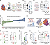 Figure 5
FN1+ pericardial macrophages inhibit cardiac fibroblast activity via cystatin C. (A and B) Schematic of in vitro coculture setup (A) and quantification of cardiac fibroblast–induced (CFb-induced) collagen gel contraction in response to TGF-β1 stimulation in the absence (CFb) or presence of enriched FN1+ pericardial macrophages or enriched pericardial T cells (B). n = 10 T cell–enriched patient pericardial samples; n = 13 FN1+ macrophage–enriched patient pericardial samples, and 13 independent experiments. **P < 0.01, 1-way ANOVA with Tukey’s multiple-comparison test. (C) Quantification of CFb-induced collagen gel contraction in response to TGF-β1 stimulation in the absence (CFb) or presence of direct or indirect (Transwell) pericardial macrophages (CD14+). n = 12 patient PF samples and 11 independent experiments. *P < 0.05; ****P < 0.0001, unpaired 2-tailed t test. (D) scRNA-seq quantification of top differentially expressed secreted factors in cluster 0 (FN1+) pericardial macrophages in either NOCAD or MI patients. (E) Schematic representation of IRF2 target sequence and RNA gene expression of its downstream activated gene, CST3, overlaid on the gene regulatory network clustering for pericardial macrophages. (F) Representative histogram plots of CST3 protein expression and quantification of isotype-normalized median fluorescence intensity in FN1+CD163hi and FN1–CD163lo human pericardial macrophages. *P < 0.05. (G) Quantification of CST3 release from conditioned medium of total human PFcs, human pericardial macrophages (CD14+), and pericardial T cells (CD3+). n = 16. ****P < 0.0001, 1-way ANOVA with Tukey’s multiple-comparison test. (H) Quantification of CST3 release from conditioned medium of human pericardial macrophages (CD14+) from NOCAD and MI patients. *P < 0.05. (I) Quantification of CFb-induced collagen gel contraction in response to TGF-β1 stimulation in the absence (CFb) or presence of PFc (CFb:PFc) with CST3 or isotype antibody blockade. n = 11 patient pericardial samples and 6 independent experiments. *P < 0.05, **P < 0.01, ***P < 0.001, 1-way ANOVA with Tukey’s multiple-comparison test.
To identify potential soluble mediators that may be responsible for the antifibrotic effects, we explored secreted factors that were differentially expressed between disease settings in cluster 0 (FN1+) macrophages. Using a 2-fold change cutoff, we identified 12 factors that were enriched in the NOCAD samples and 13 mediators that were enhanced in the MI samples (Figure 5D). Cystatin C (CST3), the second highest differentially expressed mediator in the NOCAD samples, was an intriguing candidate to pursue as the antifibrotic mediator. It is traditionally known as a cysteine protease inhibitor intracellularly but has been reported to inhibit TGF-β signaling through the engagement of TGF-β receptor 1 extracellularly (20). CST3 was more highly expressed transcriptionally by macrophage cluster 0 (FN1+) and is regulated by IRF2, identified by the SCENIC analysis as a key driver of this macrophage phenotype (Figure 4B and Figure 5E). Higher protein expression of CST3 in FN1+ pericardial macrophages relative to FN1– pericardial macrophages was also confirmed by flow cytometry (Figure 5F). To further validate its active release, we analyzed conditioned media from cultured total PFcs, enriched FN1+CD163hi pericardial macrophages, or enriched pericardial T cells. Total PFcs and FN1+ pericardial macrophages showed higher release of CST3 than the pericardial T cells (Figure 5G). We also found that FN1+ pericardial macrophages from NOCAD patients secreted a significantly higher amount of CST3 compared with MI patient samples (Figure 5H), which is in line with the greater antifibrotic action observed from NOCAD PFcs (Figure 1C). Finally, CST3 antibody blockade in the coculture system abrogated the antifibrotic capacity of PFcs, confirming CST3 as a key mediator of this inhibitory action of pericardial macrophages (Figure 5I).
To determine whether FN1+ pericardial macrophages could play a more direct role in the injured heart, we explored whether the observed decrease in FN1+ macrophages in PF (Figure 2D) is complemented by the presence of FN1+ macrophages in the human heart acutely following MI. For this, we utilized the single-nucleus RNA-seq (snRNA-seq) dataset from Kuppe et al. (21), who sampled left ventricular (LV) cardiac tissue from healthy (control) and ischemic patients at both early (ischemic, <12 days after MI) and late time points (fibrotic, >30 days after MI). FN1+ macrophages were enriched as part of the SPP1+ macrophage cluster and were primarily found in the ischemic regions of the heart relative to the healthy myogenic or fibrotic tissue (Supplemental Figure 6, A–C). Consistent with the downregulation in CST3 observed in the FN1+ pericardial macrophages, FN1+ macrophages in the heart displayed lower expression of CST3 when compared with resident cardiac macrophage and monocyte/DC clusters.
Myeloid-derived Cst3 alters fibrotic activity in vivo. The observed antifibrotic property of pericardial macrophage–derived CST3 in vitro prompted the exploration of CST3 function in vivo following MI. We first determined whether pericardial macrophages in the mouse system shared features with those described in patient samples. The mouse system has 2 pericardial macrophage populations; the major population is defined by its expression of the transcription factor Gata6. Gata6-expressing pericardial macrophages (GPCMs) denote high protein expression of FN1 and TIM4, and to a lesser extent LYVE1 (Figure 6A). Further, Fn1 and Cst3 were among the top-expressed genes from an RNA-seq dataset of sorted GPCMs (Figure 6B) (15).
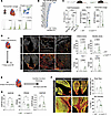 Figure 6
Myeloid-derived Cst3 alters cardiac fibrosis. (A) Representative flow cytometry analysis of mouse GPCM expression of FN1, TIM4, and LYVE1. Representative of n = 4. (B) RNA-seq quantification of top-expressed genes by sorted mouse GPCMs. n = 4. (C) qPCR quantification of Cst3 mRNA expression in pericardial macrophages, cardiac macrophages, and Ly6Chi monocytes from WT (Cst3fl/fl) and Cst3ΔLyz2 mice (left), and ELISA quantification of serum (middle) and pericardial lavage (right) CST3 levels from WT and Cst3ΔLyz2 mice. n = 3 for qPCR; n = 8 for serum samples; n = 6 and 4 for pericardial lavage samples from WT and Cst3ΔLyz2 mice, respectively. *P < 0.05, **P < 0.01, unpaired 2-tailed t test for ELISA; ****P < 0.0001, 1-way ANOVA with Tukey’s multiple-comparison test for qPCR. (D) Immunohistochemistry staining and quantification for α-smooth muscle actin (α-SMA) and collagen I (Col I) in the infarct and border zones from WT (Cst3fl/fl) and Cst3ΔLyz2 mice at 7 days after MI. Scale bars: 1,000 μm. Data are represented as mean ± SEM; n = 6 and 8 for WT and Cst3ΔLyz2 mice, respectively. Unpaired 2-tailed t test, non-parametric (Mann-Whitney) for α-SMA and parametric (Welch’s correction) for Col I. (E) Schematic of experimental timeline for cardiac function and fibrosis analysis at 28 days after MI. (F) Representative confocal composite stitch images of the LV and border zone (BZ) with Picrosirius red (PSR) staining and quantification of total LV scar size and BZ fibrosis indicated by PSR staining in cardiac cross sections at 28 days after MI for WT and Cst3ΔLyz2 mice. Scale bars: 1,000 μm. Data are represented as mean ± SEM; n = 8 and 10 for WT and Cst3ΔLyz2 mice, respectively. *P < 0.05, 2-tailed t test. (G) LV functional parameters (end systolic pressure volume relationship [ESPVR], preload recruitable stroke work [PRSW], and end diastolic pressure volume relationship [EDPVR]) at 28 days after MI for WT and Cst3ΔLyz2 mice measured by pressure-volume assessment. Data are represented as mean ± SEM; n = 14 for WT and n = 15 for Cst3ΔLyz2 mice. *P < 0.05, unpaired 2-tailed t test.
To investigate a role of CST3 in vivo, a mouse with conditional deletion of Cst3 under the control of the lysozyme 2 promoter–driven Cre recombinase (Cst3ΔLyz2) was generated. This construct contributed to a decrease in Cst3 expression in pericardial macrophages in addition to cardiac macrophages in Cst3ΔLyz2 mice compared with Cst3fl/fl (WT) littermate controls (Figure 6C). Ly6Chi monocytes demonstrated a reduced basal expression of Cst3 relative to these macrophage populations and remained unchanged in the conditional knockout system (Figure 6C). CST3 protein levels in circulation and pericardial lavage fluid were also decreased in Cst3ΔLyz2 mice relative to WT mice (Figure 6C). Baseline cardiac function was not altered between genotypes (Supplemental Table 4). The loss of Cst3 in myeloid cells did not impact infarct size after MI induction (48 hours after MI) nor myeloid cell recruitment (day 5 after MI) in comparison with WT littermate mice (Supplemental Figure 7, A and B). To assess whether myeloid-derived Cst3 could influence myofibroblast generation in vivo, α-smooth muscle actin (α-SMA) and collagen I staining was performed on infarcted cardiac tissue sections collected at day 7 after MI (Figure 6D). Both markers displayed a numerical increase in the infarcted and border zone regions of Cst3ΔLyz2 mice compared with WT controls, although these differences did not reach statistical significance (Figure 6D). To determine the downstream impact of these myofibroblast-linked features, fibrosis and cardiac function were further assessed at 28 days after MI (Figure 6E). The overall scar size did not change; however, increased border zone fibrosis was noted in Cst3ΔLyz2 mice compared with WT littermates (Figure 6F). This was combined with worse cardiac function in Cst3ΔLyz2 mice, including an increase in LV stiffness as indicated by augmented end diastolic pressure volume relationship values and a decrease in the systolic parameters end systolic pressure volume relationship and preload recruitable stroke work (Figure 6G and Supplemental Table 5). Collectively, this establishes a new role for myeloid cell–derived CST3 in modulating cardiac fibrosis in vivo.


