
Research ArticleAIDS/HIVImmunologyInfectious disease
Open Access |  10.1172/jci.insight.201268
The pleural tuberculosis–associated microenvironment promotes HIV-1 persistence by impairing CD8+ T cell–mediated viral control
Samantha Cronin,1,2 Jennifer Simpson,1,2 Andrea Pereyra-Casanova,1,2 Yuchen Li,1,2 Josefina Marín-Rojas,1,2 Freja A. Warner van Dijk,1,2 Katie Fisher,2 Daniel J. Buffa,1,2 Hafsa Rana,1,2 Zoï Vahlas,3 Joaquina Barros,4,5 Mariano Maio,4,5 Thomas R. O’Neil,1,2 Kirstie M. Bertram,1,2 Eunok Lee,2 Najla Nasr,1,2 Andrew N. Harman,1,2 Gabriela Turk,4,5 Maria Florencia Quiroga,4,5 Anthony D. Kelleher,6 Christel Vérollet,3,7 Luciana Balboa,4,5,7 Sarah Palmer,1,2 and Gabriel Duette1,2
1The University of Sydney, Faculty of Medicine and Health, Sydney, New South Wales, Australia.
2The Westmead Institute for Medical Rese arch, Centre for Virus Research, Sydney, New South Wales, Australia.
3Université de Toulouse, CNRS, Institut de Pharmacologie et de Biologie Structurale (IPBS), Toulouse, France.
4Universidad de Buenos Aires, Facultad de Medicina, Departamento de Microbiología, Parasitología e Inmunología, Buenos Aires, Argentina.
5Consejo Nacional de Investigaciones Científicas y Técnicas (CONICET) – Universidad de Buenos Aires. Instituto de Investigaciones Biomédicas en Retrovirus y SIDA (INBIRS). Buenos Aires. Argentina.
6Kirby Institute, University of New South Wales, Sydney, New South Wales, Australia.
7International Research Project CNRS “MAC-TB/HIV,” Toulouse, France, and Buenos Aires, Argentina.
Address correspondence to: Samantha Cronin or Gabriel Duette, The Westmead Institute for Medical Research, 176 Hawkesbury Road, Westmead, New South Wales 2145, Australia. Email: samantha.cronin@sydney.edu.au (SC); gabriel.duette@sydney.edu.au (GD).
Find articles by Cronin, S. in: PubMed | Google Scholar
1The University of Sydney, Faculty of Medicine and Health, Sydney, New South Wales, Australia.
2The Westmead Institute for Medical Rese arch, Centre for Virus Research, Sydney, New South Wales, Australia.
3Université de Toulouse, CNRS, Institut de Pharmacologie et de Biologie Structurale (IPBS), Toulouse, France.
4Universidad de Buenos Aires, Facultad de Medicina, Departamento de Microbiología, Parasitología e Inmunología, Buenos Aires, Argentina.
5Consejo Nacional de Investigaciones Científicas y Técnicas (CONICET) – Universidad de Buenos Aires. Instituto de Investigaciones Biomédicas en Retrovirus y SIDA (INBIRS). Buenos Aires. Argentina.
6Kirby Institute, University of New South Wales, Sydney, New South Wales, Australia.
7International Research Project CNRS “MAC-TB/HIV,” Toulouse, France, and Buenos Aires, Argentina.
Address correspondence to: Samantha Cronin or Gabriel Duette, The Westmead Institute for Medical Research, 176 Hawkesbury Road, Westmead, New South Wales 2145, Australia. Email: samantha.cronin@sydney.edu.au (SC); gabriel.duette@sydney.edu.au (GD).
Find articles by Simpson, J. in: PubMed | Google Scholar
1The University of Sydney, Faculty of Medicine and Health, Sydney, New South Wales, Australia.
2The Westmead Institute for Medical Rese arch, Centre for Virus Research, Sydney, New South Wales, Australia.
3Université de Toulouse, CNRS, Institut de Pharmacologie et de Biologie Structurale (IPBS), Toulouse, France.
4Universidad de Buenos Aires, Facultad de Medicina, Departamento de Microbiología, Parasitología e Inmunología, Buenos Aires, Argentina.
5Consejo Nacional de Investigaciones Científicas y Técnicas (CONICET) – Universidad de Buenos Aires. Instituto de Investigaciones Biomédicas en Retrovirus y SIDA (INBIRS). Buenos Aires. Argentina.
6Kirby Institute, University of New South Wales, Sydney, New South Wales, Australia.
7International Research Project CNRS “MAC-TB/HIV,” Toulouse, France, and Buenos Aires, Argentina.
Address correspondence to: Samantha Cronin or Gabriel Duette, The Westmead Institute for Medical Research, 176 Hawkesbury Road, Westmead, New South Wales 2145, Australia. Email: samantha.cronin@sydney.edu.au (SC); gabriel.duette@sydney.edu.au (GD).
Find articles by Pereyra-Casanova, A. in: PubMed | Google Scholar
1The University of Sydney, Faculty of Medicine and Health, Sydney, New South Wales, Australia.
2The Westmead Institute for Medical Rese arch, Centre for Virus Research, Sydney, New South Wales, Australia.
3Université de Toulouse, CNRS, Institut de Pharmacologie et de Biologie Structurale (IPBS), Toulouse, France.
4Universidad de Buenos Aires, Facultad de Medicina, Departamento de Microbiología, Parasitología e Inmunología, Buenos Aires, Argentina.
5Consejo Nacional de Investigaciones Científicas y Técnicas (CONICET) – Universidad de Buenos Aires. Instituto de Investigaciones Biomédicas en Retrovirus y SIDA (INBIRS). Buenos Aires. Argentina.
6Kirby Institute, University of New South Wales, Sydney, New South Wales, Australia.
7International Research Project CNRS “MAC-TB/HIV,” Toulouse, France, and Buenos Aires, Argentina.
Address correspondence to: Samantha Cronin or Gabriel Duette, The Westmead Institute for Medical Research, 176 Hawkesbury Road, Westmead, New South Wales 2145, Australia. Email: samantha.cronin@sydney.edu.au (SC); gabriel.duette@sydney.edu.au (GD).
Find articles by Li, Y. in: PubMed | Google Scholar
1The University of Sydney, Faculty of Medicine and Health, Sydney, New South Wales, Australia.
2The Westmead Institute for Medical Rese arch, Centre for Virus Research, Sydney, New South Wales, Australia.
3Université de Toulouse, CNRS, Institut de Pharmacologie et de Biologie Structurale (IPBS), Toulouse, France.
4Universidad de Buenos Aires, Facultad de Medicina, Departamento de Microbiología, Parasitología e Inmunología, Buenos Aires, Argentina.
5Consejo Nacional de Investigaciones Científicas y Técnicas (CONICET) – Universidad de Buenos Aires. Instituto de Investigaciones Biomédicas en Retrovirus y SIDA (INBIRS). Buenos Aires. Argentina.
6Kirby Institute, University of New South Wales, Sydney, New South Wales, Australia.
7International Research Project CNRS “MAC-TB/HIV,” Toulouse, France, and Buenos Aires, Argentina.
Address correspondence to: Samantha Cronin or Gabriel Duette, The Westmead Institute for Medical Research, 176 Hawkesbury Road, Westmead, New South Wales 2145, Australia. Email: samantha.cronin@sydney.edu.au (SC); gabriel.duette@sydney.edu.au (GD).
Find articles by Marín-Rojas, J. in: PubMed | Google Scholar
1The University of Sydney, Faculty of Medicine and Health, Sydney, New South Wales, Australia.
2The Westmead Institute for Medical Rese arch, Centre for Virus Research, Sydney, New South Wales, Australia.
3Université de Toulouse, CNRS, Institut de Pharmacologie et de Biologie Structurale (IPBS), Toulouse, France.
4Universidad de Buenos Aires, Facultad de Medicina, Departamento de Microbiología, Parasitología e Inmunología, Buenos Aires, Argentina.
5Consejo Nacional de Investigaciones Científicas y Técnicas (CONICET) – Universidad de Buenos Aires. Instituto de Investigaciones Biomédicas en Retrovirus y SIDA (INBIRS). Buenos Aires. Argentina.
6Kirby Institute, University of New South Wales, Sydney, New South Wales, Australia.
7International Research Project CNRS “MAC-TB/HIV,” Toulouse, France, and Buenos Aires, Argentina.
Address correspondence to: Samantha Cronin or Gabriel Duette, The Westmead Institute for Medical Research, 176 Hawkesbury Road, Westmead, New South Wales 2145, Australia. Email: samantha.cronin@sydney.edu.au (SC); gabriel.duette@sydney.edu.au (GD).
Find articles by Warner van Dijk, F. in: PubMed | Google Scholar
1The University of Sydney, Faculty of Medicine and Health, Sydney, New South Wales, Australia.
2The Westmead Institute for Medical Rese arch, Centre for Virus Research, Sydney, New South Wales, Australia.
3Université de Toulouse, CNRS, Institut de Pharmacologie et de Biologie Structurale (IPBS), Toulouse, France.
4Universidad de Buenos Aires, Facultad de Medicina, Departamento de Microbiología, Parasitología e Inmunología, Buenos Aires, Argentina.
5Consejo Nacional de Investigaciones Científicas y Técnicas (CONICET) – Universidad de Buenos Aires. Instituto de Investigaciones Biomédicas en Retrovirus y SIDA (INBIRS). Buenos Aires. Argentina.
6Kirby Institute, University of New South Wales, Sydney, New South Wales, Australia.
7International Research Project CNRS “MAC-TB/HIV,” Toulouse, France, and Buenos Aires, Argentina.
Address correspondence to: Samantha Cronin or Gabriel Duette, The Westmead Institute for Medical Research, 176 Hawkesbury Road, Westmead, New South Wales 2145, Australia. Email: samantha.cronin@sydney.edu.au (SC); gabriel.duette@sydney.edu.au (GD).
Find articles by Fisher, K. in: PubMed | Google Scholar
1The University of Sydney, Faculty of Medicine and Health, Sydney, New South Wales, Australia.
2The Westmead Institute for Medical Rese arch, Centre for Virus Research, Sydney, New South Wales, Australia.
3Université de Toulouse, CNRS, Institut de Pharmacologie et de Biologie Structurale (IPBS), Toulouse, France.
4Universidad de Buenos Aires, Facultad de Medicina, Departamento de Microbiología, Parasitología e Inmunología, Buenos Aires, Argentina.
5Consejo Nacional de Investigaciones Científicas y Técnicas (CONICET) – Universidad de Buenos Aires. Instituto de Investigaciones Biomédicas en Retrovirus y SIDA (INBIRS). Buenos Aires. Argentina.
6Kirby Institute, University of New South Wales, Sydney, New South Wales, Australia.
7International Research Project CNRS “MAC-TB/HIV,” Toulouse, France, and Buenos Aires, Argentina.
Address correspondence to: Samantha Cronin or Gabriel Duette, The Westmead Institute for Medical Research, 176 Hawkesbury Road, Westmead, New South Wales 2145, Australia. Email: samantha.cronin@sydney.edu.au (SC); gabriel.duette@sydney.edu.au (GD).
Find articles by Buffa, D. in: PubMed | Google Scholar
1The University of Sydney, Faculty of Medicine and Health, Sydney, New South Wales, Australia.
2The Westmead Institute for Medical Rese arch, Centre for Virus Research, Sydney, New South Wales, Australia.
3Université de Toulouse, CNRS, Institut de Pharmacologie et de Biologie Structurale (IPBS), Toulouse, France.
4Universidad de Buenos Aires, Facultad de Medicina, Departamento de Microbiología, Parasitología e Inmunología, Buenos Aires, Argentina.
5Consejo Nacional de Investigaciones Científicas y Técnicas (CONICET) – Universidad de Buenos Aires. Instituto de Investigaciones Biomédicas en Retrovirus y SIDA (INBIRS). Buenos Aires. Argentina.
6Kirby Institute, University of New South Wales, Sydney, New South Wales, Australia.
7International Research Project CNRS “MAC-TB/HIV,” Toulouse, France, and Buenos Aires, Argentina.
Address correspondence to: Samantha Cronin or Gabriel Duette, The Westmead Institute for Medical Research, 176 Hawkesbury Road, Westmead, New South Wales 2145, Australia. Email: samantha.cronin@sydney.edu.au (SC); gabriel.duette@sydney.edu.au (GD).
Find articles by Rana, H. in: PubMed | Google Scholar
1The University of Sydney, Faculty of Medicine and Health, Sydney, New South Wales, Australia.
2The Westmead Institute for Medical Rese arch, Centre for Virus Research, Sydney, New South Wales, Australia.
3Université de Toulouse, CNRS, Institut de Pharmacologie et de Biologie Structurale (IPBS), Toulouse, France.
4Universidad de Buenos Aires, Facultad de Medicina, Departamento de Microbiología, Parasitología e Inmunología, Buenos Aires, Argentina.
5Consejo Nacional de Investigaciones Científicas y Técnicas (CONICET) – Universidad de Buenos Aires. Instituto de Investigaciones Biomédicas en Retrovirus y SIDA (INBIRS). Buenos Aires. Argentina.
6Kirby Institute, University of New South Wales, Sydney, New South Wales, Australia.
7International Research Project CNRS “MAC-TB/HIV,” Toulouse, France, and Buenos Aires, Argentina.
Address correspondence to: Samantha Cronin or Gabriel Duette, The Westmead Institute for Medical Research, 176 Hawkesbury Road, Westmead, New South Wales 2145, Australia. Email: samantha.cronin@sydney.edu.au (SC); gabriel.duette@sydney.edu.au (GD).
Find articles by Vahlas, Z. in: PubMed | Google Scholar
1The University of Sydney, Faculty of Medicine and Health, Sydney, New South Wales, Australia.
2The Westmead Institute for Medical Rese arch, Centre for Virus Research, Sydney, New South Wales, Australia.
3Université de Toulouse, CNRS, Institut de Pharmacologie et de Biologie Structurale (IPBS), Toulouse, France.
4Universidad de Buenos Aires, Facultad de Medicina, Departamento de Microbiología, Parasitología e Inmunología, Buenos Aires, Argentina.
5Consejo Nacional de Investigaciones Científicas y Técnicas (CONICET) – Universidad de Buenos Aires. Instituto de Investigaciones Biomédicas en Retrovirus y SIDA (INBIRS). Buenos Aires. Argentina.
6Kirby Institute, University of New South Wales, Sydney, New South Wales, Australia.
7International Research Project CNRS “MAC-TB/HIV,” Toulouse, France, and Buenos Aires, Argentina.
Address correspondence to: Samantha Cronin or Gabriel Duette, The Westmead Institute for Medical Research, 176 Hawkesbury Road, Westmead, New South Wales 2145, Australia. Email: samantha.cronin@sydney.edu.au (SC); gabriel.duette@sydney.edu.au (GD).
Find articles by Barros, J. in: PubMed | Google Scholar
1The University of Sydney, Faculty of Medicine and Health, Sydney, New South Wales, Australia.
2The Westmead Institute for Medical Rese arch, Centre for Virus Research, Sydney, New South Wales, Australia.
3Université de Toulouse, CNRS, Institut de Pharmacologie et de Biologie Structurale (IPBS), Toulouse, France.
4Universidad de Buenos Aires, Facultad de Medicina, Departamento de Microbiología, Parasitología e Inmunología, Buenos Aires, Argentina.
5Consejo Nacional de Investigaciones Científicas y Técnicas (CONICET) – Universidad de Buenos Aires. Instituto de Investigaciones Biomédicas en Retrovirus y SIDA (INBIRS). Buenos Aires. Argentina.
6Kirby Institute, University of New South Wales, Sydney, New South Wales, Australia.
7International Research Project CNRS “MAC-TB/HIV,” Toulouse, France, and Buenos Aires, Argentina.
Address correspondence to: Samantha Cronin or Gabriel Duette, The Westmead Institute for Medical Research, 176 Hawkesbury Road, Westmead, New South Wales 2145, Australia. Email: samantha.cronin@sydney.edu.au (SC); gabriel.duette@sydney.edu.au (GD).
Find articles by Maio, M. in: PubMed | Google Scholar
1The University of Sydney, Faculty of Medicine and Health, Sydney, New South Wales, Australia.
2The Westmead Institute for Medical Rese arch, Centre for Virus Research, Sydney, New South Wales, Australia.
3Université de Toulouse, CNRS, Institut de Pharmacologie et de Biologie Structurale (IPBS), Toulouse, France.
4Universidad de Buenos Aires, Facultad de Medicina, Departamento de Microbiología, Parasitología e Inmunología, Buenos Aires, Argentina.
5Consejo Nacional de Investigaciones Científicas y Técnicas (CONICET) – Universidad de Buenos Aires. Instituto de Investigaciones Biomédicas en Retrovirus y SIDA (INBIRS). Buenos Aires. Argentina.
6Kirby Institute, University of New South Wales, Sydney, New South Wales, Australia.
7International Research Project CNRS “MAC-TB/HIV,” Toulouse, France, and Buenos Aires, Argentina.
Address correspondence to: Samantha Cronin or Gabriel Duette, The Westmead Institute for Medical Research, 176 Hawkesbury Road, Westmead, New South Wales 2145, Australia. Email: samantha.cronin@sydney.edu.au (SC); gabriel.duette@sydney.edu.au (GD).
Find articles by O’Neil, T. in: PubMed | Google Scholar
1The University of Sydney, Faculty of Medicine and Health, Sydney, New South Wales, Australia.
2The Westmead Institute for Medical Rese arch, Centre for Virus Research, Sydney, New South Wales, Australia.
3Université de Toulouse, CNRS, Institut de Pharmacologie et de Biologie Structurale (IPBS), Toulouse, France.
4Universidad de Buenos Aires, Facultad de Medicina, Departamento de Microbiología, Parasitología e Inmunología, Buenos Aires, Argentina.
5Consejo Nacional de Investigaciones Científicas y Técnicas (CONICET) – Universidad de Buenos Aires. Instituto de Investigaciones Biomédicas en Retrovirus y SIDA (INBIRS). Buenos Aires. Argentina.
6Kirby Institute, University of New South Wales, Sydney, New South Wales, Australia.
7International Research Project CNRS “MAC-TB/HIV,” Toulouse, France, and Buenos Aires, Argentina.
Address correspondence to: Samantha Cronin or Gabriel Duette, The Westmead Institute for Medical Research, 176 Hawkesbury Road, Westmead, New South Wales 2145, Australia. Email: samantha.cronin@sydney.edu.au (SC); gabriel.duette@sydney.edu.au (GD).
Find articles by Bertram, K. in: PubMed | Google Scholar
1The University of Sydney, Faculty of Medicine and Health, Sydney, New South Wales, Australia.
2The Westmead Institute for Medical Rese arch, Centre for Virus Research, Sydney, New South Wales, Australia.
3Université de Toulouse, CNRS, Institut de Pharmacologie et de Biologie Structurale (IPBS), Toulouse, France.
4Universidad de Buenos Aires, Facultad de Medicina, Departamento de Microbiología, Parasitología e Inmunología, Buenos Aires, Argentina.
5Consejo Nacional de Investigaciones Científicas y Técnicas (CONICET) – Universidad de Buenos Aires. Instituto de Investigaciones Biomédicas en Retrovirus y SIDA (INBIRS). Buenos Aires. Argentina.
6Kirby Institute, University of New South Wales, Sydney, New South Wales, Australia.
7International Research Project CNRS “MAC-TB/HIV,” Toulouse, France, and Buenos Aires, Argentina.
Address correspondence to: Samantha Cronin or Gabriel Duette, The Westmead Institute for Medical Research, 176 Hawkesbury Road, Westmead, New South Wales 2145, Australia. Email: samantha.cronin@sydney.edu.au (SC); gabriel.duette@sydney.edu.au (GD).
Find articles by Lee, E. in: PubMed | Google Scholar
1The University of Sydney, Faculty of Medicine and Health, Sydney, New South Wales, Australia.
2The Westmead Institute for Medical Rese arch, Centre for Virus Research, Sydney, New South Wales, Australia.
3Université de Toulouse, CNRS, Institut de Pharmacologie et de Biologie Structurale (IPBS), Toulouse, France.
4Universidad de Buenos Aires, Facultad de Medicina, Departamento de Microbiología, Parasitología e Inmunología, Buenos Aires, Argentina.
5Consejo Nacional de Investigaciones Científicas y Técnicas (CONICET) – Universidad de Buenos Aires. Instituto de Investigaciones Biomédicas en Retrovirus y SIDA (INBIRS). Buenos Aires. Argentina.
6Kirby Institute, University of New South Wales, Sydney, New South Wales, Australia.
7International Research Project CNRS “MAC-TB/HIV,” Toulouse, France, and Buenos Aires, Argentina.
Address correspondence to: Samantha Cronin or Gabriel Duette, The Westmead Institute for Medical Research, 176 Hawkesbury Road, Westmead, New South Wales 2145, Australia. Email: samantha.cronin@sydney.edu.au (SC); gabriel.duette@sydney.edu.au (GD).
Find articles by Nasr, N. in: PubMed | Google Scholar
1The University of Sydney, Faculty of Medicine and Health, Sydney, New South Wales, Australia.
2The Westmead Institute for Medical Rese arch, Centre for Virus Research, Sydney, New South Wales, Australia.
3Université de Toulouse, CNRS, Institut de Pharmacologie et de Biologie Structurale (IPBS), Toulouse, France.
4Universidad de Buenos Aires, Facultad de Medicina, Departamento de Microbiología, Parasitología e Inmunología, Buenos Aires, Argentina.
5Consejo Nacional de Investigaciones Científicas y Técnicas (CONICET) – Universidad de Buenos Aires. Instituto de Investigaciones Biomédicas en Retrovirus y SIDA (INBIRS). Buenos Aires. Argentina.
6Kirby Institute, University of New South Wales, Sydney, New South Wales, Australia.
7International Research Project CNRS “MAC-TB/HIV,” Toulouse, France, and Buenos Aires, Argentina.
Address correspondence to: Samantha Cronin or Gabriel Duette, The Westmead Institute for Medical Research, 176 Hawkesbury Road, Westmead, New South Wales 2145, Australia. Email: samantha.cronin@sydney.edu.au (SC); gabriel.duette@sydney.edu.au (GD).
Find articles by
Harman, A.
in:
PubMed
|
Google Scholar
|

1The University of Sydney, Faculty of Medicine and Health, Sydney, New South Wales, Australia.
2The Westmead Institute for Medical Rese arch, Centre for Virus Research, Sydney, New South Wales, Australia.
3Université de Toulouse, CNRS, Institut de Pharmacologie et de Biologie Structurale (IPBS), Toulouse, France.
4Universidad de Buenos Aires, Facultad de Medicina, Departamento de Microbiología, Parasitología e Inmunología, Buenos Aires, Argentina.
5Consejo Nacional de Investigaciones Científicas y Técnicas (CONICET) – Universidad de Buenos Aires. Instituto de Investigaciones Biomédicas en Retrovirus y SIDA (INBIRS). Buenos Aires. Argentina.
6Kirby Institute, University of New South Wales, Sydney, New South Wales, Australia.
7International Research Project CNRS “MAC-TB/HIV,” Toulouse, France, and Buenos Aires, Argentina.
Address correspondence to: Samantha Cronin or Gabriel Duette, The Westmead Institute for Medical Research, 176 Hawkesbury Road, Westmead, New South Wales 2145, Australia. Email: samantha.cronin@sydney.edu.au (SC); gabriel.duette@sydney.edu.au (GD).
Find articles by Turk, G. in: PubMed | Google Scholar
1The University of Sydney, Faculty of Medicine and Health, Sydney, New South Wales, Australia.
2The Westmead Institute for Medical Rese arch, Centre for Virus Research, Sydney, New South Wales, Australia.
3Université de Toulouse, CNRS, Institut de Pharmacologie et de Biologie Structurale (IPBS), Toulouse, France.
4Universidad de Buenos Aires, Facultad de Medicina, Departamento de Microbiología, Parasitología e Inmunología, Buenos Aires, Argentina.
5Consejo Nacional de Investigaciones Científicas y Técnicas (CONICET) – Universidad de Buenos Aires. Instituto de Investigaciones Biomédicas en Retrovirus y SIDA (INBIRS). Buenos Aires. Argentina.
6Kirby Institute, University of New South Wales, Sydney, New South Wales, Australia.
7International Research Project CNRS “MAC-TB/HIV,” Toulouse, France, and Buenos Aires, Argentina.
Address correspondence to: Samantha Cronin or Gabriel Duette, The Westmead Institute for Medical Research, 176 Hawkesbury Road, Westmead, New South Wales 2145, Australia. Email: samantha.cronin@sydney.edu.au (SC); gabriel.duette@sydney.edu.au (GD).
Find articles by
Quiroga, M.
in:
PubMed
|
Google Scholar
|
1The University of Sydney, Faculty of Medicine and Health, Sydney, New South Wales, Australia.
2The Westmead Institute for Medical Rese arch, Centre for Virus Research, Sydney, New South Wales, Australia.
3Université de Toulouse, CNRS, Institut de Pharmacologie et de Biologie Structurale (IPBS), Toulouse, France.
4Universidad de Buenos Aires, Facultad de Medicina, Departamento de Microbiología, Parasitología e Inmunología, Buenos Aires, Argentina.
5Consejo Nacional de Investigaciones Científicas y Técnicas (CONICET) – Universidad de Buenos Aires. Instituto de Investigaciones Biomédicas en Retrovirus y SIDA (INBIRS). Buenos Aires. Argentina.
6Kirby Institute, University of New South Wales, Sydney, New South Wales, Australia.
7International Research Project CNRS “MAC-TB/HIV,” Toulouse, France, and Buenos Aires, Argentina.
Address correspondence to: Samantha Cronin or Gabriel Duette, The Westmead Institute for Medical Research, 176 Hawkesbury Road, Westmead, New South Wales 2145, Australia. Email: samantha.cronin@sydney.edu.au (SC); gabriel.duette@sydney.edu.au (GD).
Find articles by Kelleher, A. in: PubMed | Google Scholar
1The University of Sydney, Faculty of Medicine and Health, Sydney, New South Wales, Australia.
2The Westmead Institute for Medical Rese arch, Centre for Virus Research, Sydney, New South Wales, Australia.
3Université de Toulouse, CNRS, Institut de Pharmacologie et de Biologie Structurale (IPBS), Toulouse, France.
4Universidad de Buenos Aires, Facultad de Medicina, Departamento de Microbiología, Parasitología e Inmunología, Buenos Aires, Argentina.
5Consejo Nacional de Investigaciones Científicas y Técnicas (CONICET) – Universidad de Buenos Aires. Instituto de Investigaciones Biomédicas en Retrovirus y SIDA (INBIRS). Buenos Aires. Argentina.
6Kirby Institute, University of New South Wales, Sydney, New South Wales, Australia.
7International Research Project CNRS “MAC-TB/HIV,” Toulouse, France, and Buenos Aires, Argentina.
Address correspondence to: Samantha Cronin or Gabriel Duette, The Westmead Institute for Medical Research, 176 Hawkesbury Road, Westmead, New South Wales 2145, Australia. Email: samantha.cronin@sydney.edu.au (SC); gabriel.duette@sydney.edu.au (GD).
Find articles by Vérollet, C. in: PubMed | Google Scholar
1The University of Sydney, Faculty of Medicine and Health, Sydney, New South Wales, Australia.
2The Westmead Institute for Medical Rese arch, Centre for Virus Research, Sydney, New South Wales, Australia.
3Université de Toulouse, CNRS, Institut de Pharmacologie et de Biologie Structurale (IPBS), Toulouse, France.
4Universidad de Buenos Aires, Facultad de Medicina, Departamento de Microbiología, Parasitología e Inmunología, Buenos Aires, Argentina.
5Consejo Nacional de Investigaciones Científicas y Técnicas (CONICET) – Universidad de Buenos Aires. Instituto de Investigaciones Biomédicas en Retrovirus y SIDA (INBIRS). Buenos Aires. Argentina.
6Kirby Institute, University of New South Wales, Sydney, New South Wales, Australia.
7International Research Project CNRS “MAC-TB/HIV,” Toulouse, France, and Buenos Aires, Argentina.
Address correspondence to: Samantha Cronin or Gabriel Duette, The Westmead Institute for Medical Research, 176 Hawkesbury Road, Westmead, New South Wales 2145, Australia. Email: samantha.cronin@sydney.edu.au (SC); gabriel.duette@sydney.edu.au (GD).
Find articles by Balboa, L. in: PubMed | Google Scholar
1The University of Sydney, Faculty of Medicine and Health, Sydney, New South Wales, Australia.
2The Westmead Institute for Medical Rese arch, Centre for Virus Research, Sydney, New South Wales, Australia.
3Université de Toulouse, CNRS, Institut de Pharmacologie et de Biologie Structurale (IPBS), Toulouse, France.
4Universidad de Buenos Aires, Facultad de Medicina, Departamento de Microbiología, Parasitología e Inmunología, Buenos Aires, Argentina.
5Consejo Nacional de Investigaciones Científicas y Técnicas (CONICET) – Universidad de Buenos Aires. Instituto de Investigaciones Biomédicas en Retrovirus y SIDA (INBIRS). Buenos Aires. Argentina.
6Kirby Institute, University of New South Wales, Sydney, New South Wales, Australia.
7International Research Project CNRS “MAC-TB/HIV,” Toulouse, France, and Buenos Aires, Argentina.
Address correspondence to: Samantha Cronin or Gabriel Duette, The Westmead Institute for Medical Research, 176 Hawkesbury Road, Westmead, New South Wales 2145, Australia. Email: samantha.cronin@sydney.edu.au (SC); gabriel.duette@sydney.edu.au (GD).
Find articles by Palmer, S. in: PubMed | Google Scholar
1The University of Sydney, Faculty of Medicine and Health, Sydney, New South Wales, Australia.
2The Westmead Institute for Medical Rese arch, Centre for Virus Research, Sydney, New South Wales, Australia.
3Université de Toulouse, CNRS, Institut de Pharmacologie et de Biologie Structurale (IPBS), Toulouse, France.
4Universidad de Buenos Aires, Facultad de Medicina, Departamento de Microbiología, Parasitología e Inmunología, Buenos Aires, Argentina.
5Consejo Nacional de Investigaciones Científicas y Técnicas (CONICET) – Universidad de Buenos Aires. Instituto de Investigaciones Biomédicas en Retrovirus y SIDA (INBIRS). Buenos Aires. Argentina.
6Kirby Institute, University of New South Wales, Sydney, New South Wales, Australia.
7International Research Project CNRS “MAC-TB/HIV,” Toulouse, France, and Buenos Aires, Argentina.
Address correspondence to: Samantha Cronin or Gabriel Duette, The Westmead Institute for Medical Research, 176 Hawkesbury Road, Westmead, New South Wales 2145, Australia. Email: samantha.cronin@sydney.edu.au (SC); gabriel.duette@sydney.edu.au (GD).
Find articles by
Duette, G.
in:
PubMed
|
Google Scholar
|
Published August 20, 2026 - More info
JCI Insight. 2026;11(19):e201268. https://doi.org/10.1172/jci.insight.201268.
© 2026 Cronin et al. This work is licensed under the Creative Commons Attribution 4.0 International License. To view a copy of this license, visit http://creativecommons.org/licenses/by/4.0/.
Received: November 4, 2025; Accepted: August 12, 2026

-
Results
HIV-1 proviral genetic landscape in PLWH/TB. Despite the clinical consequences of HIV-1/Mtb coinfection, the impact of active TB on the HIV-1 proviral genetic landscape within circulating HIV-1–infected cells remains unclear. To address this, we analyzed HIV-1 proviral sequences from peripheral blood mononuclear cell (PBMC) samples obtained from 5 PLWH and 5 individuals with HIV-1 and active TB (PLWH/TB) (Table 1). The duration of ART ranged from 1 to 4 months for most participants, except for one individual who had been on ART for 8 years and experienced treatment failure (Table 1). Plasma viral loads ranged from less than 50 to 1,180 copies/mL, with no significant differences observed between the 2 groups (PLWH median 285, IQR 66.5–973, vs. PLWH/TB median 823, IQR 128.5–1,132) (Figure 1A). Consistent with previous studies, CD4+ T cell counts were significantly lower in PLWH/TB than in PLWH (PLWH median 441, IQR 388.5–542.5, vs. PLWH/TB median 132, IQR 109.5–360.5; P = 0.007) (Figure 1A) (28, 29).
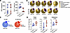 Figure 1
HIV-1 proviral genetic landscape in PLWH/TB. (A) Plasma HIV-1 RNA copies/mL and CD4+ T cell counts in 5 PLWH (blue) and 5 PLWH/TB (red). (B) Pie charts showing the proportion of genetically intact and defective proviral HIV-1 sequences for each donor. PSD, packaging signal deletion; MSD, major splice donor. (C) Averaged proportion of genetically intact and defective proviral HIV-1 sequences across groups. The total number of sequences analyzed per group is shown below each pie chart. (D) Infection frequency of total, genetically intact, and genetically defective proviruses per 106 CD4+ T cells. Frequencies were calculated based on the proportion of CD4+ T cells in each sample and log-transformed. Each data point represents a single donor. Bars represent the median with 95% CI. Statistical significance was determined by 2-tailed paired t test, with P < 0.05 considered significant.
To characterize the HIV-1 proviral genetic landscape in both groups, we used the full-length individual proviral sequencing (FLIPS) assay (30), obtaining a total of 434 HIV-1 DNA sequences from the 10 participants. As previously described in PLWH, the majority of sequences were genetically defective (65.9%–100%), with internal deletions being the most frequent defect (47.1%–94.9%) (Figure 1B) (1). Genetically intact HIV-1 sequences were identified in 9 of the 10 participants (Figure 1B). The proportion of genetically intact proviruses was 18% in PLWH/TB and 12% in PLWH (Figure 1C). When infection frequency was calculated, we observed that in PLWH/TB the frequency of infected cells per million CD4+ T cells tended to be higher for total (PLWH median 48.30, IQR 26.73–145.7, vs. PLWH/TB median 303.2, IQR 90.21–701.4), genetically defective (PLWH median 38.83, IQR 19.03–96.61, vs. PLWH/TB median 165.4, IQR 72.77–434.1), and genetically intact (PLWH median 4.20, IQR 1.92–23.41, vs. PLWH/TB median 36.82, IQR 8.36–110.1) HIV-1 DNA sequences (Figure 1D). These results suggest that concurrent TB may increase the frequency of cells infected with HIV-1.
The proportion of genetically intact HIV-1 provirus is significantly higher at the site of the coinfection. Most studies on HIV-1 infection in PLWH/TB have been conducted using blood samples. However, we and others have shown that at the site of the coinfection the TB-associated microenvironment can influence HIV-1 pathogenesis (21–24, 26, 27). For instance, in people who developed tuberculous pleural effusion (TB-PE), the number of HIV-1 RNA copies was higher in this fluid when compared with matched plasma (Figure 2A and Supplemental Figure 1; supplemental material available online with this article; https://doi.org/10.1172/jci.insight.201268DS1) (21–23). This result led us to investigate the impact of the TB-associated microenvironment on the HIV-1 genetic landscape at this site of coinfection. To this end, we accessed PBMC and pleural effusion–derived mononuclear cell (PEMC) samples from 3 PLWH/TB and performed FLIPS. One of these participants was receiving ART at the time of sample collection, with undetectable plasma viral load, but a relatively high HIV-1 RNA copy number in the TB-PE (Figure 2A and Table 2). In contrast, 2 of these participants were untreated for HIV-1 at the time of sampling (Figure 2A and Table 2). Interestingly, the proportion of genetically intact HIV-1 DNA sequences in PEMCs was higher in comparison with PBMCs across all 3 participants (0%–25.8% PBMCs; 17.8%–61.7% PEMCs) (Figure 2B). In participant 122, who was virally suppressed on ART, no intact provirus was found in the 4 sequences obtained from the blood. However, 17.8% of the proviral sequences were genetically intact in cells from the pleural space. Importantly, when HIV-1 infection frequency was calculated, we observed a significantly higher frequency of cells harboring genetically intact HIV-1 provirus in TB-PE when compared with peripheral blood (PBMC median 3.10, IQR 0–7.36, vs. PEMC median 128.2, IQR 121–183.1; P = 0.03, unpaired, 2-tailed t test on log-transformed values) (Figure 2C). Our data suggest that the TB-associated microenvironment may promote the persistence of genetically intact HIV-1 at the site of the coinfection in PLWH/TB.
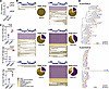 Figure 2
The site of HIV-1/Mtb coinfection is enriched in HIV-1 DNA and RNA. (A) HIV-1 RNA copies/mL in plasma (blue) and TB-PE (red) from 3 PLWH/TB. (B) Pie charts showing the proportion of genetically intact and defective proviral HIV-1 sequences derived from PBMCs and PEMCs for each donor. Horizontal lines adjacent to pie charts represent individual contig sequences aligned to HXB2. (C) Log-transformed infection frequency of total, genetically intact, and genetically defective proviruses per million cells. Each data point represents a single donor. Bars represent the median with 95% CI. Statistical significance was determined by 2-tailed paired t test, with P < 0.05 considered significant. (D) Phylogenetic tree of HIV-1 RNA sequences derived from plasma (blue) and TB-PE (red).
Genomic HIV-1 RNA derived from TB-PE is not genetically distinct from that derived from plasma. Given the increased frequency of genetically intact HIV-1 and elevated HIV-1 RNA levels in TB-PE compared with paired blood (Figure 2, A–C, and Supplemental Figure 1), we investigated whether the pleural space represents a “sanctuary” site for enhanced viral replication. If this compartment was isolated from the peripheral circulation, genetic divergence between virus in the TB-PE and virus in the plasma would be expected (23). To test this, we sequenced HIV-1 RNA from plasma and TB-PE samples obtained from 2 untreated PLWH/TB (donors 127 and 134) using our recently developed HIV-1 RNA-seq assay (31). Maximum-likelihood phylogenetic analysis of these HIV-1 RNA sequences, spanning the integrase–3′ long-terminal repeat (LTR) genomic region, revealed no compartmental clustering patterns between the HIV-1 RNA derived from the pleural space and that derived from the plasma (Figure 2D).
Effector memory CD4+ T cells are preferentially infected by HIV-1 in TB-PE. To determine what cell types are preferentially infected during active TB, we performed scRNA-seq on PEMCs from 2 PLWH/TB (donors 127 and 154). Since the cellular composition of TB-PE in PLWH/TB has not been well defined, we compared our dataset with publicly available scRNA-seq data of PEMCs from TB-PE taken from participants with Mtb mono-infection (Figure 3A) (32). Clustering patterns were similar across datasets, suggesting comparable PEMC phenotypes and supporting the reliability of our data (Figure 3A).
 Figure 3
scRNA-seq of TB-PE reveals preferential infection of effector memory CD4+ T cells. (A) UMAP of PEMCs from 2 PLWH/TB analyzed in-house compared with PEMCs from 5 TB patients reported by Cai et al. (32). (B) UMAP of immune cell populations from integrated PEMCs. (C) UMAP showing clusters of CD4+ T cells within integrated PEMCs. Pie charts indicate the proportions of central memory (Tcm), effector memory (Tem), and naive CD4+ T cells (top) and naive, regulatory T cell (Treg) memory, Treg naive, and T helper (Th) subsets (bottom) across both donors. (D) UMAP highlighting cells expressing HIV-1 transcripts (red). Bar graphs show the distribution of HIV-1 reads among CD4+ T cell subsets. Tfh, follicular helper T cells.
Cell type identification analysis of the two PLWH/TB showed a predominance of CD4+ and CD8+ T cells (Figure 3B), as described in previous studies (33, 34). Further characterization of the CD4+ T cells identified central, effector, and naive populations as well as several T helper subsets (Figure 3C).
To characterize HIV-1–infected cells derived from TB-PE, we identified cells expressing HIV-1 transcripts. We first amplified proviral HIV-1 DNA by FLIPS in both donors and used these sequences as a reference to map HIV-1 transcripts in our dataset. HIV-1 transcripts were more prevalent in effector memory CD4+ T cells when compared with central memory and naive subsets (Figure 3D) (35). Our analysis also revealed that HIV-1 transcripts were enriched within the Th17 subset, followed by follicular helper T, Th2, and Th1/17 cells, respectively (Figure 3D).
The majority of TB-PE–derived CD8+ T cells do not express canonical markers of tissue residency and exhibit an activated/exhausted phenotype. Our results showing an increase in the proportion of genetically intact HIV-1–infected cells in TB-PE (Figure 2) suggest reduced immune pressure at this site of coinfection. Since CD8+ T cells are critical for HIV-1 control, we investigated whether CD8+ T cells derived from TB-PE are phenotypically dysfunctional. It has been previously reported that CD8+ T cells in the airways of PLWH exhibit a tissue-resident phenotype, defined by the expression of CD103 (36), which is associated with reduced cytotoxic potential (37, 38). To determine whether TB-PE is enriched for tissue-resident CD8+ T cells, we analyzed the phenotype of PEMC-derived CD8+ T cells from 2 PLWH/TB (donors 127 and 122) using a 19-color flow cytometry panel. Integration of both datasets followed by unbiased clustering identified 9 distinct clusters (Figure 4, A–D). CD103 expression was restricted to clusters 4, 8, and 9, indicating that although a fraction of the TB-PE–derived CD8+ T cells were potentially tissue resident, they did not represent the majority (Figure 4, C and D). CD69, another marker associated with tissue residency, was highly expressed in cluster 8 (Figure 4, A and D). However, this cluster also displayed high levels of the activation markers CD38 and human leukocyte antigen DR isotype (HLA-DR), suggesting that in this context CD69 primarily reflects activation rather than tissue residency (Figure 4D). In addition to cluster 8, CD38 and HLA-DR coexpression was also observed across clusters 6 and 7, together with the exhaustion markers programmed cell death protein 1 (PD-1) and T cell immunoglobulin and ITIM domain (TIGIT) (Figure 4D). PD-1 and TIGIT were also detected in cluster 5. CD39, which is associated with a terminally exhausted phenotype (39), was coexpressed with CD38 and HLA-DR in cluster 7 (Figure 4D). These data suggest the presence of an expanded pool of chronically activated CD8+ T cells with an exhausted phenotype. Notably, CCR5 was widely expressed across most clusters, consistent with migration of effector cells from the blood into the inflamed pleural space (Figure 4, B and D) (33). Clusters 1, 2, and 4 were characterized by the expression of CCR7, the costimulatory molecules CD27 and CD28, and the homeostatic cytokine receptor CD127, indicating a less differentiated, circulating phenotype (Figure 4D).
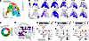 Figure 4
Immunophenotype of TB-PE–derived CD8+ T cells. (A) UMAP showing unbiased clustering of CD8+ T cells from 2 PLWH/TB based on the immune markers CD103, CD69, CD27, CCR7, CD127, CCR5, CD28, HLA-DR, CD38, PD-1, CD39, and TIGIT. (B) UMAP of expression levels of selected immune markers in TB-PE–derived CD8+ T cells from both donors. (C) Proportions of CD8+ T cell clusters identified by UMAP analysis. (D) Heatmap of average expression of 12 immune markers across clusters. (E–J) scRNA/TCR-seq analysis of TB-PE–derived virus-specific CD8+ T cells from 2 PLWH/TB. (E) Cytokine signature analysis. (F) Dot plot showing relative expression of IFNG and TNF. (G) Cytotoxicity signature analysis. (H) Dot plot showing relative expression of GZMA, GZMB, GZMK, and PRF1. (I) Exhaustion signature analysis. (J) Dot plot showing relative expression of TIGIT, LAG3, CTLA4, and HAVCR2.
Together, these data show that TB-PE–derived CD8+ T cells are phenotypically diverse and that the majority do not exhibit a tissue-resident phenotype. Moreover, we identified CD8+ T cells displaying features of chronic activation and exhaustion, consistent with an inflammatory microenvironment and suggesting impaired antiviral function at the site of coinfection.
HIV-1–specific CD8+ T cells exhibit a functionally exhausted phenotype in TB-PE. Our analysis suggested the presence of chronically activated and exhausted CD8+ T cells at the site of the coinfection. However, this analysis did not allow us to determine whether the CD8+ T cells exhibiting this phenotype were specific for HIV-1. To address this, we combined the scRNA-seq data from donors 127 and 154 (Figure 3A) with paired single-cell T cell receptor (TCR) sequencing data to identify and characterize virus-specific CD8+ T cells. Comparing the TCRs from both donors with data from publicly available TCR databases (40), we identified CD8+ T cell–derived TCRs with specificity for HIV-1, EBV, HBV, influenza A, SARS-CoV-2, and CMV (Figure 4, E–J). We then analyzed the transcriptomic profile of these cells and compared the cytokine and cytotoxic signatures across the CD8+ T cells with different specificities to assess effector functionality. HIV-1–specific CD8+ T cells showed a strong cytokine signature relative to CD8+ T cells of other specificities, mainly driven by the expression of IFNG, indicating recent contact with the antigen (Figure 4, E and F). However, their cytotoxic signature was considerably lower than that of other virus-specific CD8+ T cells (Figure 4G). We observed that HIV-1–specific cells exhibited relatively lower expression of the cytotoxic genes granzyme A (GZMA), granzyme K (GZMK), and perforin 1 (PRF1) (Figure 4H). To further understand this result, we also assessed the relative exhaustion signature of HIV-1–specific CD8+ T cells. Notably, HIV-1–specific CD8+ T cells exhibited an exhausted signature, driven by high expression levels of TIGIT, cytotoxic T lymphocyte–associated protein 4 (CTLA4), and HAVCR2, which encodes the T cell immunoglobulin and mucin domain containing-3 (TIM-3) protein (Figure 4J). Collectively, these results indicate that HIV-1–specific CD8+ T cells are phenotypically exhausted, potentially contributing to reduced immune pressure at the site of the coinfection.
The TB-associated microenvironment inhibits effector CD8+ T cell functionality. Exhaustion of CD8+ T cells is a well-established hallmark of HIV-1 pathogenesis (5, 6). Therefore, our finding that HIV-1–specific CD8+ T cells in TB-PE are functionally exhausted is not unique to coinfection with Mtb. To better understand the specific contribution of the TB-associated microenvironment to CD8+ T cell functionality, we developed an in vitro model using pooled acellular fractions of TB-PE from individuals with active pleural TB (Figure 5A) (27). In this model, we stimulated CD8+ T cells isolated from healthy donors with anti-CD3/CD28 antibodies to mimic antigenic stimulation. Stimulated and unstimulated CD8+ T cells were cultured in the presence or absence of TB-PE to determine whether the inflammatory TB-associated microenvironment affected the CD8+ T cell effector response (Figure 5A). We first performed bulk RNA-seq to characterize the transcriptomic profile of cells stimulated with anti-CD3/CD28 antibodies exposed to TB-PE. Differential gene expression analysis identified 228 upregulated genes and 237 downregulated genes between controls and cells exposed to TB-PE (Figure 5, B and C, and Supplemental Figure 2). Gene Ontology pathway analysis revealed that cell pathways and genes associated with T cell effector functions were significantly downmodulated by TB-PE (Figure 5, D–F). Moreover, cell pathways related to chemotaxis were upregulated, reflective of the inflammatory nature of the TB-associated microenvironment (Figure 5E). We further validated these results by measuring the cell-surface protein expression of the activation markers CD69, CD25, and HLA-DR. Consistent with the transcriptomic profile shaped by TB-PE, the presence of TB-PE impaired the upregulation of these activation markers upon T cell stimulation (Figure 5G). To confirm whether the TB-PE–mediated inhibition of T cell activation is a common feature of pleural TB, we tested additional pooled and individual TB-PE samples. In addition, to test that this immunosuppressive phenotype was specific to TB-PE rather than a feature of the pleural environment, we also included pleural effusion from individuals with heart failure (HF-PE). Unlike HF-PE, both the additional pool and the individual TB-PE samples reproduced the inhibitory effect (Figure 5H). These findings suggest that the impairment of CD8+ T cell activation is a consistent feature of TB-PE.
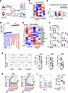 Figure 5
TB-PE treatment downmodulates T cell effector functionality. (A) Schematic of the experimental model used to study the effect of TB-PE on CD8+ T cell functionality. (B) Heatmap showing hierarchical clustering of the top 100 variable genes between TB-PE–treated and control CD8+ T cells stimulated with anti-CD3/CD28 antibodies. (C) Volcano plot of differentially expressed genes (DEGs). (D) Gene set enrichment analysis (GSEA) of DEGs (P < 0.05 and absolute log fold change > 1) comparing TB-PE–treated and control activated CD8+ T cells. Dot plots illustrate the top 10 upregulated and downmodulated enriched Gene Ontology biological process terms. (E) Heatmap of normalized expression of selected genes involved in T cell activation. (F) Gene expression levels of GZMB, IFNG, TNF, IL21, and CD69 across conditions. (G) CD8+ T cells were stimulated with anti-CD3/CD28 antibodies in the presence of TB-PE; cells activated in the absence of PE served as controls. Expression of the activation markers CD69, CD25, and HLA-DR was measured by flow cytometry after 24 hours (CD69, CD25) or 48 hours (HLA-DR). Representative flow plots (left) and quantification of activated cells (right) are shown. (H) Proportions of CD69+ (left) or CD25+ (right) cells stimulated in the presence of an additional TB-PE pool, 3 independent TB-PE samples (red), or HF-PE (gray). Values are relative to the non-PE control condition (blue). Data are shown as mean ± SD. Each dot represents an independent donor. Statistical significance was determined by paired 2-tailed t test or 1-way ANOVA followed by Tukey’s honestly significant difference (HSD) post-test. *P ≤ 0.05, **P ≤ 0.01. (I) HIF-1α expression in activated CD8+ T cells determined by flow cytometry. gMFI, geometric mean fluorescence intensity. Each dot represents an independent donor. Statistical significance was determined by paired 2-tailed t test or 1-way ANOVA followed by Tukey’s HSD post-test. *P ≤ 0.05, **P ≤ 0.01.
To further characterize the impact of the TB-associated microenvironment on effector functions, we also measured the expression of hypoxia-inducible factor 1α (HIF-1α), since this transcription factor is necessary for an efficient T cell effector response (41). Cells exposed to TB-PE exhibited reduced expression of HIF-1α (Figure 5I).
Taken together, these data indicate that the TB-associated microenvironment impairs CD8+ T cell activation and effector functionality upon antigenic stimulation.
HIV-1–specific CD8+ T cell responses are inhibited by exposure to TB-PE. Our results demonstrate that the TB-associated microenvironment impairs the effector functionality of CD8+ T cells after antigen stimulation. This prompted us to investigate whether TB-PE exposure can also inhibit the antiviral function and HIV-1–specific effector responses mediated by CD8+ T cells derived from PLWH. To address this question, we stimulated PLWH-derived PBMCs with a pool of HIV-1 peptides to expand HIV-1–specific CD8+ T cells for 14 days. After expansion, cells were restimulated with the HIV-1 peptides in the presence or absence of TB-PE, to induce the expression of the degranulation markers CD107a and CD107b and the effector cytokines IFN-γ and TNF-α (Figure 6A). Importantly, cells treated with TB-PE exhibited significantly lower expression of CD107a/b, IFN-γ, and TNF-α when compared with cells expanded without TB-PE (Figure 6, B and C). Notably, we observed no significant differences between stimulated cells and cells stimulated in the presence of HF-PE (Figure 6, B and C). These results were further confirmed when cells from healthy donors were stimulated with a pool of peptides for common viruses (CMV, EBV, and influenza; CEF peptide pool), in the presence or absence of TB-PE (Supplemental Figure 3). Overall, these data demonstrate that the TB-associated microenvironment inhibits the ability of CD8+ T cells to mount an efficient antiviral effector response.
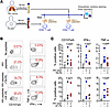 Figure 6
The effector anti–HIV-1 responses of CD8+ T cells are inhibited by TB-PE. (A) Schematic of the experimental model. (B) Representative flow cytometry of PLWH-derived CD8+ T cells expressing CD107a/b, TNF-α, and IFN-γ as markers for effector response upon restimulation with HIV-1 peptides in the presence or absence of TB-PE. Cells exposed to no peptides were used as an experimental control. Cells exposed to HF-PE were used as a control to determine the specific effect of TB-PE. (C) Quantification of expression of CD107a/b, TNF-α, and IFN-γ in PLWH-derived CD8+ T cells exposed to TB-PE (n = 5; top) or HF-PE (n = 4; bottom). Each data point represents a single donor. Statistical significance was determined by 2-tailed paired t test, with P < 0.05 considered significant.
TB-PE prevents HIV-1–specific CD8+ T cells from acquiring cytotoxic function. Considering that TB-PE inhibits efficient TCR-dependent T cell activation, we hypothesized that the TB-associated microenvironment could also impair the differentiation of CD8+ T cells into a cytotoxic phenotype upon antigen encounter, leading to inefficient killing of HIV-1–infected CD4+ T cells. To assess this, PBMCs from PLWH were expanded with HIV-1 peptides in the presence or absence of TB-PE for 14 days. After expansion, CD8+ T cells were cocultured in the absence of TB-PE with autologous CD4+ T cells superinfected with HIV-1 in vitro (Figure 7A). Notably, at the time of coculture, the number of CD8+ T cells was normalized based on the expression of CD107a/b, ensuring that equal numbers of cytotoxic CD8+ T cells were included across conditions (Figure 7A). As expected, HIV-1–specific CD8+ T cells expanded with peptides alone significantly reduced the number of HIV-1–infected CD4+ T cells when compared with the control culture of CD4+ T cells (Figure 7B). In contrast, HIV-1–specific CD8+ T cells expanded in the presence of TB-PE were unable to eliminate HIV-1–infected CD4+ T cells when compared with CD8+ T cells expanded with peptides alone (Figure 7B). This finding was further confirmed when cells from healthy donors were expanded with the CEF peptide pool in the presence or absence of TB-PE and cocultured with target CD4+ T cells pulsed with CEF peptides (Supplemental Figure 4). These results indicate that the TB-associated microenvironment hampers the differentiation of HIV-1–specific CD8+ T cells into fully cytotoxic effectors upon antigen exposure.
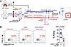 Figure 7
TB-PE inhibits the cytotoxic function of HIV-1–specific CD8+ T cells. (A) Schematic of the experimental model. (B) Representative flow cytometry plots showing p24 levels in HIV-1–infected CD4+ T cells cocultured with CD8+ T cells expanded in the presence or absence of TB-PE. As a control, CD4+ T cells were cultured without CD8+ T cells. Quantification of HIV-1–infected cell killing was performed using samples from 4 PLWH. Each data point represents a single donor. Statistical significance was determined by 2-tailed paired t test, with P < 0.05 considered significant.
CD8+ T cells exposed to TB-PE accumulate neutral lipids. To explore the mechanisms underlying the immunosuppressive effect of TB-PE on CD8+ T cells, we leveraged our bulk RNA-seq dataset generated using our in vitro model (Figure 5, B and C, and Supplemental Figure 2). Since stimulation with anti-CD3/CD28 antibodies could potentially mask direct effects of TB-PE on exposed cells, we first focused our analysis on cells treated with TB-PE in the absence of activating stimuli. Interestingly, the 2 most upregulated genes were fatty acid–binding protein 4 (FABP4) and lipoprotein lipase (LPL) (Supplemental Figure 2), which are involved in fatty acid trafficking and storage. This result prompted us to further characterize cellular pathways associated with fatty acid transport and storage. In both unstimulated and anti-CD3/CD28–stimulated conditions, TB-PE exposure resulted in the upregulation of pathways associated with lipid transport and storage (Figure 8, A and C). Consistently, TB-PE–exposed cells showed increased expression of genes involved in lipid transport and metabolism, including LPL, ATP-binding cassette transporter A1 (ABCA1), and carnitine palmitoyltransferase 1A (CPT1A), irrespective of anti-CD3/CD28 stimulation (Figure 8, B and D).
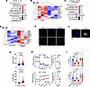 Figure 8
TB-PE–derived lipids accumulate in CD8+ T cells and inhibit T cell activation. (A) Dot plot illustrating lipid-associated pathways upregulated in TB-PE–exposed resting CD8+ T cells. (B) Heatmap presenting normalized expression values of lipid-associated genes between unstimulated control and TB-PE–exposed cells. (C) Dot plot illustrating upregulated lipid-associated pathways in CD8+ T cells stimulated with anti-CD3/CD28 antibodies exposed to TB-PE when compared with control CD8+ T cells. (D) Heatmap of normalized expression values of lipid-associated genes between anti-CD3/CD28–treated control and TB-PE–exposed CD8+ T cells. (E and F) Representative images at ×40 (E) and ×100 (F) original magnification of neutral lipid staining in CD8+ T cells in the presence and absence of TB-PE. Scale bars: 20 μm (E), 4 μm (F). (G) Quantification of BODIPY mean fluorescence intensity (MFI) between control and TB-PE–exposed CD8+ T cells. (H) Top: Oxidative phosphorylation measured as oxygen consumption rate (OCR; left) and mitochondria-dependent ATP production (right). Bottom: Glycolysis measured as proton efflux rate (PER; left) and glycolysis-dependent ATP production (right). Olig, oligomycin; Rot, rotenone; AA, antimycin A. (I) Proportion of CD69+ (top) and CD25+ (bottom) cells stimulated with anti-CD3/CD28 antibodies in the presence of either pooled TB-PE (red), the lipid fraction (Lip-PE) (red), lipid vehicle (gray), the polar fraction (PF-PE) (red), or the polar fraction vehicle (gray). Data are shown as mean ± SD. Each dot represents an independent donor. Statistical significance was determined by paired 2-tailed t test or 1-way ANOVA followed by Tukey’s HSD post-test. **P ≤ 0.01, ***P ≤ 0.001, ****P ≤ 0.0001.
To assess whether TB-PE exposure promotes intracellular lipid accumulation, we stained CD8+ T cells with BODIPY 493/503, a fluorescent dye that labels neutral lipids and can be used to visualize and quantify intracellular lipid accumulation by fluorescence microscopy. CD8+ T cells exposed to TB-PE for 24 hours exhibited higher levels of neutral lipids than unexposed controls, as shown by increased mean fluorescence intensity in TB-PE–exposed cells (P < 0.0001) (Figure 8, E–G).
Together, these results show that TB-PE exposure induces a transcriptional program associated with lipid transport and storage and promotes intracellular accumulation of neutral lipids in CD8+ T cells.
TB-PE exposure downmodulates oxidative phosphorylation and glycolysis. Efficient T cell responses require increased glycolysis and oxidative phosphorylation (OXPHOS) (42). It has recently been reported that lipid-enriched tumor microenvironments can promote lipid uptake and storage in infiltrating CD8+ T cells, leading to the downmodulation of glycolysis and OXPHOS and, ultimately, impaired effector function (43). To determine whether TB-PE exposure affects activation-induced glycolysis and OXPHOS, we assessed the metabolic profile of CD8+ T cells exposed to TB-PE using a Seahorse cell flux analyzer. Oxygen consumption rate and proton efflux rate were measured as indicators of OXPHOS and glycolysis, respectively (Figure 8H). The intracellular ATP production rate derived from glycolysis (glycoATP) or OXPHOS (mitoATP) was also quantified (Figure 8H). Both OXPHOS and glycolysis were significantly reduced in TB-PE–treated cells compared with controls (Figure 8H). These results suggest that TB-PE exposure impairs the metabolic reprogramming required for CD8+ T cell activation, potentially contributing to reduced effector function.
TB-PE–derived lipids inhibit CD8+ T cell activation. Finally, to determine whether lipids derived from TB-PE can directly affect CD8+ T cell effector functions, we separated TB-PE into 2 fractions: one enriched in polar metabolites (PF-PE) and another comprising lipids (Lip-PE). As a proxy for T cell effector function, we measured the expression of the activation markers CD69 and CD25 following stimulation with anti-CD3/CD28 antibodies. CD8+ T cells were preincubated with PF-PE or Lip-PE for 1 hour before stimulation with anti-CD3/CD28 antibodies. After 24 hours, CD8+ T cells exposed to Lip-PE, but not those exposed to PF-PE, exhibited significantly reduced expression of both CD69 and CD25 (Figure 8I). These results suggest that lipids present in the TB-associated microenvironment contribute to impaired CD8+ T cell activation.
In summary, our results suggest that the higher proportion of genetically intact HIV-1–infected cells observed at the site of coinfection may result, at least in part, from impaired CD8+ T cell–mediated control of HIV-1. This impairment is associated with reduced effector differentiation and function, potentially driven by lipids present in the TB-associated microenvironment.


