
Research ArticleMetabolismNeuroscience
Open Access |  10.1172/jci.insight.200761
Mitochonic acid 5 alleviates amyotrophic lateral sclerosis phenotypes via mitochondrial augmentation
Yoshitsugu Oikawa,1,2 Yuhan Luo,3 Naoki Suzuki,4,5 Tomoko Kasahara,1 Yoshiyasu Tongu,1 Yuki Yoshida,3 Tsukasa Tominari,6 Shogo Tanabe,7 Yoshiko Suto,1 Hitomi Kashiwagi,1 Saki Saito,4 Kensuke Ikeda,4 Chitose Suzuki,1 Arata Kuranaga,8 Tetsuya Akiyama,4 Satoru Morimoto,9,10 Yoshitsugu Aoki,6 Rieko Muramatsu,7 Tomoyoshi Soga,11 Masashi Aoki,4 Hideyuki Okano,9,10 Tetsuhiro Tanaka,12 Takaaki Abe,1,13 Erina Kuranaga,3,8 and Takafumi Toyohara1,13,14
1Department of Medical Science, Tohoku University Graduate School of Biomedical Engineering, Sendai, Japan.
2Department of Pediatrics, Tohoku University Graduate School of Medicine, Sendai, Japan.
3Laboratory for Histogenetic Dynamics, Graduate School of Life Sciences, Tohoku University, Sendai, Japan.
4Department of Neurology and
5Department of Rehabilitation Medicine, Tohoku University Graduate School of Medicine, Sendai, Japan.
6Department of Molecular Therapy and
7Department of Molecular Pharmacology, National Institute of Neuroscience, National Center of Neurology and Psychiatry, Tokyo, Japan.
8Laboratory for Histogenetic Dynamics, Graduate School of Pharmaceutical Sciences, Kyoto University, Kyoto, Japan.
9Keio University Regenerative Medicine Research Center, Kawasaki, Japan.
10Division of Neurodegenerative Disease Research, Tokyo Metropolitan Institute for Geriatrics and Gerontology, Tokyo, Japan.
11Human Biology-Microbiome-Quantum Research Center, Keio University, Tsuruoka, Yamagata, Japan.
12Department of Nephrology and Hypertension, Tohoku University Hospital, Sendai, Japan.
13Department of Clinical Biology and Hormonal Regulation, Tohoku University Graduate School of Medicine, Sendai, Japan.
14Division of Bioengineering for Renal and Circulatory Regeneration, Tohoku University Graduate School of Biomedical Engineering, Sendai, Japan.
Address correspondence to: Erina Kuranaga, Laboratory for Histogenetic Dynamics, Graduate School of Pharmaceutical Sciences, Kyoto University, 46-29 Yoshida-Shimoadachi-cho, Sakyo-ku, Kyoto 606-8501, Japan. Email: kuranaga.erina.7j@kyoto-u.ac.jp. Or to: Takafumi Toyohara, Division of Bioengineering for Renal and Circulatory Regeneration, Tohoku University Graduate School of Biomedical Engineering, 1-1 Seiryo-machi, Aoba-ku, Sendai, Miyagi 980-8574, Japan. Email: takafumi.toyohara.e6@tohoku.ac.jp.
Authorship note: YO and YL contributed equally to this work.
Find articles by Oikawa, Y. in: PubMed | Google Scholar
1Department of Medical Science, Tohoku University Graduate School of Biomedical Engineering, Sendai, Japan.
2Department of Pediatrics, Tohoku University Graduate School of Medicine, Sendai, Japan.
3Laboratory for Histogenetic Dynamics, Graduate School of Life Sciences, Tohoku University, Sendai, Japan.
4Department of Neurology and
5Department of Rehabilitation Medicine, Tohoku University Graduate School of Medicine, Sendai, Japan.
6Department of Molecular Therapy and
7Department of Molecular Pharmacology, National Institute of Neuroscience, National Center of Neurology and Psychiatry, Tokyo, Japan.
8Laboratory for Histogenetic Dynamics, Graduate School of Pharmaceutical Sciences, Kyoto University, Kyoto, Japan.
9Keio University Regenerative Medicine Research Center, Kawasaki, Japan.
10Division of Neurodegenerative Disease Research, Tokyo Metropolitan Institute for Geriatrics and Gerontology, Tokyo, Japan.
11Human Biology-Microbiome-Quantum Research Center, Keio University, Tsuruoka, Yamagata, Japan.
12Department of Nephrology and Hypertension, Tohoku University Hospital, Sendai, Japan.
13Department of Clinical Biology and Hormonal Regulation, Tohoku University Graduate School of Medicine, Sendai, Japan.
14Division of Bioengineering for Renal and Circulatory Regeneration, Tohoku University Graduate School of Biomedical Engineering, Sendai, Japan.
Address correspondence to: Erina Kuranaga, Laboratory for Histogenetic Dynamics, Graduate School of Pharmaceutical Sciences, Kyoto University, 46-29 Yoshida-Shimoadachi-cho, Sakyo-ku, Kyoto 606-8501, Japan. Email: kuranaga.erina.7j@kyoto-u.ac.jp. Or to: Takafumi Toyohara, Division of Bioengineering for Renal and Circulatory Regeneration, Tohoku University Graduate School of Biomedical Engineering, 1-1 Seiryo-machi, Aoba-ku, Sendai, Miyagi 980-8574, Japan. Email: takafumi.toyohara.e6@tohoku.ac.jp.
Authorship note: YO and YL contributed equally to this work.
Find articles by Luo, Y. in: PubMed | Google Scholar
1Department of Medical Science, Tohoku University Graduate School of Biomedical Engineering, Sendai, Japan.
2Department of Pediatrics, Tohoku University Graduate School of Medicine, Sendai, Japan.
3Laboratory for Histogenetic Dynamics, Graduate School of Life Sciences, Tohoku University, Sendai, Japan.
4Department of Neurology and
5Department of Rehabilitation Medicine, Tohoku University Graduate School of Medicine, Sendai, Japan.
6Department of Molecular Therapy and
7Department of Molecular Pharmacology, National Institute of Neuroscience, National Center of Neurology and Psychiatry, Tokyo, Japan.
8Laboratory for Histogenetic Dynamics, Graduate School of Pharmaceutical Sciences, Kyoto University, Kyoto, Japan.
9Keio University Regenerative Medicine Research Center, Kawasaki, Japan.
10Division of Neurodegenerative Disease Research, Tokyo Metropolitan Institute for Geriatrics and Gerontology, Tokyo, Japan.
11Human Biology-Microbiome-Quantum Research Center, Keio University, Tsuruoka, Yamagata, Japan.
12Department of Nephrology and Hypertension, Tohoku University Hospital, Sendai, Japan.
13Department of Clinical Biology and Hormonal Regulation, Tohoku University Graduate School of Medicine, Sendai, Japan.
14Division of Bioengineering for Renal and Circulatory Regeneration, Tohoku University Graduate School of Biomedical Engineering, Sendai, Japan.
Address correspondence to: Erina Kuranaga, Laboratory for Histogenetic Dynamics, Graduate School of Pharmaceutical Sciences, Kyoto University, 46-29 Yoshida-Shimoadachi-cho, Sakyo-ku, Kyoto 606-8501, Japan. Email: kuranaga.erina.7j@kyoto-u.ac.jp. Or to: Takafumi Toyohara, Division of Bioengineering for Renal and Circulatory Regeneration, Tohoku University Graduate School of Biomedical Engineering, 1-1 Seiryo-machi, Aoba-ku, Sendai, Miyagi 980-8574, Japan. Email: takafumi.toyohara.e6@tohoku.ac.jp.
Authorship note: YO and YL contributed equally to this work.
Find articles by Suzuki, N. in: PubMed | Google Scholar
1Department of Medical Science, Tohoku University Graduate School of Biomedical Engineering, Sendai, Japan.
2Department of Pediatrics, Tohoku University Graduate School of Medicine, Sendai, Japan.
3Laboratory for Histogenetic Dynamics, Graduate School of Life Sciences, Tohoku University, Sendai, Japan.
4Department of Neurology and
5Department of Rehabilitation Medicine, Tohoku University Graduate School of Medicine, Sendai, Japan.
6Department of Molecular Therapy and
7Department of Molecular Pharmacology, National Institute of Neuroscience, National Center of Neurology and Psychiatry, Tokyo, Japan.
8Laboratory for Histogenetic Dynamics, Graduate School of Pharmaceutical Sciences, Kyoto University, Kyoto, Japan.
9Keio University Regenerative Medicine Research Center, Kawasaki, Japan.
10Division of Neurodegenerative Disease Research, Tokyo Metropolitan Institute for Geriatrics and Gerontology, Tokyo, Japan.
11Human Biology-Microbiome-Quantum Research Center, Keio University, Tsuruoka, Yamagata, Japan.
12Department of Nephrology and Hypertension, Tohoku University Hospital, Sendai, Japan.
13Department of Clinical Biology and Hormonal Regulation, Tohoku University Graduate School of Medicine, Sendai, Japan.
14Division of Bioengineering for Renal and Circulatory Regeneration, Tohoku University Graduate School of Biomedical Engineering, Sendai, Japan.
Address correspondence to: Erina Kuranaga, Laboratory for Histogenetic Dynamics, Graduate School of Pharmaceutical Sciences, Kyoto University, 46-29 Yoshida-Shimoadachi-cho, Sakyo-ku, Kyoto 606-8501, Japan. Email: kuranaga.erina.7j@kyoto-u.ac.jp. Or to: Takafumi Toyohara, Division of Bioengineering for Renal and Circulatory Regeneration, Tohoku University Graduate School of Biomedical Engineering, 1-1 Seiryo-machi, Aoba-ku, Sendai, Miyagi 980-8574, Japan. Email: takafumi.toyohara.e6@tohoku.ac.jp.
Authorship note: YO and YL contributed equally to this work.
Find articles by Kasahara, T. in: PubMed | Google Scholar
1Department of Medical Science, Tohoku University Graduate School of Biomedical Engineering, Sendai, Japan.
2Department of Pediatrics, Tohoku University Graduate School of Medicine, Sendai, Japan.
3Laboratory for Histogenetic Dynamics, Graduate School of Life Sciences, Tohoku University, Sendai, Japan.
4Department of Neurology and
5Department of Rehabilitation Medicine, Tohoku University Graduate School of Medicine, Sendai, Japan.
6Department of Molecular Therapy and
7Department of Molecular Pharmacology, National Institute of Neuroscience, National Center of Neurology and Psychiatry, Tokyo, Japan.
8Laboratory for Histogenetic Dynamics, Graduate School of Pharmaceutical Sciences, Kyoto University, Kyoto, Japan.
9Keio University Regenerative Medicine Research Center, Kawasaki, Japan.
10Division of Neurodegenerative Disease Research, Tokyo Metropolitan Institute for Geriatrics and Gerontology, Tokyo, Japan.
11Human Biology-Microbiome-Quantum Research Center, Keio University, Tsuruoka, Yamagata, Japan.
12Department of Nephrology and Hypertension, Tohoku University Hospital, Sendai, Japan.
13Department of Clinical Biology and Hormonal Regulation, Tohoku University Graduate School of Medicine, Sendai, Japan.
14Division of Bioengineering for Renal and Circulatory Regeneration, Tohoku University Graduate School of Biomedical Engineering, Sendai, Japan.
Address correspondence to: Erina Kuranaga, Laboratory for Histogenetic Dynamics, Graduate School of Pharmaceutical Sciences, Kyoto University, 46-29 Yoshida-Shimoadachi-cho, Sakyo-ku, Kyoto 606-8501, Japan. Email: kuranaga.erina.7j@kyoto-u.ac.jp. Or to: Takafumi Toyohara, Division of Bioengineering for Renal and Circulatory Regeneration, Tohoku University Graduate School of Biomedical Engineering, 1-1 Seiryo-machi, Aoba-ku, Sendai, Miyagi 980-8574, Japan. Email: takafumi.toyohara.e6@tohoku.ac.jp.
Authorship note: YO and YL contributed equally to this work.
Find articles by Tongu, Y. in: PubMed | Google Scholar
1Department of Medical Science, Tohoku University Graduate School of Biomedical Engineering, Sendai, Japan.
2Department of Pediatrics, Tohoku University Graduate School of Medicine, Sendai, Japan.
3Laboratory for Histogenetic Dynamics, Graduate School of Life Sciences, Tohoku University, Sendai, Japan.
4Department of Neurology and
5Department of Rehabilitation Medicine, Tohoku University Graduate School of Medicine, Sendai, Japan.
6Department of Molecular Therapy and
7Department of Molecular Pharmacology, National Institute of Neuroscience, National Center of Neurology and Psychiatry, Tokyo, Japan.
8Laboratory for Histogenetic Dynamics, Graduate School of Pharmaceutical Sciences, Kyoto University, Kyoto, Japan.
9Keio University Regenerative Medicine Research Center, Kawasaki, Japan.
10Division of Neurodegenerative Disease Research, Tokyo Metropolitan Institute for Geriatrics and Gerontology, Tokyo, Japan.
11Human Biology-Microbiome-Quantum Research Center, Keio University, Tsuruoka, Yamagata, Japan.
12Department of Nephrology and Hypertension, Tohoku University Hospital, Sendai, Japan.
13Department of Clinical Biology and Hormonal Regulation, Tohoku University Graduate School of Medicine, Sendai, Japan.
14Division of Bioengineering for Renal and Circulatory Regeneration, Tohoku University Graduate School of Biomedical Engineering, Sendai, Japan.
Address correspondence to: Erina Kuranaga, Laboratory for Histogenetic Dynamics, Graduate School of Pharmaceutical Sciences, Kyoto University, 46-29 Yoshida-Shimoadachi-cho, Sakyo-ku, Kyoto 606-8501, Japan. Email: kuranaga.erina.7j@kyoto-u.ac.jp. Or to: Takafumi Toyohara, Division of Bioengineering for Renal and Circulatory Regeneration, Tohoku University Graduate School of Biomedical Engineering, 1-1 Seiryo-machi, Aoba-ku, Sendai, Miyagi 980-8574, Japan. Email: takafumi.toyohara.e6@tohoku.ac.jp.
Authorship note: YO and YL contributed equally to this work.
Find articles by Yoshida, Y. in: PubMed | Google Scholar
1Department of Medical Science, Tohoku University Graduate School of Biomedical Engineering, Sendai, Japan.
2Department of Pediatrics, Tohoku University Graduate School of Medicine, Sendai, Japan.
3Laboratory for Histogenetic Dynamics, Graduate School of Life Sciences, Tohoku University, Sendai, Japan.
4Department of Neurology and
5Department of Rehabilitation Medicine, Tohoku University Graduate School of Medicine, Sendai, Japan.
6Department of Molecular Therapy and
7Department of Molecular Pharmacology, National Institute of Neuroscience, National Center of Neurology and Psychiatry, Tokyo, Japan.
8Laboratory for Histogenetic Dynamics, Graduate School of Pharmaceutical Sciences, Kyoto University, Kyoto, Japan.
9Keio University Regenerative Medicine Research Center, Kawasaki, Japan.
10Division of Neurodegenerative Disease Research, Tokyo Metropolitan Institute for Geriatrics and Gerontology, Tokyo, Japan.
11Human Biology-Microbiome-Quantum Research Center, Keio University, Tsuruoka, Yamagata, Japan.
12Department of Nephrology and Hypertension, Tohoku University Hospital, Sendai, Japan.
13Department of Clinical Biology and Hormonal Regulation, Tohoku University Graduate School of Medicine, Sendai, Japan.
14Division of Bioengineering for Renal and Circulatory Regeneration, Tohoku University Graduate School of Biomedical Engineering, Sendai, Japan.
Address correspondence to: Erina Kuranaga, Laboratory for Histogenetic Dynamics, Graduate School of Pharmaceutical Sciences, Kyoto University, 46-29 Yoshida-Shimoadachi-cho, Sakyo-ku, Kyoto 606-8501, Japan. Email: kuranaga.erina.7j@kyoto-u.ac.jp. Or to: Takafumi Toyohara, Division of Bioengineering for Renal and Circulatory Regeneration, Tohoku University Graduate School of Biomedical Engineering, 1-1 Seiryo-machi, Aoba-ku, Sendai, Miyagi 980-8574, Japan. Email: takafumi.toyohara.e6@tohoku.ac.jp.
Authorship note: YO and YL contributed equally to this work.
Find articles by Tominari, T. in: PubMed | Google Scholar
1Department of Medical Science, Tohoku University Graduate School of Biomedical Engineering, Sendai, Japan.
2Department of Pediatrics, Tohoku University Graduate School of Medicine, Sendai, Japan.
3Laboratory for Histogenetic Dynamics, Graduate School of Life Sciences, Tohoku University, Sendai, Japan.
4Department of Neurology and
5Department of Rehabilitation Medicine, Tohoku University Graduate School of Medicine, Sendai, Japan.
6Department of Molecular Therapy and
7Department of Molecular Pharmacology, National Institute of Neuroscience, National Center of Neurology and Psychiatry, Tokyo, Japan.
8Laboratory for Histogenetic Dynamics, Graduate School of Pharmaceutical Sciences, Kyoto University, Kyoto, Japan.
9Keio University Regenerative Medicine Research Center, Kawasaki, Japan.
10Division of Neurodegenerative Disease Research, Tokyo Metropolitan Institute for Geriatrics and Gerontology, Tokyo, Japan.
11Human Biology-Microbiome-Quantum Research Center, Keio University, Tsuruoka, Yamagata, Japan.
12Department of Nephrology and Hypertension, Tohoku University Hospital, Sendai, Japan.
13Department of Clinical Biology and Hormonal Regulation, Tohoku University Graduate School of Medicine, Sendai, Japan.
14Division of Bioengineering for Renal and Circulatory Regeneration, Tohoku University Graduate School of Biomedical Engineering, Sendai, Japan.
Address correspondence to: Erina Kuranaga, Laboratory for Histogenetic Dynamics, Graduate School of Pharmaceutical Sciences, Kyoto University, 46-29 Yoshida-Shimoadachi-cho, Sakyo-ku, Kyoto 606-8501, Japan. Email: kuranaga.erina.7j@kyoto-u.ac.jp. Or to: Takafumi Toyohara, Division of Bioengineering for Renal and Circulatory Regeneration, Tohoku University Graduate School of Biomedical Engineering, 1-1 Seiryo-machi, Aoba-ku, Sendai, Miyagi 980-8574, Japan. Email: takafumi.toyohara.e6@tohoku.ac.jp.
Authorship note: YO and YL contributed equally to this work.
Find articles by Tanabe, S. in: PubMed | Google Scholar
1Department of Medical Science, Tohoku University Graduate School of Biomedical Engineering, Sendai, Japan.
2Department of Pediatrics, Tohoku University Graduate School of Medicine, Sendai, Japan.
3Laboratory for Histogenetic Dynamics, Graduate School of Life Sciences, Tohoku University, Sendai, Japan.
4Department of Neurology and
5Department of Rehabilitation Medicine, Tohoku University Graduate School of Medicine, Sendai, Japan.
6Department of Molecular Therapy and
7Department of Molecular Pharmacology, National Institute of Neuroscience, National Center of Neurology and Psychiatry, Tokyo, Japan.
8Laboratory for Histogenetic Dynamics, Graduate School of Pharmaceutical Sciences, Kyoto University, Kyoto, Japan.
9Keio University Regenerative Medicine Research Center, Kawasaki, Japan.
10Division of Neurodegenerative Disease Research, Tokyo Metropolitan Institute for Geriatrics and Gerontology, Tokyo, Japan.
11Human Biology-Microbiome-Quantum Research Center, Keio University, Tsuruoka, Yamagata, Japan.
12Department of Nephrology and Hypertension, Tohoku University Hospital, Sendai, Japan.
13Department of Clinical Biology and Hormonal Regulation, Tohoku University Graduate School of Medicine, Sendai, Japan.
14Division of Bioengineering for Renal and Circulatory Regeneration, Tohoku University Graduate School of Biomedical Engineering, Sendai, Japan.
Address correspondence to: Erina Kuranaga, Laboratory for Histogenetic Dynamics, Graduate School of Pharmaceutical Sciences, Kyoto University, 46-29 Yoshida-Shimoadachi-cho, Sakyo-ku, Kyoto 606-8501, Japan. Email: kuranaga.erina.7j@kyoto-u.ac.jp. Or to: Takafumi Toyohara, Division of Bioengineering for Renal and Circulatory Regeneration, Tohoku University Graduate School of Biomedical Engineering, 1-1 Seiryo-machi, Aoba-ku, Sendai, Miyagi 980-8574, Japan. Email: takafumi.toyohara.e6@tohoku.ac.jp.
Authorship note: YO and YL contributed equally to this work.
Find articles by Suto, Y. in: PubMed | Google Scholar
1Department of Medical Science, Tohoku University Graduate School of Biomedical Engineering, Sendai, Japan.
2Department of Pediatrics, Tohoku University Graduate School of Medicine, Sendai, Japan.
3Laboratory for Histogenetic Dynamics, Graduate School of Life Sciences, Tohoku University, Sendai, Japan.
4Department of Neurology and
5Department of Rehabilitation Medicine, Tohoku University Graduate School of Medicine, Sendai, Japan.
6Department of Molecular Therapy and
7Department of Molecular Pharmacology, National Institute of Neuroscience, National Center of Neurology and Psychiatry, Tokyo, Japan.
8Laboratory for Histogenetic Dynamics, Graduate School of Pharmaceutical Sciences, Kyoto University, Kyoto, Japan.
9Keio University Regenerative Medicine Research Center, Kawasaki, Japan.
10Division of Neurodegenerative Disease Research, Tokyo Metropolitan Institute for Geriatrics and Gerontology, Tokyo, Japan.
11Human Biology-Microbiome-Quantum Research Center, Keio University, Tsuruoka, Yamagata, Japan.
12Department of Nephrology and Hypertension, Tohoku University Hospital, Sendai, Japan.
13Department of Clinical Biology and Hormonal Regulation, Tohoku University Graduate School of Medicine, Sendai, Japan.
14Division of Bioengineering for Renal and Circulatory Regeneration, Tohoku University Graduate School of Biomedical Engineering, Sendai, Japan.
Address correspondence to: Erina Kuranaga, Laboratory for Histogenetic Dynamics, Graduate School of Pharmaceutical Sciences, Kyoto University, 46-29 Yoshida-Shimoadachi-cho, Sakyo-ku, Kyoto 606-8501, Japan. Email: kuranaga.erina.7j@kyoto-u.ac.jp. Or to: Takafumi Toyohara, Division of Bioengineering for Renal and Circulatory Regeneration, Tohoku University Graduate School of Biomedical Engineering, 1-1 Seiryo-machi, Aoba-ku, Sendai, Miyagi 980-8574, Japan. Email: takafumi.toyohara.e6@tohoku.ac.jp.
Authorship note: YO and YL contributed equally to this work.
Find articles by Kashiwagi, H. in: PubMed | Google Scholar
1Department of Medical Science, Tohoku University Graduate School of Biomedical Engineering, Sendai, Japan.
2Department of Pediatrics, Tohoku University Graduate School of Medicine, Sendai, Japan.
3Laboratory for Histogenetic Dynamics, Graduate School of Life Sciences, Tohoku University, Sendai, Japan.
4Department of Neurology and
5Department of Rehabilitation Medicine, Tohoku University Graduate School of Medicine, Sendai, Japan.
6Department of Molecular Therapy and
7Department of Molecular Pharmacology, National Institute of Neuroscience, National Center of Neurology and Psychiatry, Tokyo, Japan.
8Laboratory for Histogenetic Dynamics, Graduate School of Pharmaceutical Sciences, Kyoto University, Kyoto, Japan.
9Keio University Regenerative Medicine Research Center, Kawasaki, Japan.
10Division of Neurodegenerative Disease Research, Tokyo Metropolitan Institute for Geriatrics and Gerontology, Tokyo, Japan.
11Human Biology-Microbiome-Quantum Research Center, Keio University, Tsuruoka, Yamagata, Japan.
12Department of Nephrology and Hypertension, Tohoku University Hospital, Sendai, Japan.
13Department of Clinical Biology and Hormonal Regulation, Tohoku University Graduate School of Medicine, Sendai, Japan.
14Division of Bioengineering for Renal and Circulatory Regeneration, Tohoku University Graduate School of Biomedical Engineering, Sendai, Japan.
Address correspondence to: Erina Kuranaga, Laboratory for Histogenetic Dynamics, Graduate School of Pharmaceutical Sciences, Kyoto University, 46-29 Yoshida-Shimoadachi-cho, Sakyo-ku, Kyoto 606-8501, Japan. Email: kuranaga.erina.7j@kyoto-u.ac.jp. Or to: Takafumi Toyohara, Division of Bioengineering for Renal and Circulatory Regeneration, Tohoku University Graduate School of Biomedical Engineering, 1-1 Seiryo-machi, Aoba-ku, Sendai, Miyagi 980-8574, Japan. Email: takafumi.toyohara.e6@tohoku.ac.jp.
Authorship note: YO and YL contributed equally to this work.
Find articles by Saito, S. in: PubMed | Google Scholar
1Department of Medical Science, Tohoku University Graduate School of Biomedical Engineering, Sendai, Japan.
2Department of Pediatrics, Tohoku University Graduate School of Medicine, Sendai, Japan.
3Laboratory for Histogenetic Dynamics, Graduate School of Life Sciences, Tohoku University, Sendai, Japan.
4Department of Neurology and
5Department of Rehabilitation Medicine, Tohoku University Graduate School of Medicine, Sendai, Japan.
6Department of Molecular Therapy and
7Department of Molecular Pharmacology, National Institute of Neuroscience, National Center of Neurology and Psychiatry, Tokyo, Japan.
8Laboratory for Histogenetic Dynamics, Graduate School of Pharmaceutical Sciences, Kyoto University, Kyoto, Japan.
9Keio University Regenerative Medicine Research Center, Kawasaki, Japan.
10Division of Neurodegenerative Disease Research, Tokyo Metropolitan Institute for Geriatrics and Gerontology, Tokyo, Japan.
11Human Biology-Microbiome-Quantum Research Center, Keio University, Tsuruoka, Yamagata, Japan.
12Department of Nephrology and Hypertension, Tohoku University Hospital, Sendai, Japan.
13Department of Clinical Biology and Hormonal Regulation, Tohoku University Graduate School of Medicine, Sendai, Japan.
14Division of Bioengineering for Renal and Circulatory Regeneration, Tohoku University Graduate School of Biomedical Engineering, Sendai, Japan.
Address correspondence to: Erina Kuranaga, Laboratory for Histogenetic Dynamics, Graduate School of Pharmaceutical Sciences, Kyoto University, 46-29 Yoshida-Shimoadachi-cho, Sakyo-ku, Kyoto 606-8501, Japan. Email: kuranaga.erina.7j@kyoto-u.ac.jp. Or to: Takafumi Toyohara, Division of Bioengineering for Renal and Circulatory Regeneration, Tohoku University Graduate School of Biomedical Engineering, 1-1 Seiryo-machi, Aoba-ku, Sendai, Miyagi 980-8574, Japan. Email: takafumi.toyohara.e6@tohoku.ac.jp.
Authorship note: YO and YL contributed equally to this work.
Find articles by Ikeda, K. in: PubMed | Google Scholar
1Department of Medical Science, Tohoku University Graduate School of Biomedical Engineering, Sendai, Japan.
2Department of Pediatrics, Tohoku University Graduate School of Medicine, Sendai, Japan.
3Laboratory for Histogenetic Dynamics, Graduate School of Life Sciences, Tohoku University, Sendai, Japan.
4Department of Neurology and
5Department of Rehabilitation Medicine, Tohoku University Graduate School of Medicine, Sendai, Japan.
6Department of Molecular Therapy and
7Department of Molecular Pharmacology, National Institute of Neuroscience, National Center of Neurology and Psychiatry, Tokyo, Japan.
8Laboratory for Histogenetic Dynamics, Graduate School of Pharmaceutical Sciences, Kyoto University, Kyoto, Japan.
9Keio University Regenerative Medicine Research Center, Kawasaki, Japan.
10Division of Neurodegenerative Disease Research, Tokyo Metropolitan Institute for Geriatrics and Gerontology, Tokyo, Japan.
11Human Biology-Microbiome-Quantum Research Center, Keio University, Tsuruoka, Yamagata, Japan.
12Department of Nephrology and Hypertension, Tohoku University Hospital, Sendai, Japan.
13Department of Clinical Biology and Hormonal Regulation, Tohoku University Graduate School of Medicine, Sendai, Japan.
14Division of Bioengineering for Renal and Circulatory Regeneration, Tohoku University Graduate School of Biomedical Engineering, Sendai, Japan.
Address correspondence to: Erina Kuranaga, Laboratory for Histogenetic Dynamics, Graduate School of Pharmaceutical Sciences, Kyoto University, 46-29 Yoshida-Shimoadachi-cho, Sakyo-ku, Kyoto 606-8501, Japan. Email: kuranaga.erina.7j@kyoto-u.ac.jp. Or to: Takafumi Toyohara, Division of Bioengineering for Renal and Circulatory Regeneration, Tohoku University Graduate School of Biomedical Engineering, 1-1 Seiryo-machi, Aoba-ku, Sendai, Miyagi 980-8574, Japan. Email: takafumi.toyohara.e6@tohoku.ac.jp.
Authorship note: YO and YL contributed equally to this work.
Find articles by Suzuki, C. in: PubMed | Google Scholar
1Department of Medical Science, Tohoku University Graduate School of Biomedical Engineering, Sendai, Japan.
2Department of Pediatrics, Tohoku University Graduate School of Medicine, Sendai, Japan.
3Laboratory for Histogenetic Dynamics, Graduate School of Life Sciences, Tohoku University, Sendai, Japan.
4Department of Neurology and
5Department of Rehabilitation Medicine, Tohoku University Graduate School of Medicine, Sendai, Japan.
6Department of Molecular Therapy and
7Department of Molecular Pharmacology, National Institute of Neuroscience, National Center of Neurology and Psychiatry, Tokyo, Japan.
8Laboratory for Histogenetic Dynamics, Graduate School of Pharmaceutical Sciences, Kyoto University, Kyoto, Japan.
9Keio University Regenerative Medicine Research Center, Kawasaki, Japan.
10Division of Neurodegenerative Disease Research, Tokyo Metropolitan Institute for Geriatrics and Gerontology, Tokyo, Japan.
11Human Biology-Microbiome-Quantum Research Center, Keio University, Tsuruoka, Yamagata, Japan.
12Department of Nephrology and Hypertension, Tohoku University Hospital, Sendai, Japan.
13Department of Clinical Biology and Hormonal Regulation, Tohoku University Graduate School of Medicine, Sendai, Japan.
14Division of Bioengineering for Renal and Circulatory Regeneration, Tohoku University Graduate School of Biomedical Engineering, Sendai, Japan.
Address correspondence to: Erina Kuranaga, Laboratory for Histogenetic Dynamics, Graduate School of Pharmaceutical Sciences, Kyoto University, 46-29 Yoshida-Shimoadachi-cho, Sakyo-ku, Kyoto 606-8501, Japan. Email: kuranaga.erina.7j@kyoto-u.ac.jp. Or to: Takafumi Toyohara, Division of Bioengineering for Renal and Circulatory Regeneration, Tohoku University Graduate School of Biomedical Engineering, 1-1 Seiryo-machi, Aoba-ku, Sendai, Miyagi 980-8574, Japan. Email: takafumi.toyohara.e6@tohoku.ac.jp.
Authorship note: YO and YL contributed equally to this work.
Find articles by Kuranaga, A. in: PubMed | Google Scholar
1Department of Medical Science, Tohoku University Graduate School of Biomedical Engineering, Sendai, Japan.
2Department of Pediatrics, Tohoku University Graduate School of Medicine, Sendai, Japan.
3Laboratory for Histogenetic Dynamics, Graduate School of Life Sciences, Tohoku University, Sendai, Japan.
4Department of Neurology and
5Department of Rehabilitation Medicine, Tohoku University Graduate School of Medicine, Sendai, Japan.
6Department of Molecular Therapy and
7Department of Molecular Pharmacology, National Institute of Neuroscience, National Center of Neurology and Psychiatry, Tokyo, Japan.
8Laboratory for Histogenetic Dynamics, Graduate School of Pharmaceutical Sciences, Kyoto University, Kyoto, Japan.
9Keio University Regenerative Medicine Research Center, Kawasaki, Japan.
10Division of Neurodegenerative Disease Research, Tokyo Metropolitan Institute for Geriatrics and Gerontology, Tokyo, Japan.
11Human Biology-Microbiome-Quantum Research Center, Keio University, Tsuruoka, Yamagata, Japan.
12Department of Nephrology and Hypertension, Tohoku University Hospital, Sendai, Japan.
13Department of Clinical Biology and Hormonal Regulation, Tohoku University Graduate School of Medicine, Sendai, Japan.
14Division of Bioengineering for Renal and Circulatory Regeneration, Tohoku University Graduate School of Biomedical Engineering, Sendai, Japan.
Address correspondence to: Erina Kuranaga, Laboratory for Histogenetic Dynamics, Graduate School of Pharmaceutical Sciences, Kyoto University, 46-29 Yoshida-Shimoadachi-cho, Sakyo-ku, Kyoto 606-8501, Japan. Email: kuranaga.erina.7j@kyoto-u.ac.jp. Or to: Takafumi Toyohara, Division of Bioengineering for Renal and Circulatory Regeneration, Tohoku University Graduate School of Biomedical Engineering, 1-1 Seiryo-machi, Aoba-ku, Sendai, Miyagi 980-8574, Japan. Email: takafumi.toyohara.e6@tohoku.ac.jp.
Authorship note: YO and YL contributed equally to this work.
Find articles by Akiyama, T. in: PubMed | Google Scholar
1Department of Medical Science, Tohoku University Graduate School of Biomedical Engineering, Sendai, Japan.
2Department of Pediatrics, Tohoku University Graduate School of Medicine, Sendai, Japan.
3Laboratory for Histogenetic Dynamics, Graduate School of Life Sciences, Tohoku University, Sendai, Japan.
4Department of Neurology and
5Department of Rehabilitation Medicine, Tohoku University Graduate School of Medicine, Sendai, Japan.
6Department of Molecular Therapy and
7Department of Molecular Pharmacology, National Institute of Neuroscience, National Center of Neurology and Psychiatry, Tokyo, Japan.
8Laboratory for Histogenetic Dynamics, Graduate School of Pharmaceutical Sciences, Kyoto University, Kyoto, Japan.
9Keio University Regenerative Medicine Research Center, Kawasaki, Japan.
10Division of Neurodegenerative Disease Research, Tokyo Metropolitan Institute for Geriatrics and Gerontology, Tokyo, Japan.
11Human Biology-Microbiome-Quantum Research Center, Keio University, Tsuruoka, Yamagata, Japan.
12Department of Nephrology and Hypertension, Tohoku University Hospital, Sendai, Japan.
13Department of Clinical Biology and Hormonal Regulation, Tohoku University Graduate School of Medicine, Sendai, Japan.
14Division of Bioengineering for Renal and Circulatory Regeneration, Tohoku University Graduate School of Biomedical Engineering, Sendai, Japan.
Address correspondence to: Erina Kuranaga, Laboratory for Histogenetic Dynamics, Graduate School of Pharmaceutical Sciences, Kyoto University, 46-29 Yoshida-Shimoadachi-cho, Sakyo-ku, Kyoto 606-8501, Japan. Email: kuranaga.erina.7j@kyoto-u.ac.jp. Or to: Takafumi Toyohara, Division of Bioengineering for Renal and Circulatory Regeneration, Tohoku University Graduate School of Biomedical Engineering, 1-1 Seiryo-machi, Aoba-ku, Sendai, Miyagi 980-8574, Japan. Email: takafumi.toyohara.e6@tohoku.ac.jp.
Authorship note: YO and YL contributed equally to this work.
Find articles by Morimoto, S. in: PubMed | Google Scholar
1Department of Medical Science, Tohoku University Graduate School of Biomedical Engineering, Sendai, Japan.
2Department of Pediatrics, Tohoku University Graduate School of Medicine, Sendai, Japan.
3Laboratory for Histogenetic Dynamics, Graduate School of Life Sciences, Tohoku University, Sendai, Japan.
4Department of Neurology and
5Department of Rehabilitation Medicine, Tohoku University Graduate School of Medicine, Sendai, Japan.
6Department of Molecular Therapy and
7Department of Molecular Pharmacology, National Institute of Neuroscience, National Center of Neurology and Psychiatry, Tokyo, Japan.
8Laboratory for Histogenetic Dynamics, Graduate School of Pharmaceutical Sciences, Kyoto University, Kyoto, Japan.
9Keio University Regenerative Medicine Research Center, Kawasaki, Japan.
10Division of Neurodegenerative Disease Research, Tokyo Metropolitan Institute for Geriatrics and Gerontology, Tokyo, Japan.
11Human Biology-Microbiome-Quantum Research Center, Keio University, Tsuruoka, Yamagata, Japan.
12Department of Nephrology and Hypertension, Tohoku University Hospital, Sendai, Japan.
13Department of Clinical Biology and Hormonal Regulation, Tohoku University Graduate School of Medicine, Sendai, Japan.
14Division of Bioengineering for Renal and Circulatory Regeneration, Tohoku University Graduate School of Biomedical Engineering, Sendai, Japan.
Address correspondence to: Erina Kuranaga, Laboratory for Histogenetic Dynamics, Graduate School of Pharmaceutical Sciences, Kyoto University, 46-29 Yoshida-Shimoadachi-cho, Sakyo-ku, Kyoto 606-8501, Japan. Email: kuranaga.erina.7j@kyoto-u.ac.jp. Or to: Takafumi Toyohara, Division of Bioengineering for Renal and Circulatory Regeneration, Tohoku University Graduate School of Biomedical Engineering, 1-1 Seiryo-machi, Aoba-ku, Sendai, Miyagi 980-8574, Japan. Email: takafumi.toyohara.e6@tohoku.ac.jp.
Authorship note: YO and YL contributed equally to this work.
Find articles by Aoki, Y. in: PubMed | Google Scholar
1Department of Medical Science, Tohoku University Graduate School of Biomedical Engineering, Sendai, Japan.
2Department of Pediatrics, Tohoku University Graduate School of Medicine, Sendai, Japan.
3Laboratory for Histogenetic Dynamics, Graduate School of Life Sciences, Tohoku University, Sendai, Japan.
4Department of Neurology and
5Department of Rehabilitation Medicine, Tohoku University Graduate School of Medicine, Sendai, Japan.
6Department of Molecular Therapy and
7Department of Molecular Pharmacology, National Institute of Neuroscience, National Center of Neurology and Psychiatry, Tokyo, Japan.
8Laboratory for Histogenetic Dynamics, Graduate School of Pharmaceutical Sciences, Kyoto University, Kyoto, Japan.
9Keio University Regenerative Medicine Research Center, Kawasaki, Japan.
10Division of Neurodegenerative Disease Research, Tokyo Metropolitan Institute for Geriatrics and Gerontology, Tokyo, Japan.
11Human Biology-Microbiome-Quantum Research Center, Keio University, Tsuruoka, Yamagata, Japan.
12Department of Nephrology and Hypertension, Tohoku University Hospital, Sendai, Japan.
13Department of Clinical Biology and Hormonal Regulation, Tohoku University Graduate School of Medicine, Sendai, Japan.
14Division of Bioengineering for Renal and Circulatory Regeneration, Tohoku University Graduate School of Biomedical Engineering, Sendai, Japan.
Address correspondence to: Erina Kuranaga, Laboratory for Histogenetic Dynamics, Graduate School of Pharmaceutical Sciences, Kyoto University, 46-29 Yoshida-Shimoadachi-cho, Sakyo-ku, Kyoto 606-8501, Japan. Email: kuranaga.erina.7j@kyoto-u.ac.jp. Or to: Takafumi Toyohara, Division of Bioengineering for Renal and Circulatory Regeneration, Tohoku University Graduate School of Biomedical Engineering, 1-1 Seiryo-machi, Aoba-ku, Sendai, Miyagi 980-8574, Japan. Email: takafumi.toyohara.e6@tohoku.ac.jp.
Authorship note: YO and YL contributed equally to this work.
Find articles by Muramatsu, R. in: PubMed | Google Scholar
1Department of Medical Science, Tohoku University Graduate School of Biomedical Engineering, Sendai, Japan.
2Department of Pediatrics, Tohoku University Graduate School of Medicine, Sendai, Japan.
3Laboratory for Histogenetic Dynamics, Graduate School of Life Sciences, Tohoku University, Sendai, Japan.
4Department of Neurology and
5Department of Rehabilitation Medicine, Tohoku University Graduate School of Medicine, Sendai, Japan.
6Department of Molecular Therapy and
7Department of Molecular Pharmacology, National Institute of Neuroscience, National Center of Neurology and Psychiatry, Tokyo, Japan.
8Laboratory for Histogenetic Dynamics, Graduate School of Pharmaceutical Sciences, Kyoto University, Kyoto, Japan.
9Keio University Regenerative Medicine Research Center, Kawasaki, Japan.
10Division of Neurodegenerative Disease Research, Tokyo Metropolitan Institute for Geriatrics and Gerontology, Tokyo, Japan.
11Human Biology-Microbiome-Quantum Research Center, Keio University, Tsuruoka, Yamagata, Japan.
12Department of Nephrology and Hypertension, Tohoku University Hospital, Sendai, Japan.
13Department of Clinical Biology and Hormonal Regulation, Tohoku University Graduate School of Medicine, Sendai, Japan.
14Division of Bioengineering for Renal and Circulatory Regeneration, Tohoku University Graduate School of Biomedical Engineering, Sendai, Japan.
Address correspondence to: Erina Kuranaga, Laboratory for Histogenetic Dynamics, Graduate School of Pharmaceutical Sciences, Kyoto University, 46-29 Yoshida-Shimoadachi-cho, Sakyo-ku, Kyoto 606-8501, Japan. Email: kuranaga.erina.7j@kyoto-u.ac.jp. Or to: Takafumi Toyohara, Division of Bioengineering for Renal and Circulatory Regeneration, Tohoku University Graduate School of Biomedical Engineering, 1-1 Seiryo-machi, Aoba-ku, Sendai, Miyagi 980-8574, Japan. Email: takafumi.toyohara.e6@tohoku.ac.jp.
Authorship note: YO and YL contributed equally to this work.
Find articles by
Soga, T.
in:
PubMed
|
Google Scholar
|

1Department of Medical Science, Tohoku University Graduate School of Biomedical Engineering, Sendai, Japan.
2Department of Pediatrics, Tohoku University Graduate School of Medicine, Sendai, Japan.
3Laboratory for Histogenetic Dynamics, Graduate School of Life Sciences, Tohoku University, Sendai, Japan.
4Department of Neurology and
5Department of Rehabilitation Medicine, Tohoku University Graduate School of Medicine, Sendai, Japan.
6Department of Molecular Therapy and
7Department of Molecular Pharmacology, National Institute of Neuroscience, National Center of Neurology and Psychiatry, Tokyo, Japan.
8Laboratory for Histogenetic Dynamics, Graduate School of Pharmaceutical Sciences, Kyoto University, Kyoto, Japan.
9Keio University Regenerative Medicine Research Center, Kawasaki, Japan.
10Division of Neurodegenerative Disease Research, Tokyo Metropolitan Institute for Geriatrics and Gerontology, Tokyo, Japan.
11Human Biology-Microbiome-Quantum Research Center, Keio University, Tsuruoka, Yamagata, Japan.
12Department of Nephrology and Hypertension, Tohoku University Hospital, Sendai, Japan.
13Department of Clinical Biology and Hormonal Regulation, Tohoku University Graduate School of Medicine, Sendai, Japan.
14Division of Bioengineering for Renal and Circulatory Regeneration, Tohoku University Graduate School of Biomedical Engineering, Sendai, Japan.
Address correspondence to: Erina Kuranaga, Laboratory for Histogenetic Dynamics, Graduate School of Pharmaceutical Sciences, Kyoto University, 46-29 Yoshida-Shimoadachi-cho, Sakyo-ku, Kyoto 606-8501, Japan. Email: kuranaga.erina.7j@kyoto-u.ac.jp. Or to: Takafumi Toyohara, Division of Bioengineering for Renal and Circulatory Regeneration, Tohoku University Graduate School of Biomedical Engineering, 1-1 Seiryo-machi, Aoba-ku, Sendai, Miyagi 980-8574, Japan. Email: takafumi.toyohara.e6@tohoku.ac.jp.
Authorship note: YO and YL contributed equally to this work.
Find articles by Aoki, M. in: PubMed | Google Scholar
1Department of Medical Science, Tohoku University Graduate School of Biomedical Engineering, Sendai, Japan.
2Department of Pediatrics, Tohoku University Graduate School of Medicine, Sendai, Japan.
3Laboratory for Histogenetic Dynamics, Graduate School of Life Sciences, Tohoku University, Sendai, Japan.
4Department of Neurology and
5Department of Rehabilitation Medicine, Tohoku University Graduate School of Medicine, Sendai, Japan.
6Department of Molecular Therapy and
7Department of Molecular Pharmacology, National Institute of Neuroscience, National Center of Neurology and Psychiatry, Tokyo, Japan.
8Laboratory for Histogenetic Dynamics, Graduate School of Pharmaceutical Sciences, Kyoto University, Kyoto, Japan.
9Keio University Regenerative Medicine Research Center, Kawasaki, Japan.
10Division of Neurodegenerative Disease Research, Tokyo Metropolitan Institute for Geriatrics and Gerontology, Tokyo, Japan.
11Human Biology-Microbiome-Quantum Research Center, Keio University, Tsuruoka, Yamagata, Japan.
12Department of Nephrology and Hypertension, Tohoku University Hospital, Sendai, Japan.
13Department of Clinical Biology and Hormonal Regulation, Tohoku University Graduate School of Medicine, Sendai, Japan.
14Division of Bioengineering for Renal and Circulatory Regeneration, Tohoku University Graduate School of Biomedical Engineering, Sendai, Japan.
Address correspondence to: Erina Kuranaga, Laboratory for Histogenetic Dynamics, Graduate School of Pharmaceutical Sciences, Kyoto University, 46-29 Yoshida-Shimoadachi-cho, Sakyo-ku, Kyoto 606-8501, Japan. Email: kuranaga.erina.7j@kyoto-u.ac.jp. Or to: Takafumi Toyohara, Division of Bioengineering for Renal and Circulatory Regeneration, Tohoku University Graduate School of Biomedical Engineering, 1-1 Seiryo-machi, Aoba-ku, Sendai, Miyagi 980-8574, Japan. Email: takafumi.toyohara.e6@tohoku.ac.jp.
Authorship note: YO and YL contributed equally to this work.
Find articles by Okano, H. in: PubMed | Google Scholar
1Department of Medical Science, Tohoku University Graduate School of Biomedical Engineering, Sendai, Japan.
2Department of Pediatrics, Tohoku University Graduate School of Medicine, Sendai, Japan.
3Laboratory for Histogenetic Dynamics, Graduate School of Life Sciences, Tohoku University, Sendai, Japan.
4Department of Neurology and
5Department of Rehabilitation Medicine, Tohoku University Graduate School of Medicine, Sendai, Japan.
6Department of Molecular Therapy and
7Department of Molecular Pharmacology, National Institute of Neuroscience, National Center of Neurology and Psychiatry, Tokyo, Japan.
8Laboratory for Histogenetic Dynamics, Graduate School of Pharmaceutical Sciences, Kyoto University, Kyoto, Japan.
9Keio University Regenerative Medicine Research Center, Kawasaki, Japan.
10Division of Neurodegenerative Disease Research, Tokyo Metropolitan Institute for Geriatrics and Gerontology, Tokyo, Japan.
11Human Biology-Microbiome-Quantum Research Center, Keio University, Tsuruoka, Yamagata, Japan.
12Department of Nephrology and Hypertension, Tohoku University Hospital, Sendai, Japan.
13Department of Clinical Biology and Hormonal Regulation, Tohoku University Graduate School of Medicine, Sendai, Japan.
14Division of Bioengineering for Renal and Circulatory Regeneration, Tohoku University Graduate School of Biomedical Engineering, Sendai, Japan.
Address correspondence to: Erina Kuranaga, Laboratory for Histogenetic Dynamics, Graduate School of Pharmaceutical Sciences, Kyoto University, 46-29 Yoshida-Shimoadachi-cho, Sakyo-ku, Kyoto 606-8501, Japan. Email: kuranaga.erina.7j@kyoto-u.ac.jp. Or to: Takafumi Toyohara, Division of Bioengineering for Renal and Circulatory Regeneration, Tohoku University Graduate School of Biomedical Engineering, 1-1 Seiryo-machi, Aoba-ku, Sendai, Miyagi 980-8574, Japan. Email: takafumi.toyohara.e6@tohoku.ac.jp.
Authorship note: YO and YL contributed equally to this work.
Find articles by Tanaka, T. in: PubMed | Google Scholar
1Department of Medical Science, Tohoku University Graduate School of Biomedical Engineering, Sendai, Japan.
2Department of Pediatrics, Tohoku University Graduate School of Medicine, Sendai, Japan.
3Laboratory for Histogenetic Dynamics, Graduate School of Life Sciences, Tohoku University, Sendai, Japan.
4Department of Neurology and
5Department of Rehabilitation Medicine, Tohoku University Graduate School of Medicine, Sendai, Japan.
6Department of Molecular Therapy and
7Department of Molecular Pharmacology, National Institute of Neuroscience, National Center of Neurology and Psychiatry, Tokyo, Japan.
8Laboratory for Histogenetic Dynamics, Graduate School of Pharmaceutical Sciences, Kyoto University, Kyoto, Japan.
9Keio University Regenerative Medicine Research Center, Kawasaki, Japan.
10Division of Neurodegenerative Disease Research, Tokyo Metropolitan Institute for Geriatrics and Gerontology, Tokyo, Japan.
11Human Biology-Microbiome-Quantum Research Center, Keio University, Tsuruoka, Yamagata, Japan.
12Department of Nephrology and Hypertension, Tohoku University Hospital, Sendai, Japan.
13Department of Clinical Biology and Hormonal Regulation, Tohoku University Graduate School of Medicine, Sendai, Japan.
14Division of Bioengineering for Renal and Circulatory Regeneration, Tohoku University Graduate School of Biomedical Engineering, Sendai, Japan.
Address correspondence to: Erina Kuranaga, Laboratory for Histogenetic Dynamics, Graduate School of Pharmaceutical Sciences, Kyoto University, 46-29 Yoshida-Shimoadachi-cho, Sakyo-ku, Kyoto 606-8501, Japan. Email: kuranaga.erina.7j@kyoto-u.ac.jp. Or to: Takafumi Toyohara, Division of Bioengineering for Renal and Circulatory Regeneration, Tohoku University Graduate School of Biomedical Engineering, 1-1 Seiryo-machi, Aoba-ku, Sendai, Miyagi 980-8574, Japan. Email: takafumi.toyohara.e6@tohoku.ac.jp.
Authorship note: YO and YL contributed equally to this work.
Find articles by Abe, T. in: PubMed | Google Scholar
1Department of Medical Science, Tohoku University Graduate School of Biomedical Engineering, Sendai, Japan.
2Department of Pediatrics, Tohoku University Graduate School of Medicine, Sendai, Japan.
3Laboratory for Histogenetic Dynamics, Graduate School of Life Sciences, Tohoku University, Sendai, Japan.
4Department of Neurology and
5Department of Rehabilitation Medicine, Tohoku University Graduate School of Medicine, Sendai, Japan.
6Department of Molecular Therapy and
7Department of Molecular Pharmacology, National Institute of Neuroscience, National Center of Neurology and Psychiatry, Tokyo, Japan.
8Laboratory for Histogenetic Dynamics, Graduate School of Pharmaceutical Sciences, Kyoto University, Kyoto, Japan.
9Keio University Regenerative Medicine Research Center, Kawasaki, Japan.
10Division of Neurodegenerative Disease Research, Tokyo Metropolitan Institute for Geriatrics and Gerontology, Tokyo, Japan.
11Human Biology-Microbiome-Quantum Research Center, Keio University, Tsuruoka, Yamagata, Japan.
12Department of Nephrology and Hypertension, Tohoku University Hospital, Sendai, Japan.
13Department of Clinical Biology and Hormonal Regulation, Tohoku University Graduate School of Medicine, Sendai, Japan.
14Division of Bioengineering for Renal and Circulatory Regeneration, Tohoku University Graduate School of Biomedical Engineering, Sendai, Japan.
Address correspondence to: Erina Kuranaga, Laboratory for Histogenetic Dynamics, Graduate School of Pharmaceutical Sciences, Kyoto University, 46-29 Yoshida-Shimoadachi-cho, Sakyo-ku, Kyoto 606-8501, Japan. Email: kuranaga.erina.7j@kyoto-u.ac.jp. Or to: Takafumi Toyohara, Division of Bioengineering for Renal and Circulatory Regeneration, Tohoku University Graduate School of Biomedical Engineering, 1-1 Seiryo-machi, Aoba-ku, Sendai, Miyagi 980-8574, Japan. Email: takafumi.toyohara.e6@tohoku.ac.jp.
Authorship note: YO and YL contributed equally to this work.
Find articles by Kuranaga, E. in: PubMed | Google Scholar
1Department of Medical Science, Tohoku University Graduate School of Biomedical Engineering, Sendai, Japan.
2Department of Pediatrics, Tohoku University Graduate School of Medicine, Sendai, Japan.
3Laboratory for Histogenetic Dynamics, Graduate School of Life Sciences, Tohoku University, Sendai, Japan.
4Department of Neurology and
5Department of Rehabilitation Medicine, Tohoku University Graduate School of Medicine, Sendai, Japan.
6Department of Molecular Therapy and
7Department of Molecular Pharmacology, National Institute of Neuroscience, National Center of Neurology and Psychiatry, Tokyo, Japan.
8Laboratory for Histogenetic Dynamics, Graduate School of Pharmaceutical Sciences, Kyoto University, Kyoto, Japan.
9Keio University Regenerative Medicine Research Center, Kawasaki, Japan.
10Division of Neurodegenerative Disease Research, Tokyo Metropolitan Institute for Geriatrics and Gerontology, Tokyo, Japan.
11Human Biology-Microbiome-Quantum Research Center, Keio University, Tsuruoka, Yamagata, Japan.
12Department of Nephrology and Hypertension, Tohoku University Hospital, Sendai, Japan.
13Department of Clinical Biology and Hormonal Regulation, Tohoku University Graduate School of Medicine, Sendai, Japan.
14Division of Bioengineering for Renal and Circulatory Regeneration, Tohoku University Graduate School of Biomedical Engineering, Sendai, Japan.
Address correspondence to: Erina Kuranaga, Laboratory for Histogenetic Dynamics, Graduate School of Pharmaceutical Sciences, Kyoto University, 46-29 Yoshida-Shimoadachi-cho, Sakyo-ku, Kyoto 606-8501, Japan. Email: kuranaga.erina.7j@kyoto-u.ac.jp. Or to: Takafumi Toyohara, Division of Bioengineering for Renal and Circulatory Regeneration, Tohoku University Graduate School of Biomedical Engineering, 1-1 Seiryo-machi, Aoba-ku, Sendai, Miyagi 980-8574, Japan. Email: takafumi.toyohara.e6@tohoku.ac.jp.
Authorship note: YO and YL contributed equally to this work.
Find articles by Toyohara, T. in: PubMed | Google Scholar
Published September 22, 2026 - More info
JCI Insight. 2026;11(18):e200761. https://doi.org/10.1172/jci.insight.200761.
© 2026 Oikawa et al. This work is licensed under the Creative Commons Attribution 4.0 International License. To view a copy of this license, visit http://creativecommons.org/licenses/by/4.0/.
Received: October 3, 2025; Accepted: August 7, 2026


-
Results
MA-5 ameliorated ALS phenotypes in Drosophila by improving mitochondrial function. To investigate the therapeutic potential of MA-5 against ALS-related neuromuscular dysfunction, we used a previously established Drosophila ALS model, in which the mutant human SOD1 G85R gene is expressed in motor neurons under the control of the D42-GAL4 driver (21) (Figure 1A). This model recapitulates progressive motor decline due to the functional deterioration of motoneurons that innervate skeletal muscle. As illustrated in Figure 1A, flies were subjected to a cyclic oral administration of MA-5: filter paper soaked in 5% sucrose solution containing MA-5 was provided during the treatment period, followed by recovery on standard cornmeal food. To prevent photodegradation of the compound, flies were kept in darkness during the MA-5 treatment cycle.
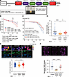 Figure 1
MA-5 ameliorates motor dysfunction and mitochondrial abnormalities in Drosophila ALS models. (A) Experimental design of MA-5 administration in Drosophila. MA-5 was delivered through filter paper soaked with MA-5–containing sucrose solution. (B) Longitudinal climbing performance in control flies and hSOD1G85R ALS-model flies with or without MA-5 (10 or 100 nM). Climbing performance was evaluated from day 10 to day 40 (n = 8 vials/group at days 10 and 20; n = 11 vials/group at days 30 and 40) and analyzed by 2-way repeated-measures ANOVA with Bonferroni correction. (C) Kaplan-Meier survival analysis of adult-onset hSOD1G85R ALS-model flies with or without MA-5 (control, n = 194; vehicle, n = 110; MA-5 10 nM, n = 121; MA-5 100 nM, n = 122). Survival curves were compared by log-rank (Mantel-Cox) test. (D) Total ATP5A-positive mitochondrial area per motor neuron (n = 40 neurons from 4 flies/group), analyzed by 1-way ANOVA followed by Tukey’s multiple-comparison test. (E) Intracellular ATP levels in motor neurons measured with the ratiometric biosensor QUEEN-7μ (52) (control, n = 3 flies; hSOD1G85R, n = 4; hSOD1G85R + MA-5, n = 4), analyzed by 1-way ANOVA followed by Tukey’s honestly significant difference (HSD) post hoc test. Scale bars: 10 μm. (F) Mitochondrial membrane potential assessed by TMRE staining in larval ventral nerve cords (control, n = 27; hSOD1G85R, n = 25; hSOD1G85R + MA-5, n = 20; 5 independent experiments/group), analyzed by 1-way ANOVA followed by Tukey’s HSD post hoc test. Scale bar: 100 μm. Data are presented as mean ± SEM, except for the Kaplan-Meier survival curve in C. *P < 0.05, ****P < 0.0001. #P < 0.05 and ##P < 0.01 for comparisons between hSOD1G85R + vehicle and hSOD1G85R + MA-5 100 nM in B.
Before assessing the therapeutic effect, we confirmed the biodistribution of MA-5 using BODIPY-MA-5, a fluorescently labeled derivative of MA-5. Fluorescence imaging revealed that MA-5 localized clearly to mitochondria within motor neurons of the thoracic ganglia (Supplemental Figure 1A; supplemental material available online with this article; https://doi.org/10.1172/jci.insight.200761DS1). These findings demonstrate that orally administered MA-5 can reach the mitochondria of motor neurons in the brain, indicating its ability to cross multiple biological barriers and directly act at the site of neurodegeneration to improve mitochondrial health.
To evaluate the functional consequences of MA-5 treatment, we performed a longitudinal negative geotaxis climbing assay, a well-established behavioral test of locomotor function in Drosophila. Climbing performance was assessed every 10 days from day 10 to day 40 in control flies, hSOD1G85R flies, and hSOD1G85R flies treated with MA-5 (10 or 100 nM) (Figure 1B and Supplemental Video 1). Two-way repeated-measures ANOVA revealed significant effects of treatment (F3,28 = 7.09, P = 0.0011) and time (P < 0.001), as well as a significant treatment × time interaction (F9,84 = 3.74, P < 0.001), indicating that the trajectory of locomotor decline differed among groups. hSOD1G85R flies exhibited progressive age-dependent locomotor decline, with a marked deficit compared with control flies at day 40 (P < 0.001). MA-5 significantly attenuated this decline: in comparison with vehicle-treated hSOD1G85R flies, 100 nM MA-5 improved climbing performance at day 30 (P = 0.0068) and day 40 (P = 0.011), whereas 10 nM MA-5 improved climbing performance at day 30 (P = 0.021). These results indicate that MA-5 significantly attenuates age-dependent motor decline in this Drosophila ALS model.
We next examined whether this preservation of motor function was accompanied by improved survival. Lifespan analysis was performed using an adult-onset Drosophila ALS model expressing hSOD1G85R under the control of the temperature-sensitive GAL80ts system. Kaplan-Meier survival analysis demonstrated that MA-5 significantly prolonged survival in comparison with vehicle-treated ALS flies (Figure 1C). Similar conclusions were obtained using 3 complementary survival tests (log rank, Wilcoxon’s, and Tarone-Ware), supporting the robustness of the survival benefit. These results indicate that MA-5 confers organismal benefits in vivo, including both preservation of locomotor function and extension of survival.
To assess whether the observed functional improvement was associated with mitochondrial integrity, we examined mitochondrial content in motor neurons of aged ALS flies by quantifying the total ATP5A-positive mitochondrial area per cell. hSOD1G85R flies showed a significant reduction in total mitochondrial area per cell, and this phenotype was markedly rescued by MA-5 administration (Figure 1D), indicating preservation of mitochondrial integrity in motor neurons. Because mitochondria in neuronal processes are highly branched and frequently overlapping in confocal images, this analysis was interpreted as a quantitative measure of mitochondrial content rather than the morphology of individual mitochondria. This observation is consistent with previous studies reporting that MA-5 enhances mitochondrial structural integrity and biogenesis in disease contexts (18, 20).
We examined whether the observed functional recovery was linked to mitochondrial energy output by using the genetically encoded ATP biosensor QUEEN (22) to visualize ATP levels in motor neurons. ATP production was significantly diminished in ALS-model flies but was restored to near-normal levels following MA-5 treatment (Figure 1E), indicating a recovery of mitochondrial energy metabolism. To complement these findings, we assessed mitochondrial membrane potential using tetramethylrhodamine ethyl ester (TMRE) staining in the adult ventral nerve cord. Compared with control flies, hSOD1G85R flies displayed impaired mitochondrial membrane potential, indicated by a significant reduction in TMRE fluorescence intensity (P < 0.0001), which was significantly restored following MA-5 administration (P < 0.0001) (Figure 1F). These results further support the role of MA-5 in restoring mitochondrial function at multiple levels.
Given that mitochondrial abnormalities in ALS are not restricted to neurons, we also examined skeletal muscle phenotypes. Previous studies have reported mitochondrial defects in skeletal muscle tissue of patients with ALS (15). We consistently observed a shift toward larger mitochondrial profiles in the indirect flight muscles of ALS-model flies, whereas MA-5 treatment suppressed this abnormal enlargement and restored the size distribution toward control levels (Supplemental Figure 1B).
We also performed a preliminary exploratory analysis of MA-5 in the SOD1G93A mouse model. Because of the difficulty in breeding this transgenic line and maintaining high-copy animals, only one mouse per group was available for analysis, precluding statistical evaluation. Nevertheless, the MA-5–treated mouse showed qualitative improvements in locomotor performance and mitochondrial morphology compared with the control mouse, consistent with the findings obtained in our Drosophila experiments (Supplemental Figure 1, C–E). These observations should therefore be interpreted as preliminary supportive data, and further validation in adequately powered mammalian in vivo studies is required.
Taken together, these results demonstrate that MA-5 localizes to the mitochondria within motor neurons and restores mitochondrial content, bioenergetic output, and membrane potential in the ALS Drosophila model. In addition, MA-5 preserves locomotor function, and prolongs survival, supporting its therapeutic potential against ALS-associated mitochondrial dysfunction.
MA-5 improved the impairment of mitochondrial function in ALS patient–derived fibroblasts. Although mitochondrial dysfunction is closely associated with ALS pathogenesis (14, 15), no biomarker for impaired mitochondria has been fully validated in patients with ALS in clinical settings. Since GDF15 has been reported as a sensitive and specific diagnostic marker for mitochondrial diseases (23, 24), we measured serum GDF15 levels in 10 patients with ALS and 4 healthy controls (patient profiles are provided in Table 1). Serum GDF15 levels were significantly higher in patients with ALS than in healthy controls (1,557.0 pg/mL vs. 487.5 pg/mL, P = 0.002) (Figure 2A), suggesting that mitochondrial function is impaired in patients with ALS.
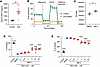 Figure 2
MA-5 restores mitochondrial function in fibroblasts derived from patients with ALS. (A) Serum GDF15 levels in patients with ALS and healthy controls (control, n = 4; ALS, n = 10). (B) Mitochondrial function was assessed in fibroblasts from individuals with SOD-ALS and healthy controls using a flux analyzer (N = 3, respectively). OCR, oxygen consumption rate. (C) ATP production was measured by an ATP measurement kit at 6 hours after DMSO or MA-5 treatment (10 μM) in fibroblasts from patients with SOD-ALS (n = 3, respectively). RLU, relative light units. (D and E) MA-5 protects against oxidative stress in fibroblasts obtained from individuals diagnosed with SOD-ALS. (D) Cell viability was measured by the MTT assay. (E) Cell damage measured by LDH assay. MA-5 was added at 0.1, 0.3, 1, 3, 10, and 30 μM (n = 4). Data are represented as mean ± SEM. *P < 0.05, **P < 0.01, ****P < 0.0001. A and B were analyzed by unpaired 2-tailed t test, while D and E were analyzed using 1-way ANOVA followed by Tukey’s multiple comparison test; each MA-5-treated group in the presence of BSO was compared with the BSO + DMSO group.
Since skin fibroblasts derived from patients with ALS are known to mimic the cellular phenotypes caused by ALS (15, 25), we isolated fibroblasts from patients with SOD1-mutated (G85R) ALS and examined their mitochondrial function using a flux analyzer, which measures key bioenergetic parameters. This analysis revealed significant mitochondrial dysfunction in the fibroblasts derived from patients with ALS (Figure 2B). Consistent with our previous findings that MA-5 upregulates ATP production by reshaping inner mitochondrial membrane supercomplexes (18, 19), MA-5 treatment significantly increased ATP production in these fibroblasts (Figure 2C).
To further evaluate the protective effects of MA-5, fibroblasts were treated with l-buthionine (S,R)-sulfoximine (BSO), a compound that induces ROS by inhibiting glutathione synthesis and mimicking mitochondrial damage (18). Under these conditions, MA-5 significantly rescued BSO-induced cell death in ALS-derived fibroblasts in a dose-dependent manner (3–30 μM) (Figure 2D). This protective effect of MA-5 was further confirmed by a reduction in lactate dehydrogenase (LDH) levels under mitochondrial stress (3–30 μM) (Figure 2E).
To validate the effectiveness of MA-5 in other types of ALS, we isolated fibroblasts from 2 patients with sporadic ALS without known ALS gene mutations. One of the fibroblasts from patients with sporadic ALS exhibited reduced mitochondrial function, similar to that of fibroblasts from patients with mutated SOD1 (G85R) ALS (Supplemental Figure 2A). MA-5 treatment increased ATP production in both fibroblast samples from patients with sporadic ALS (Supplemental Figure 2B). To further confirm the effect of MA-5 on mitochondrial ATP production, we analyzed isolated mitochondria. MA-5 treatment increased ATP production in isolated mitochondria (Supplemental Figure 2C). BSO-induced cell death and LDH release were attenuated by MA-5 in sporadic ALS–derived fibroblasts in a dose-dependent manner up to 10 μM, whereas 30 μM MA-5 appeared to be less effective or potentially harmful (Supplemental Figure 2, D and E).
These findings indicate that MA-5 ameliorates mitochondrial dysfunction in fibroblasts derived from patients with various types of ALS, including those with SOD1 mutations and sporadic ALS, highlighting its potential as a therapeutic intervention for ALS.
MA-5 restored the phenotypes of iPSC-derived motor neurons from patients with ALS. In addition to the effects of MA-5 on skin fibroblasts derived from patients with ALS, we generated iPSCs from patients with ALS carrying SOD1 mutations (SOD1-ALS-iPSCs; SOD-Pt1 and SOD-Pt2 from two ALS patients, respectively) and differentiated these iPSCs into motor neurons, as previously described, given that motor neurons are not easily procured from patients with ALS (26) (Figure 3A). Motor neurons derived from SOD1-ALS-iPSCs showed impaired mitochondrial respiratory parameters compared with those derived from healthy control iPSCs, although the extent of impairment varied between patient-derived lines (Figure 3B). Basal respiration was significantly reduced in one of the two ALS-derived motor neuron lines, whereas ATP production and maximal respiration were consistently impaired in both lines (Figure 3B). Consistent with the results observed in fibroblasts derived from patients with ALS, ATP production was significantly increased by MA-5 treatment in SOD1-ALS-iPSC–derived motor neurons (Figure 3C). We also examined multiple concentrations of MA-5 to determine the optimal dose for enhancing ATP production. These analyses suggested that 100 nM MA-5 effectively increased ATP production, whereas concentrations of 10 μM or higher appeared to be ineffective or potentially harmful in this experimental setting. Electron microscopy further revealed mitochondrial malformations in SOD1-ALS-iPSC–derived motor neurons, which were rescued by MA-5 treatment (Figure 3D). Mitochondrial dysfunction affects both mitochondrial dynamics and morphology (27). To investigate this, we compared the mitochondrial dynamics in SOD1-ALS-iPSC–derived motor neurons treated with either dimethylsulfoxide (DMSO) or MA-5. The average velocity of mitochondrial movement was significantly improved by MA-5 treatment (0.005 vs. 0.017 μm/s and 0.006 vs. 0.010 μm/s, P = 0.017 and P = 0.019, respectively) (Figure 3E and Supplemental Video 2). Similar results were observed in motor neurons derived from iPSCs of patients with ALS carrying FUSH517D/H517D gene mutations (FUS-ALS-iPSC–derived motor neurons) (28). Mitochondrial function parameters, including basal respiration, ATP production, and maximal respiration, were significantly lower in FUS-ALS-iPSC–derived motor neurons than in those derived from healthy controls (Supplemental Figure 3A). MA-5 treatment significantly improved ATP production, mitochondrial morphology, and mitochondrial dynamics, consistent with the results observed in SOD1-ALS-iPSC–derived motor neurons (Supplemental Figure 3, B–D, and Supplemental Video 3). These findings indicate that MA-5 improves mitochondrial function in motor neurons derived from patients with ALS and suggest therapeutic potential for treating ALS phenotypes.
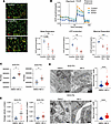 Figure 3
MA-5 alleviates mitochondrial dysfunction and structural abnormalities in motor neurons derived from SOD1-ALS-iPSCs. (A) Motor neurons differentiated from SOD1-ALS-iPSCs (2 patients; SOD-Pt1 and SOD-Pt2). Scale bars: 100 μm. Motor neuron markers: ISL1 (ISL LIM homeobox 1) and MAC2 (galectin 3). (B) A flux analyzer was used to assess mitochondrial function in motor neurons derived from SOD1-ALS-iPSCs and healthy controls (control, n = 4; SOD1, n = 4; SOD2, n = 5). Oxygen consumption rate (OCR) values were normalized to the protein concentration of the motor neurons. (C) ATP production at 6 hours after DMSO or MA-5 treatment (100 nM) in motor neurons from SOD1-ALS-iPSCs (n = 4, respectively). RLU, relative light units. (D) Mitochondrial malformation measured as the ratio of maximal length to width (100 mitochondria measured) in motor neurons from SOD1-ALS-iPSCs with or without MA-5 treatment (100 nM). (E) Mitochondrial movement velocity in motor neurons from SOD1-ALS-iPSCs (SOD-Pt1, n = 9; SOD-Pt2, n = 10) with or without MA-5 treatment (100 nM). Data are represented as mean ± SEM. *P < 0.05, **P < 0.01, ***P < 0.001, ****P < 0.0001. B was analyzed by 1-way ANOVA, while C–E were analyzed using unpaired 2-tailed t tests.
MA-5 likely treated iPSC-derived motor neurons by manipulating the expression of ALS-related genes and mitochondrial genes. We next investigated transcriptomic alterations in SOD1-ALS-iPSC–derived motor neurons to elucidate the molecular mechanisms by which MA-5 ameliorates disease-associated phenotypes. Differential gene expression analysis was performed by comparison of SOD1-ALS-iPSC–derived motor neurons treated with or without MA-5. Genes commonly regulated by MA-5 in both SOD-Pt1 and SOD-Pt2 lines were subjected to gene set enrichment analysis. The analysis revealed significant enrichment of mitochondria-related gene sets, including those associated with the mitochondrial inner membrane and the oxidative phosphorylation (OXPHOS) system (Figure 4A). These findings suggest that MA-5 exerts its therapeutic effects, at least in part, by modulating mitochondrial gene expression and restoring mitochondrial function in ALS motor neurons.
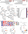 Figure 4
Effect of MA-5 on iPSC-derived motor neurons and potential clinical markers identified in ALS-iPSCs. (A) Enrichment analyses of gene expression changes caused by MA-5 treatment (100 nM) in motor neurons derived from SOD1-ALS-iPSCs. The enrichment analysis was performed using Gene Ontology Cellular Component 2023 (left) and WikiPathways 2023 (right), which are integrated in the Enrichr tool (https://maayanlab.cloud/Enrichr/). Mutual genes altered by MA-5 in SOD-Pt1 and SOD-Pt2 lines were analyzed. (B) Metabolites involved in the tricarboxylic acid (TCA) cycle in motor neurons derived from healthy controls and SOD1-ALS-iPSCs with or without MA-5 (10 μM) (N = 3). (C) Variable importance in projection (VIP) scores in motor neurons derived from SOD1-ALS-iPSCs with or without MA-5 (10 μM) (N = 6, respectively). (D) The top 10 ORF genes increased in motor neurons derived from SOD1-ALS-iPSCs and decreased by MA-5 (100 nM). Gene expression in motor neurons derived from sporadic-ALS-iPSCs with or without MA-5 (100 nM) is also shown. (E) C3orf62 and C7orf31 expression in motor neurons from SOD1-ALS-iPSCs, sporadic-ALS-iPSCs, and healthy controls (control, n = 3; SOD, n = 6; sporadic, n = 6). TPM, transcripts per kilobase million. (F) Plasma C7orf31 levels in healthy controls (n = 8), patients with SOD-ALS (n = 5), patients with sporadic ALS (n = 21), and those with other neurological diseases (n = 13; neuropathy with liability to pressure palsies, dermatomyositis, Parkinson’s disease, and spinocerebellar degeneration). Data are represented as mean ± SEM. *P < 0.05, **P < 0.01, ***P < 0.001, ****P < 0.0001. B, E, and F were analyzed using 1-way ANOVA.
We further analyzed the metabolites in SOD1-ALS-iPSC–derived motor neurons using metabolomics, which enabled the comprehensive analysis of over 600 metabolites. The results indicated that metabolites upstream of succinate in the tricarboxylic acid (TCA) cycle tended to accumulate in SOD1-ALS-iPSC–derived motor neurons compared with those in healthy controls, whereas metabolites downstream of succinate were significantly decreased (Figure 4B). Reduced activity of oxoglutarate dehydrogenase, an enzyme that converts α-ketoglutarate to succinyl-CoA, has been reported in the spinal cords of hSOD1G93A mice, a known ALS model carrying mutated human SOD1 (29, 30). We also confirmed that similar metabolic changes were observed in the cerebrospinal fluid of patients with ALS, where metabolites upstream of succinate were significantly increased compared with those in disease controls (patients with spinocerebellar degeneration) (Supplemental Figure 4A). These metabolic findings suggest that the phenotypes observed in SOD1-ALS-iPSC–derived motor neurons resemble those observed in other ALS models, specifically regarding disrupted metabolic pathways. However, MA-5 did not reverse the metabolic changes in the TCA cycle observed in ALS motor neurons, suggesting that its mechanism of action may involve pathways independent of the TCA cycle.
To identify additional metabolites affected by MA-5 treatment, we performed comparative metabolomic profiling of SOD1-ALS-iPSC–derived motor neurons treated with or without MA-5, using variable importance in projection (VIP) scores (Figure 4C). Among the top-ranked metabolites, a reduction in glycerophosphate and an elevation in NADH levels were particularly notable following MA-5 treatment. These changes suggest a potential downregulation of the glycerophosphate shuttle pathway. The glycerophosphate shuttle pathway is known to contribute to mitochondrial ROS production, which plays a pathogenic role in neurodegenerative diseases, including ALS (31–33). We further examined whether pharmacological inhibition of the glycerophosphate shuttle reduces mitochondrial ROS production in SOD1-ALS-iPSC–derived motor neurons using iGP-1, a specific inhibitor of mitochondrial glycerol-3-phosphate dehydrogenase (mGPDH), a key enzyme in this pathway (34). iGP-1 treatment significantly reduced mitochondrial ROS production in these cells (Supplemental Figure 4B). These findings imply that MA-5 may attenuate dysregulation of the glycerophosphate shuttle, thereby partially restoring mitochondrial redox balance and function.
A potential biomarker to diagnose subtypes of ALS and monitor the effect of MA-5 treatment. Based on the established in vitro disease model using ALS-iPSC–derived motor neurons, we further investigated potential clinical markers to monitor the effects of MA-5 treatment. Since ORF genes have been reported as novel clinical markers (35), we focused on ORF genes. RNA-seq analysis revealed that C3orf62 and C7orf31 were upregulated in SOD1-ALS-iPSC–derived motor neurons, following downregulation by MA-5 treatment, suggesting that these genes may serve as markers for monitoring the therapeutic effects of MA-5 (Figure 4D).
Notably, while C3orf62 was also upregulated in sporadic-ALS-iPSC–derived motor neurons, C7orf31 expression did not increase in these cells (Figure 4, D and E). These findings suggest that C7orf31 may serve as a potential clinical marker for ALS subtyping, aiding in the distinction between SOD1-mutated and sporadic forms of the disease.
To validate these findings, we measured the concentration of C7orf31 protein in the plasma of patients with SOD1-ALS and those with sporadic ALS. The plasma concentration of C7orf31 protein was significantly higher in patients with SOD1-ALS than in those with sporadic ALS (28.32 ± 12.37 ng/mL vs. 7.20 ± 4.52 ng/mL, P < 0.0001) (Figure 4F). This result indicates that plasma C7orf31 may be a promising biomarker for diagnosing ALS with SOD1 mutations and for monitoring the effect of MA-5 therapy.

-
Methods
Additional reagents and resources are listed in Table 2.
Sex as a biological variable. Human clinical samples and cell lines were derived from both female and male. ALS model mice also included male (SOD1-Ctrl) and female (SOD1-MA-5). In the Drosophila experiments, male flies were used for the climbing assay and all imaging analyses, whereas the lifespan analysis included both males and females, whose survival data were pooled. Males were used elsewhere to match the original characterization of this human SOD1 (hSOD1) model, in which the behavioral and histological analyses were performed on males (21), and to remove sex as a source of variability.
Human participants. To investigate the level of mitochondrial dysfunction in patients with ALS, serum levels of GDF15 were measured in patients with ALS and healthy controls (Figure 2A). Human skin fibroblasts were isolated from patients with ALS and healthy controls as previously described to examine the mitochondrial function in cells from patients with ALS (18) (Figure 2, B–E). The patient profiles are presented in Tables 1 and 3. iPSCs derived from patients with ALS carrying the SOD mutation, HPS0476 and HPS0485, were obtained from the RIKEN Cell Bank in Japan. iPSCs derived from patients with sporadic ALS were generated in our laboratory. In contrast, iPSCs derived from patients with ALS carrying the FUS gene mutation were described previously (28).
Drosophila stocks and MA-5 treatment. The GAL4-UAS expression system was used to direct the transgene expression in specific cell types. The D42-GAL4 driver line was used for motor neuron–specific expression, whereas the w1118 strain, a commonly used genetic background for transgenic Drosophila, served as the control. The following stocks were obtained from the Bloomington Drosophila Stock Center (Indiana University, Bloomington, Indiana, USA): w1118 (stock 3605), D42-GAL4 (stock 8816), UAS-hSOD1G85R (stock 33608), UAS-mCD8:GFP (stock 5137), elav-GAL4c155 (stock 458), and tubP-GAL80ts (stock 7019). The elav-GAL4c155; tub-GAL80ts/CyO stock used for the lifespan assay was generated in this laboratory by combining of the latter two. Flies were maintained at 25°C under standard conditions.
MA-5 was dissolved in 5% sucrose solution and administered orally via filter paper placed in vials. Flies were treated with MA-5 (10 or 100 nM) for 3 days, followed by 2 days on standard food. To minimize MA-5 photodegradation, flies were kept in darkness during drug treatment.
Drosophila climbing assay. Flies were collected within 4 days of eclosion and divided into independent vials containing 15–20 flies each. Locomotor function was assessed every 10 days from day 10 to day 40 using a negative geotaxis assay. For each trial, flies were gently tapped to the bottom of an empty vial and allowed to climb for 20 seconds. The climbing pass rate was calculated as the percentage of flies that reached or exceeded a height of 5 cm within 20 seconds. Each vial was tested 3 times per session with approximately 1-minute intervals between trials, and the mean of the 3 trials was used for subsequent analysis. Each vial was treated as 1 biological replicate. For each treatment group, a total of 11 vials were analyzed; 8 vials were tracked at all time points from day 10 to day 40, whereas 3 additional vials were included from day 30 onward. Consequently, the number of vials per group was 8 at days 10 and 20 and 11 at days 30 and 40.
Immunohistochemistry and image analysis in Drosophila. To visualize mitochondria, samples were stained with an anti-ATP5A antibody (1:500; Abcam, 14748). Neuronal nuclei were labeled using Rat-Elav-7E8A10 antibody (1:500; Developmental Studies Hybridoma Bank, 7E8A10), and muscle actin was visualized using Alexa Fluor 633 phalloidin. Samples were imaged using a Zeiss LSM 980 confocal microscope. Total ATP5A-positive mitochondrial area was quantified using Fiji/ImageJ (NIH). Biodistribution studies were performed using BODIPY-MA-5.
TMRE staining and quantification. For TMRE staining, third-instar larval ventral nerve cords (thoracic ganglia) were dissected in HL3.1 buffer and incubated with 100 nM TMRE in HL3.1 buffer for 12 minutes at 25°C. After staining, samples were washed twice (30 seconds each) in HL3.1 buffer containing 25 nM TMRE. Live imaging was performed on freshly prepared samples, with all acquisitions completed within 1 hour of dissection. Images were acquired on a Zeiss LSM980 confocal microscope with a ×20 objective (TMRE excitation ~555 nm). TMRE fluorescence intensity was quantified in Fiji from a single region of interest spanning the T1–T2 segments of each sample, and normalized to the mean of the control group within the same experimental batch to account for inter-batch variability.
Generation of transgenic QUEEN flies and QUEEN imaging and analysis. The UAS-QUEEN construct was generated from pN1-QUEEN-7μ (Addgene, 129307) and inserted into the attP40 landing site by WellGenetics. Intracellular ATP levels were measured using the genetically encoded ATP biosensor QUEEN. Thoracic ganglia were dissected in HL3.1 buffer and imaged within 1 hour using a Zeiss LSM980 confocal microscope (45). Fluorescence images were acquired at 405 nm and 488 nm excitation wavelengths, and ATP levels were estimated from the 405/488 fluorescence ratio (22). Three control flies, 4 vehicle-treated hSOD1G85R flies, and 4 MA-5–treated hSOD1G85R flies were analyzed. For each fly, regions within the T1–T2 segments of the thoracic ganglion with low background fluorescence and clear QUEEN signal were selected as regions of interest. A total of 1,144 cells from control flies, 551 cells from vehicle-treated hSOD1G85R flies, and 549 cells from MA-5–treated hSOD1G85R flies were quantified.
Drosophila lifespan assay. Adult-onset neuronal expression of hSOD1G85R was induced using the elav-GAL4c155 tub-GAL80ts driver system. UAS-hSOD1G85R flies, carrying transgene insertions on the second and third chromosomes, were introgressed into a w1118 background before use. Both male and female flies were used. Adult-onset ALS-model flies were elav-GAL4c155/+ (females) or elav-GAL4c155/Y (males); tub-GAL80ts/UAS-hSOD1G85R; UAS-hSOD1G85R/+, and the corresponding driver-only genotype, elav-GAL4c155/+ or /Y; tub-GAL80ts/+, was used as the control.
Flies were maintained at 18°C during development to suppress GAL4 activity. Newly eclosed adults were collected on day 1 after eclosion, distributed across 3 independent vials per condition (control, 194 flies in 11 vials; hSOD1G85R + vehicle, 110 in 7 vials; + MA-5 10 nM, 121 in 7 vials; + MA-5 100 nM, 122 in 7 vials, with 11–22 flies per vial), and maintained at 18°C. On day 4 after eclosion, flies were shifted to 29°C to induce pan-neuronal hSOD1G85R expression; this day was designated day 0 post-induction. MA-5 was administered at 10 nM and 100 nM in 5% sucrose containing 0.01% DMSO via filter paper placed in empty vials, and vehicle-treated flies received 5% sucrose containing 0.01% DMSO. Flies were alternately transferred between drug-treatment vials and standard food vials every other day without anesthesia. Survival was monitored daily until day 50 post-induction.
SOD1G93A mouse model studies. All animal procedures were performed in accordance with protocols approved by the Animal Experiment Committee of the National Institute of Neuroscience, National Center of Neurology and Psychiatry. SOD1G93A transgenic mice, which express a G93A mutant form of human SOD1, were obtained from The Jackson Laboratory (002726). Heterozygous (SOD1G93A) males were bred with wild-type female C57BL/6J mice (Japan SLC) as previously reported (46). SOD1G93A mice were fed standard chow with or without MA-5 (0.02%) starting at 40 days of age. Rotarod and grip strength tests were performed at 100 days of age. Mice were sacrificed at 120 days of age, and motor neurons in the lumbar spinal cord were analyzed by electron microscopy.
Measurement of serum GDF15 levels. Blood samples were collected from patients with ALS visiting Tohoku University Hospital and stored at –80°C until analysis. Serum GDF15 levels were measured in patients with ALS and healthy controls using the Human GDF-15 ELISA Kit (R&D Systems).
Isolation of human skin fibroblasts and assays. Human skin fibroblasts were isolated from patients with ALS and healthy controls as previously described (18). The cells were cultured in low-glucose DMEM (1.0 g/L) supplemented with 10% fetal bovine serum. Cell viability was assessed using Cell Count Reagent SF (Nacalai Tesque) 72 hours after treatment with BSO (Wako Pure Chemical Industries) or DMSO. Lactate dehydrogenase (LDH) levels were measured using the LDH Cytotoxicity Detection Kit (Takara). ATP production was evaluated using an ATP measurement kit (Toyo Ink) 6 hours after DMSO or MA-5 treatment (10 μM). Mitochondria were isolated using the Mitochondria Isolation Kit for Cultured Cells according to the manufacturer’s instructions (Thermo Fisher Scientific, 89874). The patient profiles are provided in Table 3.
Mitochondrial function measurement. Oxygen consumption in fibroblasts and iPSC-derived motor neurons was measured using the Flux Analyzer XF24 or XF96 (Agilent), as previously described (18). Cells were incubated in the assay medium without CO2 for 60 minutes before sequential injections of mitochondrial oxidative phosphorylation inhibitors. Because iPSC-derived motor neurons can show variability in seeding density and attachment efficiency, the data were normalized to protein concentration.
Culture and differentiation of iPSCs. iPSCs derived from patients with ALS carrying the SOD mutation, HPS0476 and HPS0485, were obtained from the RIKEN Cell Bank in Japan and maintained as described previously (47). iPSCs derived from patients with sporadic ALS were generated in our laboratory using the Epi5 Episomal iPSC Reprogramming Kit (Life Technologies). Motor neurons were differentiated using a previous protocol (26). Briefly, iPSCs were dissociated and seeded on Matrigel-coated plates. On the following day, the iPSC medium was replaced with a neuron differentiation medium (1:1 mixture of DMEM/F12 and Neurobasal medium [Thermo Fisher Scientific] with 0.5× N2 and B27, 0.1 mM ascorbic acid [Sigma Aldrich], and 1× GlutaMAX [Life Technologies]) supplemented with 3 μM CHIR99021 (Cayman), 2 μM DMH1 (Cayman), and 2 μM SB431542 (Cayman) to differentiate iPSCs into neuron epithelial progenitors (NEPs). On day 6, the cells were dissociated and split at 1:6 with the neuron differentiation medium, including 1 μM CHIR99021, 2 μM DMH1, 2 μM SB431542, 0.1 μM retinoic acid (Cayman), and 0.5 μM purmorphamine (Cayman). The cells were maintained under these conditions for 6 days so that the NEPs could differentiate into OLIG2+ motor neuron progenitors (MNPs). To produce motor neurons, OLIG2+ MNPs were dissociated with dispase and cultured in the differentiation medium with 0.5 μM retinoic acid and 0.1 μM purmorphamine. Under these conditions for 6 days, the motor neurons obtained were dissociated and plated on Matrigel-coated plates in the differentiation medium, including 0.5 μM retinoic acid, 0.1 μM purmorphamine, and 0.1 μM compound E (Calbiochem) for maturation. In contrast, iPSCs derived from patients with ALS carrying the FUS gene mutation were described previously (28) and differentiated as previously described (28). For experiments examining inhibition of the glycerophosphate shuttle, iPSCs were differentiated into motor neurons using a previously reported protocol (48).
Immunofluorescence. Immunofluorescence assays were performed as previously described (47). iPSC-derived motor neurons were fixed in 4% paraformaldehyde and stained with primary and secondary antibodies (Table 2).
Mitochondrial motility analysis. Live-cell imaging of the motor neurons stained with MitoTracker Green was performed using a KEYENCE BZ-X700 microscope. Time-lapse videos were analyzed for cell migration using the VW-H2MA software (KEYENCE) (20).
Electron microscopy analysis. Mitochondrial dimensions in iPSC-derived motor neurons were measured as previously reported (18). For Drosophila, the electron microscopy images were analyzed using size-based categorization (49).
RNA sequencing analysis. RNA sequence libraries were prepared using QIAseq Stranded RNA Library Kits (QIAGEN, catalog 180451) following the manufacturer’s instructions. The Illumina libraries were converted into circular single-stranded DNA libraries using the MGI Easy Universal Library Conversion Kit (App-A, MGI Tech). The converted libraries were sequenced on a DNBSEQ-G400RS platform (MGI Tech) using the 2 × 150 bp paired-end mode. RNA sequencing (RNA-seq) was performed using DNAFORM (Yokohama). Sequence reads in FASTQ format were evaluated for quality using FastQC (https://www.bioinformatics.babraham.ac.uk/projects/fastqc/), and low-quality reads were removed using Trimmomatic-0.39 (https://github.com/usadellab/Trimmomatic). High-quality reads were mapped to the GRCh38 reference genome using STAR (v2.7.10a; https://github.com/alexdobin/STAR), a splice-aware aligner. Gene- and transcript-level abundances were quantified using StringTie (v2.2.0; https://ccb.jhu.edu/software/stringtie/), which provides accurate expression estimates. Gene expression levels were analyzed using the edgeR package (v3.15) in Bioconductor, which normalizes the data and identifies differentially expressed genes. Functional enrichment analyses were performed using EnrichR, focusing on the mutual genes affected by MA-5 treatment in SOD-Pt1 and SOD-Pt2 cell lines to identify relevant pathways and biological processes.
Metabolomics analysis. Metabolomics analysis was performed as previously described (50, 51). Metabolites were initially separated by capillary electrophoresis based on their charge and size. This method enables high-resolution separation of small molecules under an electric field. Metabolites were then selectively detected using mass spectrometry, which monitors a broad range of mass-to-charge (m/z) values to capture a comprehensive metabolite profile. Variable importance in projection (VIP) scores were calculated and analyzed using MetaboAnalyst (https://www.metaboanalyst.ca/) to identify the metabolites that contributed most significantly to the group differences between patients with ALS and healthy controls.
Glycerophosphate shuttle inhibition. Differentiated motor neurons derived from HPS0485 iPSCs were seeded in 96-well plates and incubated with or without iGP-1 (10 μM) for 1 hour. MitoSOX Red mitochondrial superoxide indicator (Invitrogen, M36008; 0.5 μM) was then added to quantify mitochondrial ROS production. Six images were acquired from each well using a BZ-X810 fluorescence microscope (KEYENCE). MitoSOX fluorescence intensity was normalized to cell number, which was determined by Hoechst staining. The average MitoSOX fluorescence intensity per cell in each well was used as the final value for analysis.
C7orf31 measurement. Plasma C7orf31 protein levels were measured using a C7orf31 ELISA Kit (MyBioSource, catalog MB9340367).
Statistics. Statistical analyses were performed using GraphPad Prism software. For comparisons between 2 groups, unpaired 2-tailed t tests were used, while 1-way analysis of variance (ANOVA) was applied for multiple comparisons, followed by Tukey’s correction. For the Drosophila climbing assay, each vial was treated as 1 biological replicate. Differences in climbing performance over time were analyzed using 2-way repeated-measures ANOVA, with treatment group as the between-subjects factor and day as the within-subject factor, followed by Bonferroni correction for multiple comparisons. For Drosophila lifespan analysis, survival data were analyzed using Kaplan-Meier estimation. Pairwise comparisons were performed using log-rank (Mantel-Cox), Wilcoxon’s (Breslow), and Tarone-Ware tests. Kaplan-Meier estimation and survival statistics were performed using custom Python 3 scripts. Statistical significance was set at a P value of <0.05.
Study approval. All these studies were approved by the Ethics Committee of the Medical School of Tohoku University (2018-2-254, 2021-1-905, 2021-1-901, and 2022-1-823). Ethical guidelines were strictly followed to ensure the integrity and confidentiality of the patient samples.
Data availability. All unique/stable materials generated in this study are available from Takafumi Toyohara with a completed material transfer agreement. The raw RNA sequencing data generated in this study from motor neurons derived from ALS-iPSCs were deposited in the NCBI’s Sequence Read Archive under BioProject accession number PRJNA1272663 (https://www.ncbi.nlm.nih.gov/bioproject/?term=PRJNA1272663). The numerical source data underlying the figures are provided in the accompanying Supporting Data Values file. Requests for further information or for resources and reagents should be directed to and will be fulfilled by corresponding author Takafumi Toyohara.


