
Research ArticleImmunologyInflammation
Open Access |  10.1172/jci.insight.185437
The E3 ubiquitin ligase HECTD3 controls immune colonic inflammation by restricting MyD88 activation and signaling
Shamima Islam,1 Shahnewaj Mannan,1 Ashley Zuniga,2 Upasana Parthasarathy,2 Valeriu B. Cismasiu,1 Leonardo Silvane,1 Sayan Chakraborty,1 Divya Priyanka Talada,1 Raghwendra Pratap Singh,1 Tomas Zelenka,1 Amreen Naveen,1 Sephra Chyanne Vickers,1 Michael G.M. Grant,3 Jonathan J. Cho,2 Theodore Drashansky,2 Alexander J. Kwiatkowski,4 Xintong Liu,5 Ross Tomaino,6 Olga A. Guryanova,7 Mariola J. Ferraro,8 Sang Yong Kim,9 Christian Jobin,10 Benjamin G. Keselowsky,4 Hongmin Li,5 Paulo C. Rodriguez,1 Martina Molgora,1 Amer A. Beg,1 Timothy I. Shaw,11 Zheng Ruan,12 Lixin Wan,3 and Dorina Avram1
1Department of Immunology, H. Lee Moffitt Cancer Center and Research Institute, Tampa, Florida, USA.
2Department of Anatomy and Cell Biology, College of Medicine, University of Florida, Gainesville, Florida, USA.
3Department of Molecular Oncology, H. Lee Moffitt Cancer Center and Research Institute, Tampa, Florida, USA.
4J. Crayton Pruitt Family Department of Biomedical Engineering, University of Florida, Gainesville, Florida, USA.
5Department of Pharmacology and Toxicology, R. Ken Coit College of Pharmacy, Department of Chemistry and Biochemistry, College of Science & College of Medicine, The BIO5 Institute, The University of Arizona, Tucson, Arizona, USA.
6Taplin Mass Spectrometry Facility, Harvard Medical School, Boston, Massachusetts, USA.
7Department of Pharmacology and Therapeutics and
8Department of Microbiology and Cell Science, IFAS, University of Florida, Gainesville, Florida, USA.
9Department of Pathology, NYU School of Medicine, New York, New York, USA.
10Division of Gastroenterology, Hepatology, and Nutrition, Department of Medicine, University of Florida, Gainesville, Florida, USA.
11Department of Biostatistics and Bioinformatics, H. Lee Moffitt Cancer Center and Research Institute, Tampa, Florida, USA.
12Department of Biochemistry & Molecular Biology, Thomas Jefferson University, Philadelphia, Pennsylvania, USA.
Address correspondence to: Dorina Avram, 12902 Magnolia Drive, Tampa, Florida, 33612, USA. Phone: 813.745.6689; Email: Dorina.Avram@moffitt.org. BC’s present address is: Victor Babes National Institute of Pathology, Bucharest, Romania. JJC’s present address is: USS CARL VINSON CVN 70, UNIT 100111 BOX 2310, FPO AP 96629.
Authorship note: SI, SM, AZ, and UP contributed equally to this work.
Find articles by Islam, S. in: PubMed | Google Scholar
1Department of Immunology, H. Lee Moffitt Cancer Center and Research Institute, Tampa, Florida, USA.
2Department of Anatomy and Cell Biology, College of Medicine, University of Florida, Gainesville, Florida, USA.
3Department of Molecular Oncology, H. Lee Moffitt Cancer Center and Research Institute, Tampa, Florida, USA.
4J. Crayton Pruitt Family Department of Biomedical Engineering, University of Florida, Gainesville, Florida, USA.
5Department of Pharmacology and Toxicology, R. Ken Coit College of Pharmacy, Department of Chemistry and Biochemistry, College of Science & College of Medicine, The BIO5 Institute, The University of Arizona, Tucson, Arizona, USA.
6Taplin Mass Spectrometry Facility, Harvard Medical School, Boston, Massachusetts, USA.
7Department of Pharmacology and Therapeutics and
8Department of Microbiology and Cell Science, IFAS, University of Florida, Gainesville, Florida, USA.
9Department of Pathology, NYU School of Medicine, New York, New York, USA.
10Division of Gastroenterology, Hepatology, and Nutrition, Department of Medicine, University of Florida, Gainesville, Florida, USA.
11Department of Biostatistics and Bioinformatics, H. Lee Moffitt Cancer Center and Research Institute, Tampa, Florida, USA.
12Department of Biochemistry & Molecular Biology, Thomas Jefferson University, Philadelphia, Pennsylvania, USA.
Address correspondence to: Dorina Avram, 12902 Magnolia Drive, Tampa, Florida, 33612, USA. Phone: 813.745.6689; Email: Dorina.Avram@moffitt.org. BC’s present address is: Victor Babes National Institute of Pathology, Bucharest, Romania. JJC’s present address is: USS CARL VINSON CVN 70, UNIT 100111 BOX 2310, FPO AP 96629.
Authorship note: SI, SM, AZ, and UP contributed equally to this work.
Find articles by Mannan, S. in: PubMed | Google Scholar
1Department of Immunology, H. Lee Moffitt Cancer Center and Research Institute, Tampa, Florida, USA.
2Department of Anatomy and Cell Biology, College of Medicine, University of Florida, Gainesville, Florida, USA.
3Department of Molecular Oncology, H. Lee Moffitt Cancer Center and Research Institute, Tampa, Florida, USA.
4J. Crayton Pruitt Family Department of Biomedical Engineering, University of Florida, Gainesville, Florida, USA.
5Department of Pharmacology and Toxicology, R. Ken Coit College of Pharmacy, Department of Chemistry and Biochemistry, College of Science & College of Medicine, The BIO5 Institute, The University of Arizona, Tucson, Arizona, USA.
6Taplin Mass Spectrometry Facility, Harvard Medical School, Boston, Massachusetts, USA.
7Department of Pharmacology and Therapeutics and
8Department of Microbiology and Cell Science, IFAS, University of Florida, Gainesville, Florida, USA.
9Department of Pathology, NYU School of Medicine, New York, New York, USA.
10Division of Gastroenterology, Hepatology, and Nutrition, Department of Medicine, University of Florida, Gainesville, Florida, USA.
11Department of Biostatistics and Bioinformatics, H. Lee Moffitt Cancer Center and Research Institute, Tampa, Florida, USA.
12Department of Biochemistry & Molecular Biology, Thomas Jefferson University, Philadelphia, Pennsylvania, USA.
Address correspondence to: Dorina Avram, 12902 Magnolia Drive, Tampa, Florida, 33612, USA. Phone: 813.745.6689; Email: Dorina.Avram@moffitt.org. BC’s present address is: Victor Babes National Institute of Pathology, Bucharest, Romania. JJC’s present address is: USS CARL VINSON CVN 70, UNIT 100111 BOX 2310, FPO AP 96629.
Authorship note: SI, SM, AZ, and UP contributed equally to this work.
Find articles by Zuniga, A. in: PubMed | Google Scholar
1Department of Immunology, H. Lee Moffitt Cancer Center and Research Institute, Tampa, Florida, USA.
2Department of Anatomy and Cell Biology, College of Medicine, University of Florida, Gainesville, Florida, USA.
3Department of Molecular Oncology, H. Lee Moffitt Cancer Center and Research Institute, Tampa, Florida, USA.
4J. Crayton Pruitt Family Department of Biomedical Engineering, University of Florida, Gainesville, Florida, USA.
5Department of Pharmacology and Toxicology, R. Ken Coit College of Pharmacy, Department of Chemistry and Biochemistry, College of Science & College of Medicine, The BIO5 Institute, The University of Arizona, Tucson, Arizona, USA.
6Taplin Mass Spectrometry Facility, Harvard Medical School, Boston, Massachusetts, USA.
7Department of Pharmacology and Therapeutics and
8Department of Microbiology and Cell Science, IFAS, University of Florida, Gainesville, Florida, USA.
9Department of Pathology, NYU School of Medicine, New York, New York, USA.
10Division of Gastroenterology, Hepatology, and Nutrition, Department of Medicine, University of Florida, Gainesville, Florida, USA.
11Department of Biostatistics and Bioinformatics, H. Lee Moffitt Cancer Center and Research Institute, Tampa, Florida, USA.
12Department of Biochemistry & Molecular Biology, Thomas Jefferson University, Philadelphia, Pennsylvania, USA.
Address correspondence to: Dorina Avram, 12902 Magnolia Drive, Tampa, Florida, 33612, USA. Phone: 813.745.6689; Email: Dorina.Avram@moffitt.org. BC’s present address is: Victor Babes National Institute of Pathology, Bucharest, Romania. JJC’s present address is: USS CARL VINSON CVN 70, UNIT 100111 BOX 2310, FPO AP 96629.
Authorship note: SI, SM, AZ, and UP contributed equally to this work.
Find articles by Parthasarathy, U. in: PubMed | Google Scholar
1Department of Immunology, H. Lee Moffitt Cancer Center and Research Institute, Tampa, Florida, USA.
2Department of Anatomy and Cell Biology, College of Medicine, University of Florida, Gainesville, Florida, USA.
3Department of Molecular Oncology, H. Lee Moffitt Cancer Center and Research Institute, Tampa, Florida, USA.
4J. Crayton Pruitt Family Department of Biomedical Engineering, University of Florida, Gainesville, Florida, USA.
5Department of Pharmacology and Toxicology, R. Ken Coit College of Pharmacy, Department of Chemistry and Biochemistry, College of Science & College of Medicine, The BIO5 Institute, The University of Arizona, Tucson, Arizona, USA.
6Taplin Mass Spectrometry Facility, Harvard Medical School, Boston, Massachusetts, USA.
7Department of Pharmacology and Therapeutics and
8Department of Microbiology and Cell Science, IFAS, University of Florida, Gainesville, Florida, USA.
9Department of Pathology, NYU School of Medicine, New York, New York, USA.
10Division of Gastroenterology, Hepatology, and Nutrition, Department of Medicine, University of Florida, Gainesville, Florida, USA.
11Department of Biostatistics and Bioinformatics, H. Lee Moffitt Cancer Center and Research Institute, Tampa, Florida, USA.
12Department of Biochemistry & Molecular Biology, Thomas Jefferson University, Philadelphia, Pennsylvania, USA.
Address correspondence to: Dorina Avram, 12902 Magnolia Drive, Tampa, Florida, 33612, USA. Phone: 813.745.6689; Email: Dorina.Avram@moffitt.org. BC’s present address is: Victor Babes National Institute of Pathology, Bucharest, Romania. JJC’s present address is: USS CARL VINSON CVN 70, UNIT 100111 BOX 2310, FPO AP 96629.
Authorship note: SI, SM, AZ, and UP contributed equally to this work.
Find articles by Cismasiu, V. in: PubMed | Google Scholar
1Department of Immunology, H. Lee Moffitt Cancer Center and Research Institute, Tampa, Florida, USA.
2Department of Anatomy and Cell Biology, College of Medicine, University of Florida, Gainesville, Florida, USA.
3Department of Molecular Oncology, H. Lee Moffitt Cancer Center and Research Institute, Tampa, Florida, USA.
4J. Crayton Pruitt Family Department of Biomedical Engineering, University of Florida, Gainesville, Florida, USA.
5Department of Pharmacology and Toxicology, R. Ken Coit College of Pharmacy, Department of Chemistry and Biochemistry, College of Science & College of Medicine, The BIO5 Institute, The University of Arizona, Tucson, Arizona, USA.
6Taplin Mass Spectrometry Facility, Harvard Medical School, Boston, Massachusetts, USA.
7Department of Pharmacology and Therapeutics and
8Department of Microbiology and Cell Science, IFAS, University of Florida, Gainesville, Florida, USA.
9Department of Pathology, NYU School of Medicine, New York, New York, USA.
10Division of Gastroenterology, Hepatology, and Nutrition, Department of Medicine, University of Florida, Gainesville, Florida, USA.
11Department of Biostatistics and Bioinformatics, H. Lee Moffitt Cancer Center and Research Institute, Tampa, Florida, USA.
12Department of Biochemistry & Molecular Biology, Thomas Jefferson University, Philadelphia, Pennsylvania, USA.
Address correspondence to: Dorina Avram, 12902 Magnolia Drive, Tampa, Florida, 33612, USA. Phone: 813.745.6689; Email: Dorina.Avram@moffitt.org. BC’s present address is: Victor Babes National Institute of Pathology, Bucharest, Romania. JJC’s present address is: USS CARL VINSON CVN 70, UNIT 100111 BOX 2310, FPO AP 96629.
Authorship note: SI, SM, AZ, and UP contributed equally to this work.
Find articles by Silvane, L. in: PubMed | Google Scholar
1Department of Immunology, H. Lee Moffitt Cancer Center and Research Institute, Tampa, Florida, USA.
2Department of Anatomy and Cell Biology, College of Medicine, University of Florida, Gainesville, Florida, USA.
3Department of Molecular Oncology, H. Lee Moffitt Cancer Center and Research Institute, Tampa, Florida, USA.
4J. Crayton Pruitt Family Department of Biomedical Engineering, University of Florida, Gainesville, Florida, USA.
5Department of Pharmacology and Toxicology, R. Ken Coit College of Pharmacy, Department of Chemistry and Biochemistry, College of Science & College of Medicine, The BIO5 Institute, The University of Arizona, Tucson, Arizona, USA.
6Taplin Mass Spectrometry Facility, Harvard Medical School, Boston, Massachusetts, USA.
7Department of Pharmacology and Therapeutics and
8Department of Microbiology and Cell Science, IFAS, University of Florida, Gainesville, Florida, USA.
9Department of Pathology, NYU School of Medicine, New York, New York, USA.
10Division of Gastroenterology, Hepatology, and Nutrition, Department of Medicine, University of Florida, Gainesville, Florida, USA.
11Department of Biostatistics and Bioinformatics, H. Lee Moffitt Cancer Center and Research Institute, Tampa, Florida, USA.
12Department of Biochemistry & Molecular Biology, Thomas Jefferson University, Philadelphia, Pennsylvania, USA.
Address correspondence to: Dorina Avram, 12902 Magnolia Drive, Tampa, Florida, 33612, USA. Phone: 813.745.6689; Email: Dorina.Avram@moffitt.org. BC’s present address is: Victor Babes National Institute of Pathology, Bucharest, Romania. JJC’s present address is: USS CARL VINSON CVN 70, UNIT 100111 BOX 2310, FPO AP 96629.
Authorship note: SI, SM, AZ, and UP contributed equally to this work.
Find articles by Chakraborty, S. in: PubMed | Google Scholar
1Department of Immunology, H. Lee Moffitt Cancer Center and Research Institute, Tampa, Florida, USA.
2Department of Anatomy and Cell Biology, College of Medicine, University of Florida, Gainesville, Florida, USA.
3Department of Molecular Oncology, H. Lee Moffitt Cancer Center and Research Institute, Tampa, Florida, USA.
4J. Crayton Pruitt Family Department of Biomedical Engineering, University of Florida, Gainesville, Florida, USA.
5Department of Pharmacology and Toxicology, R. Ken Coit College of Pharmacy, Department of Chemistry and Biochemistry, College of Science & College of Medicine, The BIO5 Institute, The University of Arizona, Tucson, Arizona, USA.
6Taplin Mass Spectrometry Facility, Harvard Medical School, Boston, Massachusetts, USA.
7Department of Pharmacology and Therapeutics and
8Department of Microbiology and Cell Science, IFAS, University of Florida, Gainesville, Florida, USA.
9Department of Pathology, NYU School of Medicine, New York, New York, USA.
10Division of Gastroenterology, Hepatology, and Nutrition, Department of Medicine, University of Florida, Gainesville, Florida, USA.
11Department of Biostatistics and Bioinformatics, H. Lee Moffitt Cancer Center and Research Institute, Tampa, Florida, USA.
12Department of Biochemistry & Molecular Biology, Thomas Jefferson University, Philadelphia, Pennsylvania, USA.
Address correspondence to: Dorina Avram, 12902 Magnolia Drive, Tampa, Florida, 33612, USA. Phone: 813.745.6689; Email: Dorina.Avram@moffitt.org. BC’s present address is: Victor Babes National Institute of Pathology, Bucharest, Romania. JJC’s present address is: USS CARL VINSON CVN 70, UNIT 100111 BOX 2310, FPO AP 96629.
Authorship note: SI, SM, AZ, and UP contributed equally to this work.
Find articles by Talada, D. in: PubMed | Google Scholar
1Department of Immunology, H. Lee Moffitt Cancer Center and Research Institute, Tampa, Florida, USA.
2Department of Anatomy and Cell Biology, College of Medicine, University of Florida, Gainesville, Florida, USA.
3Department of Molecular Oncology, H. Lee Moffitt Cancer Center and Research Institute, Tampa, Florida, USA.
4J. Crayton Pruitt Family Department of Biomedical Engineering, University of Florida, Gainesville, Florida, USA.
5Department of Pharmacology and Toxicology, R. Ken Coit College of Pharmacy, Department of Chemistry and Biochemistry, College of Science & College of Medicine, The BIO5 Institute, The University of Arizona, Tucson, Arizona, USA.
6Taplin Mass Spectrometry Facility, Harvard Medical School, Boston, Massachusetts, USA.
7Department of Pharmacology and Therapeutics and
8Department of Microbiology and Cell Science, IFAS, University of Florida, Gainesville, Florida, USA.
9Department of Pathology, NYU School of Medicine, New York, New York, USA.
10Division of Gastroenterology, Hepatology, and Nutrition, Department of Medicine, University of Florida, Gainesville, Florida, USA.
11Department of Biostatistics and Bioinformatics, H. Lee Moffitt Cancer Center and Research Institute, Tampa, Florida, USA.
12Department of Biochemistry & Molecular Biology, Thomas Jefferson University, Philadelphia, Pennsylvania, USA.
Address correspondence to: Dorina Avram, 12902 Magnolia Drive, Tampa, Florida, 33612, USA. Phone: 813.745.6689; Email: Dorina.Avram@moffitt.org. BC’s present address is: Victor Babes National Institute of Pathology, Bucharest, Romania. JJC’s present address is: USS CARL VINSON CVN 70, UNIT 100111 BOX 2310, FPO AP 96629.
Authorship note: SI, SM, AZ, and UP contributed equally to this work.
Find articles by Singh, R. in: PubMed | Google Scholar
1Department of Immunology, H. Lee Moffitt Cancer Center and Research Institute, Tampa, Florida, USA.
2Department of Anatomy and Cell Biology, College of Medicine, University of Florida, Gainesville, Florida, USA.
3Department of Molecular Oncology, H. Lee Moffitt Cancer Center and Research Institute, Tampa, Florida, USA.
4J. Crayton Pruitt Family Department of Biomedical Engineering, University of Florida, Gainesville, Florida, USA.
5Department of Pharmacology and Toxicology, R. Ken Coit College of Pharmacy, Department of Chemistry and Biochemistry, College of Science & College of Medicine, The BIO5 Institute, The University of Arizona, Tucson, Arizona, USA.
6Taplin Mass Spectrometry Facility, Harvard Medical School, Boston, Massachusetts, USA.
7Department of Pharmacology and Therapeutics and
8Department of Microbiology and Cell Science, IFAS, University of Florida, Gainesville, Florida, USA.
9Department of Pathology, NYU School of Medicine, New York, New York, USA.
10Division of Gastroenterology, Hepatology, and Nutrition, Department of Medicine, University of Florida, Gainesville, Florida, USA.
11Department of Biostatistics and Bioinformatics, H. Lee Moffitt Cancer Center and Research Institute, Tampa, Florida, USA.
12Department of Biochemistry & Molecular Biology, Thomas Jefferson University, Philadelphia, Pennsylvania, USA.
Address correspondence to: Dorina Avram, 12902 Magnolia Drive, Tampa, Florida, 33612, USA. Phone: 813.745.6689; Email: Dorina.Avram@moffitt.org. BC’s present address is: Victor Babes National Institute of Pathology, Bucharest, Romania. JJC’s present address is: USS CARL VINSON CVN 70, UNIT 100111 BOX 2310, FPO AP 96629.
Authorship note: SI, SM, AZ, and UP contributed equally to this work.
Find articles by Zelenka, T. in: PubMed | Google Scholar
1Department of Immunology, H. Lee Moffitt Cancer Center and Research Institute, Tampa, Florida, USA.
2Department of Anatomy and Cell Biology, College of Medicine, University of Florida, Gainesville, Florida, USA.
3Department of Molecular Oncology, H. Lee Moffitt Cancer Center and Research Institute, Tampa, Florida, USA.
4J. Crayton Pruitt Family Department of Biomedical Engineering, University of Florida, Gainesville, Florida, USA.
5Department of Pharmacology and Toxicology, R. Ken Coit College of Pharmacy, Department of Chemistry and Biochemistry, College of Science & College of Medicine, The BIO5 Institute, The University of Arizona, Tucson, Arizona, USA.
6Taplin Mass Spectrometry Facility, Harvard Medical School, Boston, Massachusetts, USA.
7Department of Pharmacology and Therapeutics and
8Department of Microbiology and Cell Science, IFAS, University of Florida, Gainesville, Florida, USA.
9Department of Pathology, NYU School of Medicine, New York, New York, USA.
10Division of Gastroenterology, Hepatology, and Nutrition, Department of Medicine, University of Florida, Gainesville, Florida, USA.
11Department of Biostatistics and Bioinformatics, H. Lee Moffitt Cancer Center and Research Institute, Tampa, Florida, USA.
12Department of Biochemistry & Molecular Biology, Thomas Jefferson University, Philadelphia, Pennsylvania, USA.
Address correspondence to: Dorina Avram, 12902 Magnolia Drive, Tampa, Florida, 33612, USA. Phone: 813.745.6689; Email: Dorina.Avram@moffitt.org. BC’s present address is: Victor Babes National Institute of Pathology, Bucharest, Romania. JJC’s present address is: USS CARL VINSON CVN 70, UNIT 100111 BOX 2310, FPO AP 96629.
Authorship note: SI, SM, AZ, and UP contributed equally to this work.
Find articles by Naveen, A. in: PubMed | Google Scholar
1Department of Immunology, H. Lee Moffitt Cancer Center and Research Institute, Tampa, Florida, USA.
2Department of Anatomy and Cell Biology, College of Medicine, University of Florida, Gainesville, Florida, USA.
3Department of Molecular Oncology, H. Lee Moffitt Cancer Center and Research Institute, Tampa, Florida, USA.
4J. Crayton Pruitt Family Department of Biomedical Engineering, University of Florida, Gainesville, Florida, USA.
5Department of Pharmacology and Toxicology, R. Ken Coit College of Pharmacy, Department of Chemistry and Biochemistry, College of Science & College of Medicine, The BIO5 Institute, The University of Arizona, Tucson, Arizona, USA.
6Taplin Mass Spectrometry Facility, Harvard Medical School, Boston, Massachusetts, USA.
7Department of Pharmacology and Therapeutics and
8Department of Microbiology and Cell Science, IFAS, University of Florida, Gainesville, Florida, USA.
9Department of Pathology, NYU School of Medicine, New York, New York, USA.
10Division of Gastroenterology, Hepatology, and Nutrition, Department of Medicine, University of Florida, Gainesville, Florida, USA.
11Department of Biostatistics and Bioinformatics, H. Lee Moffitt Cancer Center and Research Institute, Tampa, Florida, USA.
12Department of Biochemistry & Molecular Biology, Thomas Jefferson University, Philadelphia, Pennsylvania, USA.
Address correspondence to: Dorina Avram, 12902 Magnolia Drive, Tampa, Florida, 33612, USA. Phone: 813.745.6689; Email: Dorina.Avram@moffitt.org. BC’s present address is: Victor Babes National Institute of Pathology, Bucharest, Romania. JJC’s present address is: USS CARL VINSON CVN 70, UNIT 100111 BOX 2310, FPO AP 96629.
Authorship note: SI, SM, AZ, and UP contributed equally to this work.
Find articles by Vickers, S. in: PubMed | Google Scholar
1Department of Immunology, H. Lee Moffitt Cancer Center and Research Institute, Tampa, Florida, USA.
2Department of Anatomy and Cell Biology, College of Medicine, University of Florida, Gainesville, Florida, USA.
3Department of Molecular Oncology, H. Lee Moffitt Cancer Center and Research Institute, Tampa, Florida, USA.
4J. Crayton Pruitt Family Department of Biomedical Engineering, University of Florida, Gainesville, Florida, USA.
5Department of Pharmacology and Toxicology, R. Ken Coit College of Pharmacy, Department of Chemistry and Biochemistry, College of Science & College of Medicine, The BIO5 Institute, The University of Arizona, Tucson, Arizona, USA.
6Taplin Mass Spectrometry Facility, Harvard Medical School, Boston, Massachusetts, USA.
7Department of Pharmacology and Therapeutics and
8Department of Microbiology and Cell Science, IFAS, University of Florida, Gainesville, Florida, USA.
9Department of Pathology, NYU School of Medicine, New York, New York, USA.
10Division of Gastroenterology, Hepatology, and Nutrition, Department of Medicine, University of Florida, Gainesville, Florida, USA.
11Department of Biostatistics and Bioinformatics, H. Lee Moffitt Cancer Center and Research Institute, Tampa, Florida, USA.
12Department of Biochemistry & Molecular Biology, Thomas Jefferson University, Philadelphia, Pennsylvania, USA.
Address correspondence to: Dorina Avram, 12902 Magnolia Drive, Tampa, Florida, 33612, USA. Phone: 813.745.6689; Email: Dorina.Avram@moffitt.org. BC’s present address is: Victor Babes National Institute of Pathology, Bucharest, Romania. JJC’s present address is: USS CARL VINSON CVN 70, UNIT 100111 BOX 2310, FPO AP 96629.
Authorship note: SI, SM, AZ, and UP contributed equally to this work.
Find articles by Grant, M. in: PubMed | Google Scholar
1Department of Immunology, H. Lee Moffitt Cancer Center and Research Institute, Tampa, Florida, USA.
2Department of Anatomy and Cell Biology, College of Medicine, University of Florida, Gainesville, Florida, USA.
3Department of Molecular Oncology, H. Lee Moffitt Cancer Center and Research Institute, Tampa, Florida, USA.
4J. Crayton Pruitt Family Department of Biomedical Engineering, University of Florida, Gainesville, Florida, USA.
5Department of Pharmacology and Toxicology, R. Ken Coit College of Pharmacy, Department of Chemistry and Biochemistry, College of Science & College of Medicine, The BIO5 Institute, The University of Arizona, Tucson, Arizona, USA.
6Taplin Mass Spectrometry Facility, Harvard Medical School, Boston, Massachusetts, USA.
7Department of Pharmacology and Therapeutics and
8Department of Microbiology and Cell Science, IFAS, University of Florida, Gainesville, Florida, USA.
9Department of Pathology, NYU School of Medicine, New York, New York, USA.
10Division of Gastroenterology, Hepatology, and Nutrition, Department of Medicine, University of Florida, Gainesville, Florida, USA.
11Department of Biostatistics and Bioinformatics, H. Lee Moffitt Cancer Center and Research Institute, Tampa, Florida, USA.
12Department of Biochemistry & Molecular Biology, Thomas Jefferson University, Philadelphia, Pennsylvania, USA.
Address correspondence to: Dorina Avram, 12902 Magnolia Drive, Tampa, Florida, 33612, USA. Phone: 813.745.6689; Email: Dorina.Avram@moffitt.org. BC’s present address is: Victor Babes National Institute of Pathology, Bucharest, Romania. JJC’s present address is: USS CARL VINSON CVN 70, UNIT 100111 BOX 2310, FPO AP 96629.
Authorship note: SI, SM, AZ, and UP contributed equally to this work.
Find articles by Cho, J. in: PubMed | Google Scholar
1Department of Immunology, H. Lee Moffitt Cancer Center and Research Institute, Tampa, Florida, USA.
2Department of Anatomy and Cell Biology, College of Medicine, University of Florida, Gainesville, Florida, USA.
3Department of Molecular Oncology, H. Lee Moffitt Cancer Center and Research Institute, Tampa, Florida, USA.
4J. Crayton Pruitt Family Department of Biomedical Engineering, University of Florida, Gainesville, Florida, USA.
5Department of Pharmacology and Toxicology, R. Ken Coit College of Pharmacy, Department of Chemistry and Biochemistry, College of Science & College of Medicine, The BIO5 Institute, The University of Arizona, Tucson, Arizona, USA.
6Taplin Mass Spectrometry Facility, Harvard Medical School, Boston, Massachusetts, USA.
7Department of Pharmacology and Therapeutics and
8Department of Microbiology and Cell Science, IFAS, University of Florida, Gainesville, Florida, USA.
9Department of Pathology, NYU School of Medicine, New York, New York, USA.
10Division of Gastroenterology, Hepatology, and Nutrition, Department of Medicine, University of Florida, Gainesville, Florida, USA.
11Department of Biostatistics and Bioinformatics, H. Lee Moffitt Cancer Center and Research Institute, Tampa, Florida, USA.
12Department of Biochemistry & Molecular Biology, Thomas Jefferson University, Philadelphia, Pennsylvania, USA.
Address correspondence to: Dorina Avram, 12902 Magnolia Drive, Tampa, Florida, 33612, USA. Phone: 813.745.6689; Email: Dorina.Avram@moffitt.org. BC’s present address is: Victor Babes National Institute of Pathology, Bucharest, Romania. JJC’s present address is: USS CARL VINSON CVN 70, UNIT 100111 BOX 2310, FPO AP 96629.
Authorship note: SI, SM, AZ, and UP contributed equally to this work.
Find articles by Drashansky, T. in: PubMed | Google Scholar
1Department of Immunology, H. Lee Moffitt Cancer Center and Research Institute, Tampa, Florida, USA.
2Department of Anatomy and Cell Biology, College of Medicine, University of Florida, Gainesville, Florida, USA.
3Department of Molecular Oncology, H. Lee Moffitt Cancer Center and Research Institute, Tampa, Florida, USA.
4J. Crayton Pruitt Family Department of Biomedical Engineering, University of Florida, Gainesville, Florida, USA.
5Department of Pharmacology and Toxicology, R. Ken Coit College of Pharmacy, Department of Chemistry and Biochemistry, College of Science & College of Medicine, The BIO5 Institute, The University of Arizona, Tucson, Arizona, USA.
6Taplin Mass Spectrometry Facility, Harvard Medical School, Boston, Massachusetts, USA.
7Department of Pharmacology and Therapeutics and
8Department of Microbiology and Cell Science, IFAS, University of Florida, Gainesville, Florida, USA.
9Department of Pathology, NYU School of Medicine, New York, New York, USA.
10Division of Gastroenterology, Hepatology, and Nutrition, Department of Medicine, University of Florida, Gainesville, Florida, USA.
11Department of Biostatistics and Bioinformatics, H. Lee Moffitt Cancer Center and Research Institute, Tampa, Florida, USA.
12Department of Biochemistry & Molecular Biology, Thomas Jefferson University, Philadelphia, Pennsylvania, USA.
Address correspondence to: Dorina Avram, 12902 Magnolia Drive, Tampa, Florida, 33612, USA. Phone: 813.745.6689; Email: Dorina.Avram@moffitt.org. BC’s present address is: Victor Babes National Institute of Pathology, Bucharest, Romania. JJC’s present address is: USS CARL VINSON CVN 70, UNIT 100111 BOX 2310, FPO AP 96629.
Authorship note: SI, SM, AZ, and UP contributed equally to this work.
Find articles by Kwiatkowski, A. in: PubMed | Google Scholar
1Department of Immunology, H. Lee Moffitt Cancer Center and Research Institute, Tampa, Florida, USA.
2Department of Anatomy and Cell Biology, College of Medicine, University of Florida, Gainesville, Florida, USA.
3Department of Molecular Oncology, H. Lee Moffitt Cancer Center and Research Institute, Tampa, Florida, USA.
4J. Crayton Pruitt Family Department of Biomedical Engineering, University of Florida, Gainesville, Florida, USA.
5Department of Pharmacology and Toxicology, R. Ken Coit College of Pharmacy, Department of Chemistry and Biochemistry, College of Science & College of Medicine, The BIO5 Institute, The University of Arizona, Tucson, Arizona, USA.
6Taplin Mass Spectrometry Facility, Harvard Medical School, Boston, Massachusetts, USA.
7Department of Pharmacology and Therapeutics and
8Department of Microbiology and Cell Science, IFAS, University of Florida, Gainesville, Florida, USA.
9Department of Pathology, NYU School of Medicine, New York, New York, USA.
10Division of Gastroenterology, Hepatology, and Nutrition, Department of Medicine, University of Florida, Gainesville, Florida, USA.
11Department of Biostatistics and Bioinformatics, H. Lee Moffitt Cancer Center and Research Institute, Tampa, Florida, USA.
12Department of Biochemistry & Molecular Biology, Thomas Jefferson University, Philadelphia, Pennsylvania, USA.
Address correspondence to: Dorina Avram, 12902 Magnolia Drive, Tampa, Florida, 33612, USA. Phone: 813.745.6689; Email: Dorina.Avram@moffitt.org. BC’s present address is: Victor Babes National Institute of Pathology, Bucharest, Romania. JJC’s present address is: USS CARL VINSON CVN 70, UNIT 100111 BOX 2310, FPO AP 96629.
Authorship note: SI, SM, AZ, and UP contributed equally to this work.
Find articles by Liu, X. in: PubMed | Google Scholar
1Department of Immunology, H. Lee Moffitt Cancer Center and Research Institute, Tampa, Florida, USA.
2Department of Anatomy and Cell Biology, College of Medicine, University of Florida, Gainesville, Florida, USA.
3Department of Molecular Oncology, H. Lee Moffitt Cancer Center and Research Institute, Tampa, Florida, USA.
4J. Crayton Pruitt Family Department of Biomedical Engineering, University of Florida, Gainesville, Florida, USA.
5Department of Pharmacology and Toxicology, R. Ken Coit College of Pharmacy, Department of Chemistry and Biochemistry, College of Science & College of Medicine, The BIO5 Institute, The University of Arizona, Tucson, Arizona, USA.
6Taplin Mass Spectrometry Facility, Harvard Medical School, Boston, Massachusetts, USA.
7Department of Pharmacology and Therapeutics and
8Department of Microbiology and Cell Science, IFAS, University of Florida, Gainesville, Florida, USA.
9Department of Pathology, NYU School of Medicine, New York, New York, USA.
10Division of Gastroenterology, Hepatology, and Nutrition, Department of Medicine, University of Florida, Gainesville, Florida, USA.
11Department of Biostatistics and Bioinformatics, H. Lee Moffitt Cancer Center and Research Institute, Tampa, Florida, USA.
12Department of Biochemistry & Molecular Biology, Thomas Jefferson University, Philadelphia, Pennsylvania, USA.
Address correspondence to: Dorina Avram, 12902 Magnolia Drive, Tampa, Florida, 33612, USA. Phone: 813.745.6689; Email: Dorina.Avram@moffitt.org. BC’s present address is: Victor Babes National Institute of Pathology, Bucharest, Romania. JJC’s present address is: USS CARL VINSON CVN 70, UNIT 100111 BOX 2310, FPO AP 96629.
Authorship note: SI, SM, AZ, and UP contributed equally to this work.
Find articles by Tomaino, R. in: PubMed | Google Scholar
1Department of Immunology, H. Lee Moffitt Cancer Center and Research Institute, Tampa, Florida, USA.
2Department of Anatomy and Cell Biology, College of Medicine, University of Florida, Gainesville, Florida, USA.
3Department of Molecular Oncology, H. Lee Moffitt Cancer Center and Research Institute, Tampa, Florida, USA.
4J. Crayton Pruitt Family Department of Biomedical Engineering, University of Florida, Gainesville, Florida, USA.
5Department of Pharmacology and Toxicology, R. Ken Coit College of Pharmacy, Department of Chemistry and Biochemistry, College of Science & College of Medicine, The BIO5 Institute, The University of Arizona, Tucson, Arizona, USA.
6Taplin Mass Spectrometry Facility, Harvard Medical School, Boston, Massachusetts, USA.
7Department of Pharmacology and Therapeutics and
8Department of Microbiology and Cell Science, IFAS, University of Florida, Gainesville, Florida, USA.
9Department of Pathology, NYU School of Medicine, New York, New York, USA.
10Division of Gastroenterology, Hepatology, and Nutrition, Department of Medicine, University of Florida, Gainesville, Florida, USA.
11Department of Biostatistics and Bioinformatics, H. Lee Moffitt Cancer Center and Research Institute, Tampa, Florida, USA.
12Department of Biochemistry & Molecular Biology, Thomas Jefferson University, Philadelphia, Pennsylvania, USA.
Address correspondence to: Dorina Avram, 12902 Magnolia Drive, Tampa, Florida, 33612, USA. Phone: 813.745.6689; Email: Dorina.Avram@moffitt.org. BC’s present address is: Victor Babes National Institute of Pathology, Bucharest, Romania. JJC’s present address is: USS CARL VINSON CVN 70, UNIT 100111 BOX 2310, FPO AP 96629.
Authorship note: SI, SM, AZ, and UP contributed equally to this work.
Find articles by Guryanova, O. in: PubMed | Google Scholar
1Department of Immunology, H. Lee Moffitt Cancer Center and Research Institute, Tampa, Florida, USA.
2Department of Anatomy and Cell Biology, College of Medicine, University of Florida, Gainesville, Florida, USA.
3Department of Molecular Oncology, H. Lee Moffitt Cancer Center and Research Institute, Tampa, Florida, USA.
4J. Crayton Pruitt Family Department of Biomedical Engineering, University of Florida, Gainesville, Florida, USA.
5Department of Pharmacology and Toxicology, R. Ken Coit College of Pharmacy, Department of Chemistry and Biochemistry, College of Science & College of Medicine, The BIO5 Institute, The University of Arizona, Tucson, Arizona, USA.
6Taplin Mass Spectrometry Facility, Harvard Medical School, Boston, Massachusetts, USA.
7Department of Pharmacology and Therapeutics and
8Department of Microbiology and Cell Science, IFAS, University of Florida, Gainesville, Florida, USA.
9Department of Pathology, NYU School of Medicine, New York, New York, USA.
10Division of Gastroenterology, Hepatology, and Nutrition, Department of Medicine, University of Florida, Gainesville, Florida, USA.
11Department of Biostatistics and Bioinformatics, H. Lee Moffitt Cancer Center and Research Institute, Tampa, Florida, USA.
12Department of Biochemistry & Molecular Biology, Thomas Jefferson University, Philadelphia, Pennsylvania, USA.
Address correspondence to: Dorina Avram, 12902 Magnolia Drive, Tampa, Florida, 33612, USA. Phone: 813.745.6689; Email: Dorina.Avram@moffitt.org. BC’s present address is: Victor Babes National Institute of Pathology, Bucharest, Romania. JJC’s present address is: USS CARL VINSON CVN 70, UNIT 100111 BOX 2310, FPO AP 96629.
Authorship note: SI, SM, AZ, and UP contributed equally to this work.
Find articles by Ferraro, M. in: PubMed | Google Scholar
1Department of Immunology, H. Lee Moffitt Cancer Center and Research Institute, Tampa, Florida, USA.
2Department of Anatomy and Cell Biology, College of Medicine, University of Florida, Gainesville, Florida, USA.
3Department of Molecular Oncology, H. Lee Moffitt Cancer Center and Research Institute, Tampa, Florida, USA.
4J. Crayton Pruitt Family Department of Biomedical Engineering, University of Florida, Gainesville, Florida, USA.
5Department of Pharmacology and Toxicology, R. Ken Coit College of Pharmacy, Department of Chemistry and Biochemistry, College of Science & College of Medicine, The BIO5 Institute, The University of Arizona, Tucson, Arizona, USA.
6Taplin Mass Spectrometry Facility, Harvard Medical School, Boston, Massachusetts, USA.
7Department of Pharmacology and Therapeutics and
8Department of Microbiology and Cell Science, IFAS, University of Florida, Gainesville, Florida, USA.
9Department of Pathology, NYU School of Medicine, New York, New York, USA.
10Division of Gastroenterology, Hepatology, and Nutrition, Department of Medicine, University of Florida, Gainesville, Florida, USA.
11Department of Biostatistics and Bioinformatics, H. Lee Moffitt Cancer Center and Research Institute, Tampa, Florida, USA.
12Department of Biochemistry & Molecular Biology, Thomas Jefferson University, Philadelphia, Pennsylvania, USA.
Address correspondence to: Dorina Avram, 12902 Magnolia Drive, Tampa, Florida, 33612, USA. Phone: 813.745.6689; Email: Dorina.Avram@moffitt.org. BC’s present address is: Victor Babes National Institute of Pathology, Bucharest, Romania. JJC’s present address is: USS CARL VINSON CVN 70, UNIT 100111 BOX 2310, FPO AP 96629.
Authorship note: SI, SM, AZ, and UP contributed equally to this work.
Find articles by Kim, S. in: PubMed | Google Scholar
1Department of Immunology, H. Lee Moffitt Cancer Center and Research Institute, Tampa, Florida, USA.
2Department of Anatomy and Cell Biology, College of Medicine, University of Florida, Gainesville, Florida, USA.
3Department of Molecular Oncology, H. Lee Moffitt Cancer Center and Research Institute, Tampa, Florida, USA.
4J. Crayton Pruitt Family Department of Biomedical Engineering, University of Florida, Gainesville, Florida, USA.
5Department of Pharmacology and Toxicology, R. Ken Coit College of Pharmacy, Department of Chemistry and Biochemistry, College of Science & College of Medicine, The BIO5 Institute, The University of Arizona, Tucson, Arizona, USA.
6Taplin Mass Spectrometry Facility, Harvard Medical School, Boston, Massachusetts, USA.
7Department of Pharmacology and Therapeutics and
8Department of Microbiology and Cell Science, IFAS, University of Florida, Gainesville, Florida, USA.
9Department of Pathology, NYU School of Medicine, New York, New York, USA.
10Division of Gastroenterology, Hepatology, and Nutrition, Department of Medicine, University of Florida, Gainesville, Florida, USA.
11Department of Biostatistics and Bioinformatics, H. Lee Moffitt Cancer Center and Research Institute, Tampa, Florida, USA.
12Department of Biochemistry & Molecular Biology, Thomas Jefferson University, Philadelphia, Pennsylvania, USA.
Address correspondence to: Dorina Avram, 12902 Magnolia Drive, Tampa, Florida, 33612, USA. Phone: 813.745.6689; Email: Dorina.Avram@moffitt.org. BC’s present address is: Victor Babes National Institute of Pathology, Bucharest, Romania. JJC’s present address is: USS CARL VINSON CVN 70, UNIT 100111 BOX 2310, FPO AP 96629.
Authorship note: SI, SM, AZ, and UP contributed equally to this work.
Find articles by
Jobin, C.
in:
PubMed
|
Google Scholar
|

1Department of Immunology, H. Lee Moffitt Cancer Center and Research Institute, Tampa, Florida, USA.
2Department of Anatomy and Cell Biology, College of Medicine, University of Florida, Gainesville, Florida, USA.
3Department of Molecular Oncology, H. Lee Moffitt Cancer Center and Research Institute, Tampa, Florida, USA.
4J. Crayton Pruitt Family Department of Biomedical Engineering, University of Florida, Gainesville, Florida, USA.
5Department of Pharmacology and Toxicology, R. Ken Coit College of Pharmacy, Department of Chemistry and Biochemistry, College of Science & College of Medicine, The BIO5 Institute, The University of Arizona, Tucson, Arizona, USA.
6Taplin Mass Spectrometry Facility, Harvard Medical School, Boston, Massachusetts, USA.
7Department of Pharmacology and Therapeutics and
8Department of Microbiology and Cell Science, IFAS, University of Florida, Gainesville, Florida, USA.
9Department of Pathology, NYU School of Medicine, New York, New York, USA.
10Division of Gastroenterology, Hepatology, and Nutrition, Department of Medicine, University of Florida, Gainesville, Florida, USA.
11Department of Biostatistics and Bioinformatics, H. Lee Moffitt Cancer Center and Research Institute, Tampa, Florida, USA.
12Department of Biochemistry & Molecular Biology, Thomas Jefferson University, Philadelphia, Pennsylvania, USA.
Address correspondence to: Dorina Avram, 12902 Magnolia Drive, Tampa, Florida, 33612, USA. Phone: 813.745.6689; Email: Dorina.Avram@moffitt.org. BC’s present address is: Victor Babes National Institute of Pathology, Bucharest, Romania. JJC’s present address is: USS CARL VINSON CVN 70, UNIT 100111 BOX 2310, FPO AP 96629.
Authorship note: SI, SM, AZ, and UP contributed equally to this work.
Find articles by Keselowsky, B. in: PubMed | Google Scholar
1Department of Immunology, H. Lee Moffitt Cancer Center and Research Institute, Tampa, Florida, USA.
2Department of Anatomy and Cell Biology, College of Medicine, University of Florida, Gainesville, Florida, USA.
3Department of Molecular Oncology, H. Lee Moffitt Cancer Center and Research Institute, Tampa, Florida, USA.
4J. Crayton Pruitt Family Department of Biomedical Engineering, University of Florida, Gainesville, Florida, USA.
5Department of Pharmacology and Toxicology, R. Ken Coit College of Pharmacy, Department of Chemistry and Biochemistry, College of Science & College of Medicine, The BIO5 Institute, The University of Arizona, Tucson, Arizona, USA.
6Taplin Mass Spectrometry Facility, Harvard Medical School, Boston, Massachusetts, USA.
7Department of Pharmacology and Therapeutics and
8Department of Microbiology and Cell Science, IFAS, University of Florida, Gainesville, Florida, USA.
9Department of Pathology, NYU School of Medicine, New York, New York, USA.
10Division of Gastroenterology, Hepatology, and Nutrition, Department of Medicine, University of Florida, Gainesville, Florida, USA.
11Department of Biostatistics and Bioinformatics, H. Lee Moffitt Cancer Center and Research Institute, Tampa, Florida, USA.
12Department of Biochemistry & Molecular Biology, Thomas Jefferson University, Philadelphia, Pennsylvania, USA.
Address correspondence to: Dorina Avram, 12902 Magnolia Drive, Tampa, Florida, 33612, USA. Phone: 813.745.6689; Email: Dorina.Avram@moffitt.org. BC’s present address is: Victor Babes National Institute of Pathology, Bucharest, Romania. JJC’s present address is: USS CARL VINSON CVN 70, UNIT 100111 BOX 2310, FPO AP 96629.
Authorship note: SI, SM, AZ, and UP contributed equally to this work.
Find articles by Li, H. in: PubMed | Google Scholar
1Department of Immunology, H. Lee Moffitt Cancer Center and Research Institute, Tampa, Florida, USA.
2Department of Anatomy and Cell Biology, College of Medicine, University of Florida, Gainesville, Florida, USA.
3Department of Molecular Oncology, H. Lee Moffitt Cancer Center and Research Institute, Tampa, Florida, USA.
4J. Crayton Pruitt Family Department of Biomedical Engineering, University of Florida, Gainesville, Florida, USA.
5Department of Pharmacology and Toxicology, R. Ken Coit College of Pharmacy, Department of Chemistry and Biochemistry, College of Science & College of Medicine, The BIO5 Institute, The University of Arizona, Tucson, Arizona, USA.
6Taplin Mass Spectrometry Facility, Harvard Medical School, Boston, Massachusetts, USA.
7Department of Pharmacology and Therapeutics and
8Department of Microbiology and Cell Science, IFAS, University of Florida, Gainesville, Florida, USA.
9Department of Pathology, NYU School of Medicine, New York, New York, USA.
10Division of Gastroenterology, Hepatology, and Nutrition, Department of Medicine, University of Florida, Gainesville, Florida, USA.
11Department of Biostatistics and Bioinformatics, H. Lee Moffitt Cancer Center and Research Institute, Tampa, Florida, USA.
12Department of Biochemistry & Molecular Biology, Thomas Jefferson University, Philadelphia, Pennsylvania, USA.
Address correspondence to: Dorina Avram, 12902 Magnolia Drive, Tampa, Florida, 33612, USA. Phone: 813.745.6689; Email: Dorina.Avram@moffitt.org. BC’s present address is: Victor Babes National Institute of Pathology, Bucharest, Romania. JJC’s present address is: USS CARL VINSON CVN 70, UNIT 100111 BOX 2310, FPO AP 96629.
Authorship note: SI, SM, AZ, and UP contributed equally to this work.
Find articles by
Rodriguez, P.
in:
PubMed
|
Google Scholar
|
1Department of Immunology, H. Lee Moffitt Cancer Center and Research Institute, Tampa, Florida, USA.
2Department of Anatomy and Cell Biology, College of Medicine, University of Florida, Gainesville, Florida, USA.
3Department of Molecular Oncology, H. Lee Moffitt Cancer Center and Research Institute, Tampa, Florida, USA.
4J. Crayton Pruitt Family Department of Biomedical Engineering, University of Florida, Gainesville, Florida, USA.
5Department of Pharmacology and Toxicology, R. Ken Coit College of Pharmacy, Department of Chemistry and Biochemistry, College of Science & College of Medicine, The BIO5 Institute, The University of Arizona, Tucson, Arizona, USA.
6Taplin Mass Spectrometry Facility, Harvard Medical School, Boston, Massachusetts, USA.
7Department of Pharmacology and Therapeutics and
8Department of Microbiology and Cell Science, IFAS, University of Florida, Gainesville, Florida, USA.
9Department of Pathology, NYU School of Medicine, New York, New York, USA.
10Division of Gastroenterology, Hepatology, and Nutrition, Department of Medicine, University of Florida, Gainesville, Florida, USA.
11Department of Biostatistics and Bioinformatics, H. Lee Moffitt Cancer Center and Research Institute, Tampa, Florida, USA.
12Department of Biochemistry & Molecular Biology, Thomas Jefferson University, Philadelphia, Pennsylvania, USA.
Address correspondence to: Dorina Avram, 12902 Magnolia Drive, Tampa, Florida, 33612, USA. Phone: 813.745.6689; Email: Dorina.Avram@moffitt.org. BC’s present address is: Victor Babes National Institute of Pathology, Bucharest, Romania. JJC’s present address is: USS CARL VINSON CVN 70, UNIT 100111 BOX 2310, FPO AP 96629.
Authorship note: SI, SM, AZ, and UP contributed equally to this work.
Find articles by Molgora, M. in: PubMed | Google Scholar
1Department of Immunology, H. Lee Moffitt Cancer Center and Research Institute, Tampa, Florida, USA.
2Department of Anatomy and Cell Biology, College of Medicine, University of Florida, Gainesville, Florida, USA.
3Department of Molecular Oncology, H. Lee Moffitt Cancer Center and Research Institute, Tampa, Florida, USA.
4J. Crayton Pruitt Family Department of Biomedical Engineering, University of Florida, Gainesville, Florida, USA.
5Department of Pharmacology and Toxicology, R. Ken Coit College of Pharmacy, Department of Chemistry and Biochemistry, College of Science & College of Medicine, The BIO5 Institute, The University of Arizona, Tucson, Arizona, USA.
6Taplin Mass Spectrometry Facility, Harvard Medical School, Boston, Massachusetts, USA.
7Department of Pharmacology and Therapeutics and
8Department of Microbiology and Cell Science, IFAS, University of Florida, Gainesville, Florida, USA.
9Department of Pathology, NYU School of Medicine, New York, New York, USA.
10Division of Gastroenterology, Hepatology, and Nutrition, Department of Medicine, University of Florida, Gainesville, Florida, USA.
11Department of Biostatistics and Bioinformatics, H. Lee Moffitt Cancer Center and Research Institute, Tampa, Florida, USA.
12Department of Biochemistry & Molecular Biology, Thomas Jefferson University, Philadelphia, Pennsylvania, USA.
Address correspondence to: Dorina Avram, 12902 Magnolia Drive, Tampa, Florida, 33612, USA. Phone: 813.745.6689; Email: Dorina.Avram@moffitt.org. BC’s present address is: Victor Babes National Institute of Pathology, Bucharest, Romania. JJC’s present address is: USS CARL VINSON CVN 70, UNIT 100111 BOX 2310, FPO AP 96629.
Authorship note: SI, SM, AZ, and UP contributed equally to this work.
Find articles by Beg, A. in: PubMed | Google Scholar
1Department of Immunology, H. Lee Moffitt Cancer Center and Research Institute, Tampa, Florida, USA.
2Department of Anatomy and Cell Biology, College of Medicine, University of Florida, Gainesville, Florida, USA.
3Department of Molecular Oncology, H. Lee Moffitt Cancer Center and Research Institute, Tampa, Florida, USA.
4J. Crayton Pruitt Family Department of Biomedical Engineering, University of Florida, Gainesville, Florida, USA.
5Department of Pharmacology and Toxicology, R. Ken Coit College of Pharmacy, Department of Chemistry and Biochemistry, College of Science & College of Medicine, The BIO5 Institute, The University of Arizona, Tucson, Arizona, USA.
6Taplin Mass Spectrometry Facility, Harvard Medical School, Boston, Massachusetts, USA.
7Department of Pharmacology and Therapeutics and
8Department of Microbiology and Cell Science, IFAS, University of Florida, Gainesville, Florida, USA.
9Department of Pathology, NYU School of Medicine, New York, New York, USA.
10Division of Gastroenterology, Hepatology, and Nutrition, Department of Medicine, University of Florida, Gainesville, Florida, USA.
11Department of Biostatistics and Bioinformatics, H. Lee Moffitt Cancer Center and Research Institute, Tampa, Florida, USA.
12Department of Biochemistry & Molecular Biology, Thomas Jefferson University, Philadelphia, Pennsylvania, USA.
Address correspondence to: Dorina Avram, 12902 Magnolia Drive, Tampa, Florida, 33612, USA. Phone: 813.745.6689; Email: Dorina.Avram@moffitt.org. BC’s present address is: Victor Babes National Institute of Pathology, Bucharest, Romania. JJC’s present address is: USS CARL VINSON CVN 70, UNIT 100111 BOX 2310, FPO AP 96629.
Authorship note: SI, SM, AZ, and UP contributed equally to this work.
Find articles by
Shaw, T.
in:
PubMed
|
Google Scholar
|
1Department of Immunology, H. Lee Moffitt Cancer Center and Research Institute, Tampa, Florida, USA.
2Department of Anatomy and Cell Biology, College of Medicine, University of Florida, Gainesville, Florida, USA.
3Department of Molecular Oncology, H. Lee Moffitt Cancer Center and Research Institute, Tampa, Florida, USA.
4J. Crayton Pruitt Family Department of Biomedical Engineering, University of Florida, Gainesville, Florida, USA.
5Department of Pharmacology and Toxicology, R. Ken Coit College of Pharmacy, Department of Chemistry and Biochemistry, College of Science & College of Medicine, The BIO5 Institute, The University of Arizona, Tucson, Arizona, USA.
6Taplin Mass Spectrometry Facility, Harvard Medical School, Boston, Massachusetts, USA.
7Department of Pharmacology and Therapeutics and
8Department of Microbiology and Cell Science, IFAS, University of Florida, Gainesville, Florida, USA.
9Department of Pathology, NYU School of Medicine, New York, New York, USA.
10Division of Gastroenterology, Hepatology, and Nutrition, Department of Medicine, University of Florida, Gainesville, Florida, USA.
11Department of Biostatistics and Bioinformatics, H. Lee Moffitt Cancer Center and Research Institute, Tampa, Florida, USA.
12Department of Biochemistry & Molecular Biology, Thomas Jefferson University, Philadelphia, Pennsylvania, USA.
Address correspondence to: Dorina Avram, 12902 Magnolia Drive, Tampa, Florida, 33612, USA. Phone: 813.745.6689; Email: Dorina.Avram@moffitt.org. BC’s present address is: Victor Babes National Institute of Pathology, Bucharest, Romania. JJC’s present address is: USS CARL VINSON CVN 70, UNIT 100111 BOX 2310, FPO AP 96629.
Authorship note: SI, SM, AZ, and UP contributed equally to this work.
Find articles by Ruan, Z. in: PubMed | Google Scholar
1Department of Immunology, H. Lee Moffitt Cancer Center and Research Institute, Tampa, Florida, USA.
2Department of Anatomy and Cell Biology, College of Medicine, University of Florida, Gainesville, Florida, USA.
3Department of Molecular Oncology, H. Lee Moffitt Cancer Center and Research Institute, Tampa, Florida, USA.
4J. Crayton Pruitt Family Department of Biomedical Engineering, University of Florida, Gainesville, Florida, USA.
5Department of Pharmacology and Toxicology, R. Ken Coit College of Pharmacy, Department of Chemistry and Biochemistry, College of Science & College of Medicine, The BIO5 Institute, The University of Arizona, Tucson, Arizona, USA.
6Taplin Mass Spectrometry Facility, Harvard Medical School, Boston, Massachusetts, USA.
7Department of Pharmacology and Therapeutics and
8Department of Microbiology and Cell Science, IFAS, University of Florida, Gainesville, Florida, USA.
9Department of Pathology, NYU School of Medicine, New York, New York, USA.
10Division of Gastroenterology, Hepatology, and Nutrition, Department of Medicine, University of Florida, Gainesville, Florida, USA.
11Department of Biostatistics and Bioinformatics, H. Lee Moffitt Cancer Center and Research Institute, Tampa, Florida, USA.
12Department of Biochemistry & Molecular Biology, Thomas Jefferson University, Philadelphia, Pennsylvania, USA.
Address correspondence to: Dorina Avram, 12902 Magnolia Drive, Tampa, Florida, 33612, USA. Phone: 813.745.6689; Email: Dorina.Avram@moffitt.org. BC’s present address is: Victor Babes National Institute of Pathology, Bucharest, Romania. JJC’s present address is: USS CARL VINSON CVN 70, UNIT 100111 BOX 2310, FPO AP 96629.
Authorship note: SI, SM, AZ, and UP contributed equally to this work.
Find articles by
Wan, L.
in:
PubMed
|
Google Scholar
|
1Department of Immunology, H. Lee Moffitt Cancer Center and Research Institute, Tampa, Florida, USA.
2Department of Anatomy and Cell Biology, College of Medicine, University of Florida, Gainesville, Florida, USA.
3Department of Molecular Oncology, H. Lee Moffitt Cancer Center and Research Institute, Tampa, Florida, USA.
4J. Crayton Pruitt Family Department of Biomedical Engineering, University of Florida, Gainesville, Florida, USA.
5Department of Pharmacology and Toxicology, R. Ken Coit College of Pharmacy, Department of Chemistry and Biochemistry, College of Science & College of Medicine, The BIO5 Institute, The University of Arizona, Tucson, Arizona, USA.
6Taplin Mass Spectrometry Facility, Harvard Medical School, Boston, Massachusetts, USA.
7Department of Pharmacology and Therapeutics and
8Department of Microbiology and Cell Science, IFAS, University of Florida, Gainesville, Florida, USA.
9Department of Pathology, NYU School of Medicine, New York, New York, USA.
10Division of Gastroenterology, Hepatology, and Nutrition, Department of Medicine, University of Florida, Gainesville, Florida, USA.
11Department of Biostatistics and Bioinformatics, H. Lee Moffitt Cancer Center and Research Institute, Tampa, Florida, USA.
12Department of Biochemistry & Molecular Biology, Thomas Jefferson University, Philadelphia, Pennsylvania, USA.
Address correspondence to: Dorina Avram, 12902 Magnolia Drive, Tampa, Florida, 33612, USA. Phone: 813.745.6689; Email: Dorina.Avram@moffitt.org. BC’s present address is: Victor Babes National Institute of Pathology, Bucharest, Romania. JJC’s present address is: USS CARL VINSON CVN 70, UNIT 100111 BOX 2310, FPO AP 96629.
Authorship note: SI, SM, AZ, and UP contributed equally to this work.
Find articles by Avram, D. in: PubMed | Google Scholar
Published September 24, 2026 - More info
JCI Insight. 2026;11(20):e185437. https://doi.org/10.1172/jci.insight.185437.
© 2026 Islam et al. This work is licensed under the Creative Commons Attribution 4.0 International License. To view a copy of this license, visit http://creativecommons.org/licenses/by/4.0/.
Received: October 29, 2024; Accepted: September 10, 2026


-
Results
HECTD3 mRNA is reduced in inflamed colonic mucosa of patients with UC and in several innate immune populations in mice with DSS colitis. Analysis of publicly available data of patients with UC (23) pointed to a reduction of HECTD3 mRNA levels in inflamed UC tissue versus normal tissue (Supplemental Figure 1A; supplemental material available online with this article; https://doi.org/10.1172/jci.insight.185437DS1). Further analysis of publicly available mouse transcriptomics datasets from mice with colitis following DSS treatment versus healthy mice (24) show that Hectd3 mRNA is broadly expressed across multiple immune cell populations, including T cells, B cells, monocytes, macrophages, cDC1, inflammatory cDC2 (inf-cDC2) (25) and monocyte-derived dendritic cells (moDCs) (Supplemental Figure 1B). Interestingly, Hectd3 expression was reduced in inf-cDC2, moDCs, and monocytes but not in T or B cells of mice with colitis (Supplemental Figure 1B), pointing to a potential role of HECTD3 in these innate populations in the context of intestinal inflammation. We conducted RNA-seq on Cd11c+ cells from intraepithelial (IE) compartment of HECTD3-KO versus WT mice with DSS-induced colitis. We did not include the myeloid cells, also with reduced Hectd3 mRNA in colitis, given that this population was diminished in DSS colitis versus normal conditions (Supplemental Figure 1B). Pathway analyses show that IE Cd11c+ cells from HECTD3-KO mice have enrichment of gene sets associated with proinflammatory cytokines, leukocyte activation, innate immunity, TLR signaling, and pathways associated with positive regulation of NF-κB activity, similarly to pathways upregulated in patients with UC (Supplemental Figure 1C).
HECTD3-KO mice develop severe DSS-induced colitis driven by the hematopoietic compartment. To test the hypothesis that reduction of HECTD3 potentially contributes to colonic inflammation, we investigated the effect of HECTD3 deletion in DSS-induced colitis, which has been used as a murine model for the human UC (26, 27). Following DSS treatment (Figure 1A), HECTD3-KO mice showed significantly more weight loss and worsened histopathological scores compared with identically treated WT mice (Figure 1, B–D). To demonstrate the implication of the hematopoietic compartment in the more severe disease of HECTD3-KO mice, we conducted BM chimeras with donors derived from HECTD3-KO mice versus WT mice (Supplemental Figure 2). Our results show that WT recipients reconstituted with HECTD3-KO BMs display more weight loss and worsened histopathological scores compared with WT recipients reconstituted with WT BMs (Figure 2, A–C). Further demonstrating the contribution of the hematopoietic compartment, WT mice reconstituted with BMs from HECTD3fl/flVav-iCre mice showed more weight loss and worsened histopathological scores compared with mice reconstituted with BMs from Vav-iCre mice (Figure 2, D–F). HECTD3 was efficiently deleted in splenic germline and Hectdfl/flVav-iCre mice (Supplemental Figure 3, A and B). Thus, these experiments together demonstrate that germline deletion of HECTD3 causes increased colitis in the DSS model, and the hematopoietic compartment plays a central role in the disease.
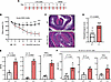 Figure 1
HECTD3-KO mice develop more severe disease in the DSS-induced colitis model. (A) Experimental model for acute DSS-induced colitis. Mice are treated with 1.5% DSS for 7 days followed by 3 days of recovery. On day 10, mice are euthanized for analysis. (B) Percent body weight changes from the initial weight of HECTD3-KO and WT mice following DSS treatment (n = 7/group). **P < 0.001; ***P < 0.0005. (C and D) Representative H&E staining (C) and histopathological scores based on criteria presented in Methods (n = 7/group) (D). Scale bar: 300 μm. (E) Multiplex cytokine (IL-1b, IL-6, TNF-α) analysis of colonic samples from HECTD3-KO and WT mice treated with DSS (n = 4). (F) Mean fluorescence intensity (MFI) of the indicated cytokines (IL-1b, IL-6, TNF-α) by FACS analysis in the Cd11c+Cd11b+ cells from the colon intraepithelial (IE) fraction of HECTD3-KO and WT mice treated with DSS (n = 4/group). In B, P values were determined by 2-way ANOVA followed by Šídák’s multiple-comparison test. For D–F, P values were determined by unpaired 2-tailed Student’s t test. Data are shown as mean ± SEM.
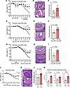 Figure 2
HECTD3 deficiency in hematopoietic system including in CD11c+ cells exacerbates DSS-induced colitis. (A–L) Percent body weight changes from the initial weight and histopathological scores for the indicated groups of mice treated with DSS colitis. (A–I) Bone marrow chimeras. (A–C) Donors: germline Hectd3 KO and WT. (D–F) Donors: Hecdt3fl/flVav-iCre and Vav-iCre. (G–I) Donors: HECTD3fl/flCd11c-Cre and CD11c-Cre. (A, D, and G) Percent body weight changes from initial body weight in the indicated chimera groups treated with 1.5% DSS for 7 days, followed by regular drinking water (n = 5/group) (*P < 0.05, ***P < 0.0005; ****P < 0.0001). P values by 2-way ANOVA followed by Šídák’s multiple-comparison test. (B, E, and H) Representative H&E-stained colon sections from each group. Scale bars: 300 μm. (C, F, and I) Histopathological scores based on criteria presented in Methods (n = 5/group). P values by Student’s unpaired t test. Data are presented as mean ± SEM. (J–L) Acute DSS-induced colitis in HECTD3fl/flCd11c-Cre and control Cd11c-Cre mice. (J) Percent body weight changes from the initial weight following 1.5% DSS treatment (n = 4/group). *P < 0.05; **P < 0.001; ****P < 0.0001. P values by 2-way ANOVA followed by Šídák’s multiple-comparison test. (K) Representative H&E-stained colon sections from each group. Scale bars: 300 μm. (L) Histopathological scores based on criteria presented in Methods (n = 4/group). (M) MFI for the indicated cytokines (IL-1b, IL-6, TNF-α) by FACS analysis in the Cd11c+Cd11b+ cells from the colonic IE fraction (n = 4/group). (L and M) P values by Student’s unpaired t test. Data are presented as mean ± SEM.
Proinflammatory cytokines are elevated in the colon of HECTD3-KO mice with DSS-induced colitis, predominantly in IE Cd11b+Cd11c+ cells. We further evaluated proinflammatory cytokines in the colons of DSS-treated HECTD3-KO mice compared with WT and found that IL-1b, IL-6, and TNF-α were elevated in HECTD3-KO mice colons compared with WT (Figure 1E). To identify the immune cell populations responsible for increased cytokine production, we performed FACS analysis of immune cells isolated from the IE compartment. This analysis showed that the HECTD3-KO IE Cd11b+Cd11c+ cells exhibited increased levels of the proinflammatory cytokines IL-b, IL-6, and TNF-α compared with WT (Figure 1F). It was particularly the MHCIIhiCD26hiCD64– population, previously defined as inf-cDC2s (25), that shows consistent increase of the proinflammatory cytokines in the HECTD3-KO mice, and to a less extent the CD26medCD64hi moDCs (Supplemental Figure 4, A–D). The frequencies of these populations, as well as of other immune populations, including B and T cells, were similar in the colon of HECTD3-KO and WT mice with colitis (Supplemental Figure 4E).
Deletion of HECTD3 in Cd11c+ cells worsens DSS-induced colitis. Given the elevated proinflammatory cytokine production observed in colonic Cd11c+ cells — particularly in inf-cDC2s and, at some extent, in moDCs — we next investigated whether Cd11c-dependent deletion of HECTD3, contributes to the aggravated colitis. We generated BM chimeras with BMs derived from Hectd3fl/flCd11c-Cre or Cd11c-Cre donor mice. Following DSS treatment, mice reconstituted with Hectd3fl/flCd11c-Cre BMs showed significantly greater body weight loss and more severe histopathological damage compared with those reconstituted with Cd11c-Cre BMs (Figure 2, G–I). In addition, Hectd3fl/flCd11c-Cre mice showed increased body weight loss and disease severity compared with Cd11c-Cre WT controls (Figure 2, J–L). Removal of HECTD3 was efficient in Hectd3fl/flCd11c-Cre BMD cells, ex vivo differentiated with GM-CSF (Supplemental Figure 3C).
FACS analysis further showed increased levels of the proinflammatory cytokines IL-1b, IL-6, and TNF-α in the colonic IE Cd11b+Cd11c+ cells from Hectd3fl/flCd11c-Cre mice with DSS-induced colitis, compared with Cd11c-Cre WT control mice — including in the inf-cDC2s population but not in CD26medCD64hi moDCs (Figure 2M and Supplemental Figure 5, A–D) — similarly to the germline Hectd3-KO mice. These results demonstrate that deletion of HECTD3 in CD11c+ cells exacerbates DSS-induced colitis, and it is the inf-cDC2s population that shows elevated proinflammatory cytokines.
Identification of MyD88 as a potential target for HECTD3. Given the link of the disease phenotype with Cd11c-dependent depletion of HECTD3 and the increase in proinflammatory cytokines, we set up a system to generate ex vivo Cd11c+ cells derived from BM and then exposed them to inflammatory conditions to identify potential targets for HECTD3 ubiquitination in an unbiased manner, focusing on proteins associated with inflammatory pathways. Specifically, we differentiated BM cells in the presence of GM-CSF (GM-CSF-BMD cells). Such treatment results in enrichment of Cd11c+ cells, believed to resemble inflammatory DCs (28). Further GM-CSF-BMD cells were treated with fecal extracts from mice with DSS-induced colitis to expose them to the inflammatory conditions associated with the disease.
The protein extracts from HECTD3 KO and WT GM-CSF-BMD cells were digested by trypsin and were then enriched for peptides with the K-ε-GG remnants using anti-K-ε-GG antibodies before being subjected to mass spectrometry to identify differentially ubiquitinated peptides.
Following LC-MS/MS analysis and a global-scaling normalization, fold-change of normalized intensities were calculated. The ubiquitinated peptides were further filtered in WT-treated cells versus HECTD3-KO–treated cells, and further versus WT- and KO-untreated cells. The enriched ubiquitinated peptide-matched proteins were further subjected to pathway enrichment analysis using EnrichR (29). Ten pathways were identified in REACTOME database (Figure 3A), which were further clustered based on a Jaccard distance matrix using the pathway connectivity R shiny application (30). The analysis revealed 4 major clustered pathways: Innate Immune System, Infectious Disease, Cytokine Signaling, and Stress Response (Figure 3B). MyD88 was present in 3 of the 4 major pathway clusters: Innate Immune System, Infectious Disease, and Cytokine Signaling. Three peptides belonging to MyD88 were identified, one with the remnant K-ε-GG at K103 (R.LLELLALLDREDILK#ELK.S, identified 2 times) and located in the Myd88 death domain (DD), and a peptide with 2 K-ε-GG remnants at K119 and K127 (K.YLGK#QQNQESEK#PLQVAR.V), located in the intermediate domain (Figure 3, C and D). Given the central role of MyD88 in innate inflammatory response as a critical hub of signal integration and transduction (31–33), and the link with the 3 proinflammatory cytokines elevated in the colon of HECTD3-KO mice with colitis, we decided to focus on Myd88 and investigate the effect of HECTD3 on MyD88 ubiquitination.
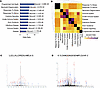 Figure 3
Ubiquitin remnant profiling and mass spectrometry analysis of extracts from HECTD3-KO and WT GM-CSF-BMD cells treated with fecal extracts from mice with colitis. (A) Bar plot showing the odds ratio of enriched REACTOME pathways performed by EnrichR. (B) Heatmap clustering performed via the pathway connectivity. Jaccard similarity index was calculated based on shared proteins enriched in each of the top 10 REACTOME pathways. (C and D) Fragmentation spectra of ubiquitinated LLELLALLDREDILK#ELKS peptide (K103 residue) and ubiquitinated KYLGK#QQNQESEK#PLQVARV peptide (K119 and K127 residues) of MyD88. Parent ions corresponding to LLELLALLDREDILK#ELKS and KYLGKQQNQESEKPLQVARV peptide masses have been subjected to higher-energy collision dissociation in mass spectrometer. The detected b-type (N-terminal) and y-type (C-terminal) fragment ion series generated by peptide backbone cleavage have been annotated.
HECTD3 interacts with MyD88 in GM-CSF-BMD cells and promotes its polyubiquitination. Given that MyD88 plays a critical role in signaling downstream of TLR4 and identification of enriched ubiquitinated MyD88 peptides in samples treated with fecal extracts, likely enriched in LPS, we treated GM-CSF-BMD cells with the TLR4 ligand LPS and conducted affinity purification of ubiquitinated proteins with Tandem Ubiquitin Binding Entities–agarose (TUBE-agarose). The results show enrichment of ubiquitinated MyD88 in LPS-treated GM-CSF-BMD WT versus HECTD3-KO cells (Figure 4A). Despite reduction in MyD88 ubiquitination in HECTD3-KO GM-CSF-BMD cells, levels of total MyD88 were the same between HECTD3-KO and WT GM-CSF-BMD cells and increased equally following LPS treatment (Figure 4B). Coimmunoprecipitation experiments show that endogenous HECTD3 and MyD88 associate in LPS-treated GM-CSF-BMD cells (Figure 4C). Thus, these results demonstrate that HECTD3 associates with MyD88 in LPS-treated GM-CSF-BMD cells and that MyD88 is polyubiquitinated in a HECTD3-dependent manner. Absence of HECTD3 in GM-CSF-BMD cells increases NF-κB activation and proinflammatory cytokine production. MyD88 serves as an adaptor that transmits signals initiated through TLRs, including TLR4, promoting NF-κB activation (19–22). We thus evaluated the phosphorylation of NF-κB p65 (P-Ser536) and found that LPS-treated HECTD3-KO GM-CSF-BMD cells exhibited higher p65 phosphorylation compared with WT control (Figure 4D). Additionally, the proinflammatory cytokines IL-1b, IL-6, and TNF-α were elevated in the LPS-treated HECTD3-KO GM-CSF-BMD cells compared with WT controls (Figure 4E), consistent with our observations in the colons of HECTD3-KO mice, including in the IE Cd11c+ cells. Thus, these results demonstrate that the absence of HECTD3 results in elevated activation of NF-κB and increased proinflammatory cytokine production in LPS-treated GM-CSF-BMD cells.
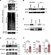 Figure 4
HECTD3 interacts with MyD88 and promotes its polyubiquitination dampening NF-κB activation and proinflammatory cytokine production in GM-CSF-BMD cells treated with LPS. (A) Immunoblot with anti-MyD88 and anti-ubiquitin antibodies following enrichment of polyubiquitinated proteins with TUBE-agarose. Protein extracts were derived from HECTD3-KO and WT GM-CSF-BMD cells treated with LPS. (B) Immunoblot for MyD88 and GAPDH in extracts from GM-CSF BMD cells treated with LPS. GM-CSF BMD cells derived from WT and HECTD3-KO mice. Data are representative of 2 independent experiments. (C) Immunoblot for HECTD3 and MyD88 following immunoprecipitation with anti-MyD88 or IgG antibodies of total protein extracts from HECTD3-KO and WT GM-CSF-BMD cells treated with LPS. (D) Immunoblot of p-NF-κB (p65), NF-κB (p65), and Lamin A/C in nuclear extracts from LPS-treated HECTD3-KO and WT GM-CSF-BMD cells. (E) Multiplex cytokines analysis for IL-1b, IL-6, and TNF-α (n = 4/group) in media of cultured GM-CSF-BMD cells treated with LPS (n = 4). (A–D) Data are representative of 2 independent experiments. Data are presented as mean ± SEM. P values by Student’s unpaired t test.
HECTD3 directly ubiquitinates MyD88. To examine in detail the HECTD3-dependent MyD88 polyubiquitination, we first generated HECTD3-KO HEK293T cells by CRISPR editing (Supplemental Figure 6). These cells were then transfected with Flag-MyD88 alone, Xpress-HECTD3 alone, or both Flag-MyD88 and Xpress-HECTD3. Western blot analysis demonstrated that anti-Xpress antibody specifically detected the ~100 kDa Xpress-HECTD3 protein and did not recognize the Flag-MyD88 (of ~35 kDa), whereas the anti-Flag antibody detected only the ~35 kDa Flag-MyD88 protein, but not the Xpress-HECTD3 (of ~100 kDa) (Supplemental Figure 7, A and B), demonstrating the absence of antibody cross-reactivity, similarly to what has been published with these antibodies (34-38).
Next HECTD3-KO and WT HEK293T cells were transfected with Flag-MyD88 and His-Ub, and the results show that, in the absence of HECTD3, MyD88 ubiquitination was reduced (Figure 5A). However, ectopic expression of WT HECTD3 in HECTD3-KO HEK293T cells restored MyD88 polyubiquitination, whereas the catalytically inactive HECTD3 C823A had a reduced effect (Figure 5, B and C). In addition, HECTD3 WT, but not the C823A mutant, promoted MyD88 ubiquitination in an in vitro ubiquitination system (Figure 5D).
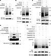 Figure 5
Molecular details of HECTD3-dependent ubiquitination of MyD88. (A) Anti-Flag immunoblot of Ni-NTA pulled-down (PD) protein extracts from WT and HECTD3 KO HEK293T cells cotransfected with HIS-Ub and Flag-MyD88. (B and C) Anti-Flag immunoblot of Ni-NTA PD protein extracts from HECTD3 KO HEK293T cells cotransfected with HIS-Ub, Flag-MyD88, and Xpress-HECTD3 WT (B) or Xpress-HECTD3 C823A (C). Input is shown for Flag-MyD88 and Xpress-HECTD3 (WT in B and HECTD3 WT and HECTD3 C823A mutant in C). (D) Anti-Flag immunoblot of in vitro ubiquitinated Flag-MyD88. Flag-MyD88 was incubated with E1 enzyme, E2 enzyme, HA-Ub and GST-HECTD3 WT, or GST-HECTD3 C823A mutant. (E) Anti-Flag immunoblot of Ni-NTA PD protein extracts from HECTD3 KO HEK293T cells cotransfected with HIS-Ub, Xpress-HECTD3, Flag-MyD88 WT, Flag-MyD88 K103R, Flag-MyD88 K119R, or Flag-MyD88 K127R. Data are the representative of the 2 independent experiments.
In the ubiquitin remnant profiling, we identified K-ε-GG remnants at MyD88 K103, K119, and K127. We thus generated K→R mutants at each of these lysine residues to further evaluate HECTD3-dependent ubiquitination of MyD88. The results show reduction in ubiquitination for all K→R mutants, with the most pronounced effect at K103R (Figure 5E). Thus, these results collectively demonstrate that HECTD3 directly promotes ubiquitination of MyD88.
HECTD3 promotes nondegradative polyubiquitination of MyD88 via K27 chains. We first asked whether HECTD3-dependent polyubiquitination of MyD88 is associated with proteasomal degradation. Treatment with the proteasomal inhibitor MG132 did not affect MyD88 levels or its ubiquitination in cells transfected with HECTD3 (Supplemental Figure 8, A and B); however, it did promote an increase in MCL1, known to be ubiquitinated and targeted for proteasomal degradation (Supplemental Figure 8A). Thus, these results suggest that HECTD3-dependent ubiquitination does not result in MyD88 targeting for proteasomal degradation.
We next sought to determine the type of polyubiquitin chains that HECTD3 promotes on MyD88. Employing ubiquitin mutants that allow only specific lysine linkages, we found that the K27-only mutant was sufficient to promote ubiquitination of MyD88 in the presence of HECTD3 at similar levels as WT construct (Figure 6A). Conversely, HECTD3-mediated polyubiquitination was remarkably decreased in transfection with Ub-K27R mutant (Figure 6B). Collectively, these results show that HECTD3 mediates nondegradative polyubiquitination of MyD88 through K27-linked polyubiquitin chains.
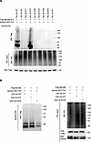 Figure 6
HECTD3 promotes polyubiquitination of MyD88 through K27-linked chains. (A) Anti-Flag immunoblot of Ni-NTA PD protein extracts from HECTD3 KO HEK293T cells cotransfected with Flag-MyD88, Xpress-HECTD3 and HIS-Ub WT or HIS-Ub K only mutants. HIS-Ub K only mutants denote that the only K in the HIS-Ub is at the indicated residue, and all other K residues are mutated to R. (B) Anti-Flag immunoblot of Ni-NTA PD protein extracts from HECTD3-KO HEK293T cells cotransfected with Flag-MyD88, Xpress-HECTD3 and HIS-Ub WT or HIS-Ub K27 ONLY or HIS-Ub K27R mutant. Data are representative of the 2 independent experiments. Input is shown for His-Ub, Flag-MyD88, and Xpress-HECTD3.
MyD88 and HECTD3 interact through the MyD88 DD and HECTD3 DOC domain. We further investigated the association between MyD88 and HECTD3 in HEK293 cells following their transfection. The transfected full-length Xpress-HECTD3 associated with full-length Flag-MyD88 (Figure 7A), similarly to what we observed for the endogenous proteins (Figure 4C). MyD88 has 3 main domains: the N terminal domain, which comprises the DD (aa 54–109), important for the recruitment of IRAK family members and signal transduction; the C-terminal Toll–IL-1 receptor domain (TIR) (aa 159–296), and the intermediate domain (ID) (aa 110–155), which separates them (20, 39–42) (Figure 7B). Removal of the MyD88 DD domain, but not of the ID or of the TIR domains, abrogated the association with full-length HECTD3 (Figure 7C), while removal of the HECTD3 DOC domain, but not of the other domains, abolished the association of HECTD3 with MyD88 (Figure 7D). Thus, these results demonstrate that MyD88 and HECTD3 interact, and the interaction occurs through the MyD88 DD domain and the HECTD3 DOC domain.
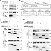 Figure 7
MyD88 death domain and HECTD3 DOC domain mediate the interaction between MyD88 and HECTD3. (A) Xpress and anti-Flag immunoblots of anti-Flag immunoprecipitated proteins from HECTD3-KO HEK293T cells cotransfected with Flag-MyD88 and Xpress-HECTD3. (B) MyD88 and HECTD3 domain representation. (C) Anti-Xpress and anti-Flag immunoblots of anti-Flag immunoprecipitated proteins from HECTD3-KO HEK293T cells cotransfected with Xpress-HECTD3 and Flag-MyD88 or Flag-MyD88 ΔDD, Flag-MyD88 ΔID, and Flag-MyD88 ΔTIR mutants (D) Anti-Xpress and anti-Flag immunoblots of anti-Flag–immunoprecipitated proteins from HECTD3-KO HEK293T cells cotransfected with Flag-MyD88 and either Xpress-HECTD3 or the Xpress-HECTD3 ΔN, ΔDOC, ΔLD, or ΔHECT mutants. Data are the representative of the 2 independent experiments.
Absence of HECTD3 increases MyD88-IRAK4 association and IRAK4 phosphorylation. MyD88 downstream signaling is initiated by MyD88 association with IRAK4 through interactions predominantly mediated by their DD domains. IRAK4 is activated by autophosphorylation, which further promotes activation of IRAK1 and IRAK2 and the assembly of the Myddosome, which further promotes the downstream signal transduction, ultimately activating NF-κB (43–49). A crystal structure of the heteromeric human MyD88-IRAK4-IRAK2 DD domain complex is available, consisting of 6 MyD88, 4 IRAK4, and 4 IRAK2 DDs (50). Directly mapping the identified mouse MyD88 ubiquitylation sites (K103, K119, and K127) onto this structure is challenging because K103 is not conserved between mouse and human, and K127 is outside of the resolved region of the structure. To address this, we generated a structural model by superimposing the AlphaFold-predicted mouse MyD88 DD domain onto the human MyD88 in Protein Data Bank (PDB) (ID: 3MOP) (50, 51). The 3 lysine residues (K103, K119, and K127) ubiquitylated by HECTD3 are highlighted (Figure 8A). MyD88 DD forms 2 types of interfaces in the helical assembly: the MyD88-MyD88 interface and the MyD88-IRAK4 interface. K103 is involved in both interfaces. Ubiquitylation of K103 by HECTD3 is anticipated to disrupt complex formation and diminish signaling output. K119 and K127, located within the MyD88-MyD88 interface, are also critical. Since the MyD88 helical assembly serves as a scaffold for IRAK4 and IRAK2 recruitment, ubiquitylation at K119 and K127 by HECTD3 is expected to negatively affect IRAK4/2 kinase recruitment and activation. Therefore, our structural modeling suggests that K103, K119, and K127 are crucial within the DD domain interface of the MyD88/IRAK4/IRAK2 complex, with their ubiquitylation likely leading to reduced NF-κB activation. In support of this hypothesis, coimmunoprecipitation experiments show that MyD88-IRAK4 interaction was more pronounced and the phosphorylation of IRAK4 was increased in LPS-treated HECTD3-KO GM-CSF-BMD cells compared with WT (Figure 8, B–D).
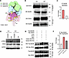 Figure 8
Absence of HECTD3 causes increased MyD88-IRAK4 interaction and IRAK4 phosphorylation. (A) A structural model of MyD88-IRAK4-IRAK2 DD domain complex based on PDBID 3MOP. Individual DD domain of MyD88 (pink), IRAK4 (blue), and IRAK2 (green) are shown in surface representation. Two illustrated models of mouse MyD88 DD domain are superimposed into the M1 and M5 units in the helical assembly. The location of K103, K119, and K127 are indicated using yellow spheres. (B) Immunoblot for IRAK4 and MyD88 following MyD88 immunoprecipitation of protein extracts from HECTD3-KO and WT GM-CSF-BMD cells treated with LPS. (C) Quantification of IRAK4 from B. (D) Immunoblot of p-IRAK4, IRAK4, and GAPDH from HECTD3-KO and WT GM-CSF-BMD cells treated with LPS. (E) HECTD3 ubiquitin ligase activity restricts constitutive MyD88-IRAK4 complex formation. HEK293T HECTD3-KO cells were cotransfected with Flag-MyD88, Myc-IRAK4, TLR4, CD14, and MD2, together with either WT HECTD3 (first lane) or the catalytically inactive HECTD3 mutant C823A (third lane). HECTD3-KO cells served as control (second lane). Cell lysates were subjected to immunoprecipitation with anti-Myc-IRAK4 followed by immunoblotting to assess MyD88-IRAK4 complex formation. (F) Quantification of Flag-MyD88 from E. (B–F) Data are representative of 2 independent experiments.
We further generated an experimental system in HEK293T cells to test the effect of HECTD3 on the interaction of MyD88 with IRAK4. HECTD3-KO HEK293T cells were cotransfected with Flag-MyD88 and Myc-IRAK4, as well as with TLR4 together with the adaptor MD-2 and CD14, to have all components necessary for MyD88-IRAK4 pathway activation, as previously described (52, 53). In addition, either WT HECTD3 or the catalytically inactive C823A HECTD3 mutant were transfected. Transfection with WT HECTD3, but not with the C823A HECTD3 mutant, reduced the association of IRAK4 with MyD88 (Figure 8, E and F).
These results together demonstrate that absence of HECTD3 increases MyD88 association with IRAK4 and IRAK4 phosphorylation.
Inhibition of MyD88 activity rescued increased DSS-induced colitis severity in HECTD3-KO mice, including the proinflammatory cytokines. To determine to what extent the elevated MyD88 activation is responsible for the increased DSS-induced colitis severity in HECTD3-KO mice, we treated mice with DSS-induced colitis with the MyD88 inhibitor NBP2–29328 peptide (MyD88 I) or the control antennapedia peptide (CP) (Figure 9A). The results show that MyD88 I, but not the CP, improved the weights and restored the disease scores of the HECTD3-KO mice to the level of WT (Figure 9, B–D). In addition, the levels of the proinflammatory cytokines IL-1b, IL-6, and TNF-α were dampened to the levels of WT mice in the HECTD3-KO mice treated with the MyD88 I but not with the CP (Figure 9E). These results together demonstrate that MyD88 plays a central role in the elevated colitis severity of the HECTD3-KO mice.
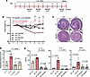 Figure 9
Inhibition of MyD88 activity rescues the exacerbation of DSS colitis in HECTD3-KO mice. (A) Experimental design. HECTD3-KO and WT mice were administered orally with 1.5% DSS for 10 days. On Days 3, 5, 7, and 10, mice were injected i.p. with the MyD88 inhibitor peptide (NBP2-29328) (MyD88 I) or control antennapedia peptide (CP). On day 11, mice were euthanized for analysis. (B) Percent body weight change from the initial weight of HECTD3-KO and WT mice following DSS treatment plus MyD88 I or CP (n = 4/group) (*P < 0.05; **P < 0.001; ***P < 0.0005). P values were calculated by 2-way ANOVA, followed by Šídák’s multiple-comparison test. (C and D) Representative H&E staining and histopathological scores (n = 4/group). Scale bar: 500 μm. (E) Multiplex cytokine analysis of IL-1b, IL-6, and TNF-α in colonic samples from HECTD3-KO and WT mice treated with MyD88 I or CP (n = 4/group). (D and E) Data are presented as mean ± SEM. P values by 2-way ANOVA followed Tukey’s multiple-comparison test.


