Genetics
Abstract
Duchenne muscular dystrophy (DMD) is a fatal genetic muscle-wasting disease characterized by loss of dystrophin protein. Therapeutic attempts to restore a functional copy of dystrophin to striated muscle are under active development, and many utilize adeno-associated viral (AAV) vectors. However, the limited cargo capacity of AAVs precludes delivery of full-length dystrophin, a 427 kDa protein, to target tissues. Recently, we developed a novel method to express large dystrophin constructs using the protein trans-splicing (PTS) mechanism mediated by split inteins and myotropic AAV vectors. The efficacy of this approach to restore muscle function in mdx4cv mice was previously assessed using histology, dystrophin immunolabeling, and western blotting. Here, we expand our molecular characterization of dystrophin constructs with variable lengths using a mass spectrometry-based proteomics approach, providing insight into unique protein expression profiles in skeletal muscles of wild-type, dystrophic mdx4cv, and AAV-treated mdx4cv. Our data reveal several affected cellular processes in mdx4cv skeletal muscles with changes in the expression profiles of key proteins to muscle homeostasis, whereas successful expression of dystrophin constructs results in an intermediate to complete restoration. This study highlights several biomarkers that could be used in future preclinical or clinical studies to evaluate the effectiveness of therapeutic strategies.
Authors
Erynn E. Johnson, Theodore R. Reyes, Jeffrey S. Chamberlain, James M. Ervasti, Hichem Tasfaout
Abstract
Over 95% of head and neck cancers are squamous cell carcinoma (HNSCC). HNSCC is mostly diagnosed late, causing a poor prognosis despite the application of invasive treatment protocols. Tumor-educated platelets (TEPs) have been shown to hold promise as a molecular tool for early cancer diagnosis. We sequenced platelet mRNA isolated from blood of 101 HNSCC patients and 101 propensity-score matched non-cancer controls. Two independent machine learning classification strategies were employed using a training and validation approach to identify a cancer predictor: a particle swarm optimized support vector machine (PSO-SVM) and a least absolute shrinkage and selection operator (LASSO) logistic regression model. The best performing PSO-SVM predictor consisted of 245 platelet transcripts and reached a maximum area under the curve (AUC) of 0.87. For the LASSO-based prediction model 1,198 mRNAs were selected, resulting in an median AUC of 0.84, independent of HPV status. Our data show that TEP RNA classification by different AI tools is promising in the diagnosis of HNSCC.
Authors
N.E. Wondergem, J.B. Poell, S.G.J.G In 't Veld, E. Post, S.W. Mes, M.G. Best, W.N. van Wieringen, T. Klausch, R.J. Baatenburg de Jong, C.H.J. Terhaard, R.P. Takes, J.A. Langendijk, I.M. Verdonck-de Leeuw, F. Lamers, C.R. Leemans, E. Bloemena, T. Würdinger, R.H Brakenhoff
Abstract
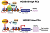
HOXB13 is a prostate-specific transcription factor best known for its role as an androgen receptor (AR) cofactor. Recent evidence suggests that HOXB13 plays critical AR-independent functions in repressing lipogenic programs and promoting prostate cancer (PCa) metastasis. However, the mechanisms linking HOXB13 loss to tumor metastasis remain unclear. Here, we show that p300 and CBP co-occupy lipogenic enhancers suppressed by HOXB13 and HDAC3 and are essential for enhancer activation and target gene expression following HOXB13 depletion. Loss of HOXB13 induces lipid-sensitive matrix metalloproteinases (MMPs), promoting increased cell motility. Importantly, pharmacological inhibition of p300 and CBP blocks HOXB13-loss-driven lipogenesis, reduces MMP expression, and decreases cell migration in vitro and tumor metastasis in vivo. Analysis of clinical samples revealed that HOXB13 expression is reduced in metastatic hormone-sensitive PCa compared with matched primary tumors, further supporting its role in tumor metastasis. These findings demonstrate that HOXB13 downregulation promotes PCa metastasis through p300- and CBP-dependent lipogenic and motility pathways, which may be targeted by p300 inhibition.
Authors
Xiaodong Lu, Liu Peng, Qi Chu, Samantha Ye, Mingyang Liu, Maha Hussain, Mehmet A. Bilen, Lara R. Harik, Jonathan Melamed, Jonathan C. Zhao, Jindan Yu

Abstract
Premature ovarian insufficiency (POI) is a complex reproductive disorder with a strong genetic component. The known POI causative genes currently account for only a small fraction of cases. In this study, we conducted whole-exome sequencing and identified a rare heterozygous missense variant in DNA helicase B (HELB) (c.349G>T, p.Asp117Tyr) in a Chinese family with POI and early menopause. To investigate the pathogenicity of this variant, a knockin mouse model carrying a heterozygous missense Helb variant (Helb+/D112Y) homologous to the human HELB c.349G>T was constructed. The Helb-mutated female mice exhibited reduced litter sizes and prolonged interlitter intervals compared with wild-type mice after reaching 10 months of age, leading to a shortened reproductive lifespan. Consistently, aged Helb+/D112Y females showed decreased ovarian weight and accelerated follicle depletion. Transcriptomic analysis of the ovaries from Helb-mutated mice revealed dysregulated expression of genes associated with impaired ovarian function and ovarian aging. Collectively, these findings in both humans and mice suggest that HELB is involved in maintaining ovarian function and regulating reproductive aging, highlighting the importance of HELB in female reproductive health.
Authors
Yuncheng Pan, Yuexin Yu, Jitong Mo, Shuting Ren, Zixue Zhou, Xi Yang, Yiqing Liu, Feng Zhang, Yanqin You, Xiaojin Zhang, Yanhua Wu

Abstract
The mechanisms underlying cyst growth and progression in Autosomal Dominant Polycystic Kidney Disease (ADPKD) remain unresolved. Since cyst expansion requires epithelial salt and water secretion likely involving basolateral membrane K+ recycling, we investigated the role of KCNN4-encoded K+ channel KCa3.1, inhibited by the potent, pharmacospecific, well-tolerated antagonist, senicapoc. We hypothesized that genetic and/or pharmacological inactivation of KCNN4/KCa3.1 would slow PKD progression. KCNN4 was upregulated in kidneys of patients with ADPKD and of mechanistically distinct PKD mouse models. Cyst expansion in Pkd1–/– murine metanephroi was stimulated by KCa3.1 agonist and was prevented/reversed by senicapoc. In rapidly and/or slowly progressive mouse Pkd1 models, Kcnn4 inactivation slowed renal cyst growth; attenuated PKD-stimulated cAMP and ERK/Myc signaling pathways; reduced PKD-associated ciliary elongation, cell proliferation, and fibrosis; improved renal function; and prolonged survival. Importantly, senicapoc treatment of Pkd1 mouse models phenocopied most effects of Kcnn4 inactivation. This first study on the efficacy of KCa3.1 inhibition in PKD progression recommends senicapoc as a clinical trial candidate for ADPKD.
Authors
Guanhan Yao, Almira Kurbegovic, Camila Parrot, William Foley, William Roman, Seth L. Alper, Marie Trudel
Abstract
Long chain fatty acid oxidation disorders (LC FAODs) cause energy deficits in heart and skeletal muscle that are only partially corrected by current medium chain lipid therapies such as triheptanoin. We find that heart and muscle lack medium chain acyl CoA synthetases, limiting the capacity for β-oxidation of medium-chain fatty acids. Instead, heart and muscle mitochondria robustly respire on medium-chain acylcarnitines. The mitochondrial matrix enzyme carnitine acetyltransferase (CrAT) efficiently converts orally delivered octanoylcarnitine (C8 carnitine) to octanoyl CoA for energy generation. C8-carnitine exhibits twice the oral bioavailability of triheptanoin and distributes to muscle and heart. A single oral dose significantly enhances grip strength and treadmill endurance while attenuating lactic acidosis in two mouse models of LC-FAODs. Thus, medium chain acylcarnitines overcome a previously unrecognized metabolic bottleneck in LC FAOD muscle and may represent an alternative to triglyceride based therapies for bioenergetic disorders.
Authors
Keaton J. Solo, Yuxun Zhang, Sivakama S. Bharathi, Bob B. Zhang, Adam C. Richert, Alexandra V. Schmidt, Clinton Van't Land, Olivia D'Annibale, Timothy C. Wood, Eric S. Goetzman
Abstract
The MTM1 gene encodes myotubularin (MTM1), a phosphatidylinositol 3-phosphate (PI(3)P) lipid phosphatase. Loss-of-function mutations in MTM1 cause X-linked myotubular myopathy (XLMTM), a severe congenital myopathy with no available cure and a poorly understood pathomechanism. The importance of MTM1 enzymatic activity and its PI(3)P substrate in physiology under normal conditions and in XLMTM is unclear. We generated the Mtm1 KI C375S mice in which the endogenous MTM1 was converted to a phosphatase-dead protein. Mutant mice survived a median of 12 weeks and demonstrated progressively impaired motor skills. Observed muscle hypotrophy and reduced force production compared to their WT littermates (~3.9-fold reduction in absolute maximal force) were responsible for these severe phenotypes. A significantly higher level of PI(3)P was found in the muscle of Mtm1 KI C375S mice. Muscle histology and molecular characterization revealed XLMTM hallmarks, with alteration of the mTOR and autophagy pathways correlating with muscle hypotrophy, and abnormal myofiber intracellular organization correlating with impaired muscle force. Overall, this study reveals the importance of MTM1 phosphatase activity and related PI(3)P substrate for postnatal muscle maintenance, and highlights the significance of MTM1 phosphatase activity in the development of X-linked myotubular myopathy.
Authors
Foteini Moschovaki-Filippidou, Christine Kretz, David Reiss, Gaetan Chicanne, Bernard Payrastre, Jocelyn Laporte
Abstract
Many patients suffering from inherited diseases do not receive a genetic diagnosis and are therefore excluded as candidates for treatments, such as gene therapies. Analyzing disease-related gene transcripts from patient cells would improve detection of mutations that have been missed or misinterpreted in terms of pathogenicity during routine genome sequencing. However, the analysis of transcripts is complicated by the fact that a biopsy of the affected tissue is often not appropriate, and many disease-associated genes are not expressed in tissues or cells that can be easily obtained from patients. Here, using CRISPR/Cas-mediated transcriptional activation (CRISPRa) we developed a robust and efficient approach to activate genes in skin-derived fibroblasts and in freshly isolated peripheral blood mononuclear cells (PBMCs) from healthy individuals. This approach was successfully applied to blood samples from patients with inherited retinal dystrophies (IRD). We were able to efficiently activate several IRD-linked genes and detect the corresponding transcripts using different diagnostically relevant methods such as RT-qPCR, RT-PCR and long- and short-read RNA sequencing. The detection and analysis of known and unknown mRNA isoforms demonstrates the potential of CRISPRa-mediated transcriptional activation in PBMCs. These results will contribute to ceasing the critical gap in the genetic diagnosis of IRD patients and other inherited diseases.
Authors
Valentin J. Weber, Alice Reschigna, Maximilian J. Gerhardt, Thomas Heigl, Klara S. Hinrichsmeyer, Sander van den Engel, Dina Y. Otify, Zoran Gavrilov, Frank Blaser, Isabelle Meneau, Christian Betz, Hanno J. Bolz, Martin Biel, Stylianos Michalakis, Elvir Becirovic
Abstract
Ischemic cardiomyopathy (ICM) is a leading cause of heart failure characterized by extensive remodeling of the cardiac extracellular matrix (ECM). While initially adaptive, ECM deposition following ischemic injury eventually turns maladaptive, promoting adverse cardiac remodeling. The strong link between the extent of fibrosis and adverse clinical outcomes has led to growing interest in ECM targeted therapies to prevent or reverse maladaptive cardiac remodeling in ICM; yet, the precise composition of the ECM in ICM remains poorly defined. In this study, we employed a sequential protein extraction enabled by the photocleavable surfactant Azo to enrich ECM proteins from left ventricular tissues of patients with end-stage ICM (n=16) and nonfailing donor hearts (n=16). High-resolution mass spectrometry-based quantitative proteomics identified and quantified over 6,000 unique protein groups, including 315 ECM proteins. We discovered significant upregulation of key ECM components, particularly glycoproteins, proteoglycans, collagens, and ECM regulators. Notably, LOXL1, FBLN1, and VCAN were among the most differentially expressed. Functional enrichment analyses revealed enhanced TGFβ signaling, integrin-mediated adhesion, and complement activation in ICM tissues, suggesting a feedback loop driving continued ECM deposition in the end-stage failing heart. Together, our findings provide a comprehensive proteomic landscape of ECM alterations in the end-stage ICM myocardium and identify promising molecular targets for therapeutic intervention.
Authors
Kevin M. Buck, Holden T. Rogers, Zachery R. Gregorich, Morgan W. Mann, Timothy J. Aballo, Zhan Gao, Emily A. Chapman, Andrew J. Perciaccante, Scott J. Price, Ienglam Lei, Paul C. Tang, Ying Ge
Abstract
Genetic diseases such as ion-channelopathies substantially burden human health. Existing treatments are limited and not genotype specific. Here, we report a two-step high-throughput approach to rapidly identify drug candidates for repurposing as genotype-specific therapy. We first screened 1,680 medicines using a new thallium-flux trafficking assay against KV11.1 gene variants causing Long QT Syndrome (LQTS), an ion-channelopathy associated with fatal cardiac arrhythmias. We identify evacetrapib as a suitable drug candidate that improves membrane trafficking and activates channels. We then use deep mutational scanning to prospectively identify all KV11.1 missense variants in a LQTS hotspot region responsive to treatment with evacetrapib. Combining high-throughput drug screens with deep mutational scanning establishes a new paradigm for mutation-specific drug discovery translatable to personalized treatment of patients with rare genetic disorders.
Authors
Christian L. Egly, Alex Shen, Tri Q. Do, Carlos Tellet Cabiya, Paxton A. Ritschel, Suah Woo, Matthew J. Ku, Brian P. Delisle, Brett Kroncke, Bjorn C. Knollmann
No posts were found with this tag.


