
Clinical Research and Public HealthClinical ResearchImmunologyOncology
Open Access |  10.1172/jci.insight.203629
Long-acting IL-7 induces distinct transcriptomic features in peripheral T cells of patients with solid tumors
Hocheol Jang,1 Jeong Yeon Kim,1 Sojeong Kim,1 Heewon Kim,2 Mi Sun Byun,2 Myung Ah Lee,3 Jong Hee Chang,4 Do-Hyun Nam,5 Tae Won Kim,6 Sin-Soo Jeun,7 Joo Hyuk Sohn,8 Su-Hyung Park,1 and Eui-Cheol Shin1
1Graduate School of Medical Science and Engineering, Korea Advanced Institute of Science and Technology (KAIST), Daejeon, Republic of Korea.
2Genexine, Inc., Seoul, Republic of Korea.
3Department of Internal Medicine, Seoul St. Mary’s Hospital, College of Medicine, The Catholic University, Seoul, Republic of Korea.
4Department of Neurosurgery, Yonsei University College of Medicine, Seoul, Republic of Korea.
5Department of Neurosurgery, Samsung Medical Center, Sungkyunkwan University School of Medicine, Seoul, Republic of Korea.
6Department of Oncology, Asan Medical Center, University of Ulsan College of Medicine, Seoul, Republic of Korea.
7Department of Neurosurgery, Seoul St. Mary’s Hospital, College of Medicine, The Catholic University of Korea, Seoul, Republic of Korea.
8Division of Medical Oncology, Department of Internal Medicine, Yonsei Cancer Center, Yonsei University College of Medicine, Seoul, Republic of Korea.
Address correspondence to: Eui-Cheol Shin, Laboratory of Immunology and Infectious Diseases, Graduate School of Medical Science and Engineering, KAIST, 193 Munji-ro, Yuseong-gu, Daejeon 34051, Republic of Korea. Email: ecshin@kaist.ac.kr. Or to: Su-Hyung Park, Laboratory of Translational Immunology & Vaccinology, Graduate School of Medical Science and Engineering, KAIST, 193 Munji-ro, Yuseong-gu, Daejeon 34051, Republic of Korea. Email: park3@kaist.ac.kr.
Authorship note: HJ, JYK, and SK contributed equally to this work. SHP and ECS are co-corresponding authors.
Find articles by Jang, H. in: PubMed | Google Scholar
1Graduate School of Medical Science and Engineering, Korea Advanced Institute of Science and Technology (KAIST), Daejeon, Republic of Korea.
2Genexine, Inc., Seoul, Republic of Korea.
3Department of Internal Medicine, Seoul St. Mary’s Hospital, College of Medicine, The Catholic University, Seoul, Republic of Korea.
4Department of Neurosurgery, Yonsei University College of Medicine, Seoul, Republic of Korea.
5Department of Neurosurgery, Samsung Medical Center, Sungkyunkwan University School of Medicine, Seoul, Republic of Korea.
6Department of Oncology, Asan Medical Center, University of Ulsan College of Medicine, Seoul, Republic of Korea.
7Department of Neurosurgery, Seoul St. Mary’s Hospital, College of Medicine, The Catholic University of Korea, Seoul, Republic of Korea.
8Division of Medical Oncology, Department of Internal Medicine, Yonsei Cancer Center, Yonsei University College of Medicine, Seoul, Republic of Korea.
Address correspondence to: Eui-Cheol Shin, Laboratory of Immunology and Infectious Diseases, Graduate School of Medical Science and Engineering, KAIST, 193 Munji-ro, Yuseong-gu, Daejeon 34051, Republic of Korea. Email: ecshin@kaist.ac.kr. Or to: Su-Hyung Park, Laboratory of Translational Immunology & Vaccinology, Graduate School of Medical Science and Engineering, KAIST, 193 Munji-ro, Yuseong-gu, Daejeon 34051, Republic of Korea. Email: park3@kaist.ac.kr.
Authorship note: HJ, JYK, and SK contributed equally to this work. SHP and ECS are co-corresponding authors.
Find articles by Kim, J. in: PubMed | Google Scholar
1Graduate School of Medical Science and Engineering, Korea Advanced Institute of Science and Technology (KAIST), Daejeon, Republic of Korea.
2Genexine, Inc., Seoul, Republic of Korea.
3Department of Internal Medicine, Seoul St. Mary’s Hospital, College of Medicine, The Catholic University, Seoul, Republic of Korea.
4Department of Neurosurgery, Yonsei University College of Medicine, Seoul, Republic of Korea.
5Department of Neurosurgery, Samsung Medical Center, Sungkyunkwan University School of Medicine, Seoul, Republic of Korea.
6Department of Oncology, Asan Medical Center, University of Ulsan College of Medicine, Seoul, Republic of Korea.
7Department of Neurosurgery, Seoul St. Mary’s Hospital, College of Medicine, The Catholic University of Korea, Seoul, Republic of Korea.
8Division of Medical Oncology, Department of Internal Medicine, Yonsei Cancer Center, Yonsei University College of Medicine, Seoul, Republic of Korea.
Address correspondence to: Eui-Cheol Shin, Laboratory of Immunology and Infectious Diseases, Graduate School of Medical Science and Engineering, KAIST, 193 Munji-ro, Yuseong-gu, Daejeon 34051, Republic of Korea. Email: ecshin@kaist.ac.kr. Or to: Su-Hyung Park, Laboratory of Translational Immunology & Vaccinology, Graduate School of Medical Science and Engineering, KAIST, 193 Munji-ro, Yuseong-gu, Daejeon 34051, Republic of Korea. Email: park3@kaist.ac.kr.
Authorship note: HJ, JYK, and SK contributed equally to this work. SHP and ECS are co-corresponding authors.
Find articles by Kim, S. in: PubMed | Google Scholar
1Graduate School of Medical Science and Engineering, Korea Advanced Institute of Science and Technology (KAIST), Daejeon, Republic of Korea.
2Genexine, Inc., Seoul, Republic of Korea.
3Department of Internal Medicine, Seoul St. Mary’s Hospital, College of Medicine, The Catholic University, Seoul, Republic of Korea.
4Department of Neurosurgery, Yonsei University College of Medicine, Seoul, Republic of Korea.
5Department of Neurosurgery, Samsung Medical Center, Sungkyunkwan University School of Medicine, Seoul, Republic of Korea.
6Department of Oncology, Asan Medical Center, University of Ulsan College of Medicine, Seoul, Republic of Korea.
7Department of Neurosurgery, Seoul St. Mary’s Hospital, College of Medicine, The Catholic University of Korea, Seoul, Republic of Korea.
8Division of Medical Oncology, Department of Internal Medicine, Yonsei Cancer Center, Yonsei University College of Medicine, Seoul, Republic of Korea.
Address correspondence to: Eui-Cheol Shin, Laboratory of Immunology and Infectious Diseases, Graduate School of Medical Science and Engineering, KAIST, 193 Munji-ro, Yuseong-gu, Daejeon 34051, Republic of Korea. Email: ecshin@kaist.ac.kr. Or to: Su-Hyung Park, Laboratory of Translational Immunology & Vaccinology, Graduate School of Medical Science and Engineering, KAIST, 193 Munji-ro, Yuseong-gu, Daejeon 34051, Republic of Korea. Email: park3@kaist.ac.kr.
Authorship note: HJ, JYK, and SK contributed equally to this work. SHP and ECS are co-corresponding authors.
Find articles by Kim, H. in: PubMed | Google Scholar
1Graduate School of Medical Science and Engineering, Korea Advanced Institute of Science and Technology (KAIST), Daejeon, Republic of Korea.
2Genexine, Inc., Seoul, Republic of Korea.
3Department of Internal Medicine, Seoul St. Mary’s Hospital, College of Medicine, The Catholic University, Seoul, Republic of Korea.
4Department of Neurosurgery, Yonsei University College of Medicine, Seoul, Republic of Korea.
5Department of Neurosurgery, Samsung Medical Center, Sungkyunkwan University School of Medicine, Seoul, Republic of Korea.
6Department of Oncology, Asan Medical Center, University of Ulsan College of Medicine, Seoul, Republic of Korea.
7Department of Neurosurgery, Seoul St. Mary’s Hospital, College of Medicine, The Catholic University of Korea, Seoul, Republic of Korea.
8Division of Medical Oncology, Department of Internal Medicine, Yonsei Cancer Center, Yonsei University College of Medicine, Seoul, Republic of Korea.
Address correspondence to: Eui-Cheol Shin, Laboratory of Immunology and Infectious Diseases, Graduate School of Medical Science and Engineering, KAIST, 193 Munji-ro, Yuseong-gu, Daejeon 34051, Republic of Korea. Email: ecshin@kaist.ac.kr. Or to: Su-Hyung Park, Laboratory of Translational Immunology & Vaccinology, Graduate School of Medical Science and Engineering, KAIST, 193 Munji-ro, Yuseong-gu, Daejeon 34051, Republic of Korea. Email: park3@kaist.ac.kr.
Authorship note: HJ, JYK, and SK contributed equally to this work. SHP and ECS are co-corresponding authors.
Find articles by Byun, M. in: PubMed | Google Scholar
1Graduate School of Medical Science and Engineering, Korea Advanced Institute of Science and Technology (KAIST), Daejeon, Republic of Korea.
2Genexine, Inc., Seoul, Republic of Korea.
3Department of Internal Medicine, Seoul St. Mary’s Hospital, College of Medicine, The Catholic University, Seoul, Republic of Korea.
4Department of Neurosurgery, Yonsei University College of Medicine, Seoul, Republic of Korea.
5Department of Neurosurgery, Samsung Medical Center, Sungkyunkwan University School of Medicine, Seoul, Republic of Korea.
6Department of Oncology, Asan Medical Center, University of Ulsan College of Medicine, Seoul, Republic of Korea.
7Department of Neurosurgery, Seoul St. Mary’s Hospital, College of Medicine, The Catholic University of Korea, Seoul, Republic of Korea.
8Division of Medical Oncology, Department of Internal Medicine, Yonsei Cancer Center, Yonsei University College of Medicine, Seoul, Republic of Korea.
Address correspondence to: Eui-Cheol Shin, Laboratory of Immunology and Infectious Diseases, Graduate School of Medical Science and Engineering, KAIST, 193 Munji-ro, Yuseong-gu, Daejeon 34051, Republic of Korea. Email: ecshin@kaist.ac.kr. Or to: Su-Hyung Park, Laboratory of Translational Immunology & Vaccinology, Graduate School of Medical Science and Engineering, KAIST, 193 Munji-ro, Yuseong-gu, Daejeon 34051, Republic of Korea. Email: park3@kaist.ac.kr.
Authorship note: HJ, JYK, and SK contributed equally to this work. SHP and ECS are co-corresponding authors.
Find articles by Lee, M. in: PubMed | Google Scholar
1Graduate School of Medical Science and Engineering, Korea Advanced Institute of Science and Technology (KAIST), Daejeon, Republic of Korea.
2Genexine, Inc., Seoul, Republic of Korea.
3Department of Internal Medicine, Seoul St. Mary’s Hospital, College of Medicine, The Catholic University, Seoul, Republic of Korea.
4Department of Neurosurgery, Yonsei University College of Medicine, Seoul, Republic of Korea.
5Department of Neurosurgery, Samsung Medical Center, Sungkyunkwan University School of Medicine, Seoul, Republic of Korea.
6Department of Oncology, Asan Medical Center, University of Ulsan College of Medicine, Seoul, Republic of Korea.
7Department of Neurosurgery, Seoul St. Mary’s Hospital, College of Medicine, The Catholic University of Korea, Seoul, Republic of Korea.
8Division of Medical Oncology, Department of Internal Medicine, Yonsei Cancer Center, Yonsei University College of Medicine, Seoul, Republic of Korea.
Address correspondence to: Eui-Cheol Shin, Laboratory of Immunology and Infectious Diseases, Graduate School of Medical Science and Engineering, KAIST, 193 Munji-ro, Yuseong-gu, Daejeon 34051, Republic of Korea. Email: ecshin@kaist.ac.kr. Or to: Su-Hyung Park, Laboratory of Translational Immunology & Vaccinology, Graduate School of Medical Science and Engineering, KAIST, 193 Munji-ro, Yuseong-gu, Daejeon 34051, Republic of Korea. Email: park3@kaist.ac.kr.
Authorship note: HJ, JYK, and SK contributed equally to this work. SHP and ECS are co-corresponding authors.
Find articles by Chang, J. in: PubMed | Google Scholar
1Graduate School of Medical Science and Engineering, Korea Advanced Institute of Science and Technology (KAIST), Daejeon, Republic of Korea.
2Genexine, Inc., Seoul, Republic of Korea.
3Department of Internal Medicine, Seoul St. Mary’s Hospital, College of Medicine, The Catholic University, Seoul, Republic of Korea.
4Department of Neurosurgery, Yonsei University College of Medicine, Seoul, Republic of Korea.
5Department of Neurosurgery, Samsung Medical Center, Sungkyunkwan University School of Medicine, Seoul, Republic of Korea.
6Department of Oncology, Asan Medical Center, University of Ulsan College of Medicine, Seoul, Republic of Korea.
7Department of Neurosurgery, Seoul St. Mary’s Hospital, College of Medicine, The Catholic University of Korea, Seoul, Republic of Korea.
8Division of Medical Oncology, Department of Internal Medicine, Yonsei Cancer Center, Yonsei University College of Medicine, Seoul, Republic of Korea.
Address correspondence to: Eui-Cheol Shin, Laboratory of Immunology and Infectious Diseases, Graduate School of Medical Science and Engineering, KAIST, 193 Munji-ro, Yuseong-gu, Daejeon 34051, Republic of Korea. Email: ecshin@kaist.ac.kr. Or to: Su-Hyung Park, Laboratory of Translational Immunology & Vaccinology, Graduate School of Medical Science and Engineering, KAIST, 193 Munji-ro, Yuseong-gu, Daejeon 34051, Republic of Korea. Email: park3@kaist.ac.kr.
Authorship note: HJ, JYK, and SK contributed equally to this work. SHP and ECS are co-corresponding authors.
Find articles by Nam, D. in: PubMed | Google Scholar
1Graduate School of Medical Science and Engineering, Korea Advanced Institute of Science and Technology (KAIST), Daejeon, Republic of Korea.
2Genexine, Inc., Seoul, Republic of Korea.
3Department of Internal Medicine, Seoul St. Mary’s Hospital, College of Medicine, The Catholic University, Seoul, Republic of Korea.
4Department of Neurosurgery, Yonsei University College of Medicine, Seoul, Republic of Korea.
5Department of Neurosurgery, Samsung Medical Center, Sungkyunkwan University School of Medicine, Seoul, Republic of Korea.
6Department of Oncology, Asan Medical Center, University of Ulsan College of Medicine, Seoul, Republic of Korea.
7Department of Neurosurgery, Seoul St. Mary’s Hospital, College of Medicine, The Catholic University of Korea, Seoul, Republic of Korea.
8Division of Medical Oncology, Department of Internal Medicine, Yonsei Cancer Center, Yonsei University College of Medicine, Seoul, Republic of Korea.
Address correspondence to: Eui-Cheol Shin, Laboratory of Immunology and Infectious Diseases, Graduate School of Medical Science and Engineering, KAIST, 193 Munji-ro, Yuseong-gu, Daejeon 34051, Republic of Korea. Email: ecshin@kaist.ac.kr. Or to: Su-Hyung Park, Laboratory of Translational Immunology & Vaccinology, Graduate School of Medical Science and Engineering, KAIST, 193 Munji-ro, Yuseong-gu, Daejeon 34051, Republic of Korea. Email: park3@kaist.ac.kr.
Authorship note: HJ, JYK, and SK contributed equally to this work. SHP and ECS are co-corresponding authors.
Find articles by Kim, T. in: PubMed | Google Scholar
1Graduate School of Medical Science and Engineering, Korea Advanced Institute of Science and Technology (KAIST), Daejeon, Republic of Korea.
2Genexine, Inc., Seoul, Republic of Korea.
3Department of Internal Medicine, Seoul St. Mary’s Hospital, College of Medicine, The Catholic University, Seoul, Republic of Korea.
4Department of Neurosurgery, Yonsei University College of Medicine, Seoul, Republic of Korea.
5Department of Neurosurgery, Samsung Medical Center, Sungkyunkwan University School of Medicine, Seoul, Republic of Korea.
6Department of Oncology, Asan Medical Center, University of Ulsan College of Medicine, Seoul, Republic of Korea.
7Department of Neurosurgery, Seoul St. Mary’s Hospital, College of Medicine, The Catholic University of Korea, Seoul, Republic of Korea.
8Division of Medical Oncology, Department of Internal Medicine, Yonsei Cancer Center, Yonsei University College of Medicine, Seoul, Republic of Korea.
Address correspondence to: Eui-Cheol Shin, Laboratory of Immunology and Infectious Diseases, Graduate School of Medical Science and Engineering, KAIST, 193 Munji-ro, Yuseong-gu, Daejeon 34051, Republic of Korea. Email: ecshin@kaist.ac.kr. Or to: Su-Hyung Park, Laboratory of Translational Immunology & Vaccinology, Graduate School of Medical Science and Engineering, KAIST, 193 Munji-ro, Yuseong-gu, Daejeon 34051, Republic of Korea. Email: park3@kaist.ac.kr.
Authorship note: HJ, JYK, and SK contributed equally to this work. SHP and ECS are co-corresponding authors.
Find articles by Jeun, S. in: PubMed | Google Scholar
1Graduate School of Medical Science and Engineering, Korea Advanced Institute of Science and Technology (KAIST), Daejeon, Republic of Korea.
2Genexine, Inc., Seoul, Republic of Korea.
3Department of Internal Medicine, Seoul St. Mary’s Hospital, College of Medicine, The Catholic University, Seoul, Republic of Korea.
4Department of Neurosurgery, Yonsei University College of Medicine, Seoul, Republic of Korea.
5Department of Neurosurgery, Samsung Medical Center, Sungkyunkwan University School of Medicine, Seoul, Republic of Korea.
6Department of Oncology, Asan Medical Center, University of Ulsan College of Medicine, Seoul, Republic of Korea.
7Department of Neurosurgery, Seoul St. Mary’s Hospital, College of Medicine, The Catholic University of Korea, Seoul, Republic of Korea.
8Division of Medical Oncology, Department of Internal Medicine, Yonsei Cancer Center, Yonsei University College of Medicine, Seoul, Republic of Korea.
Address correspondence to: Eui-Cheol Shin, Laboratory of Immunology and Infectious Diseases, Graduate School of Medical Science and Engineering, KAIST, 193 Munji-ro, Yuseong-gu, Daejeon 34051, Republic of Korea. Email: ecshin@kaist.ac.kr. Or to: Su-Hyung Park, Laboratory of Translational Immunology & Vaccinology, Graduate School of Medical Science and Engineering, KAIST, 193 Munji-ro, Yuseong-gu, Daejeon 34051, Republic of Korea. Email: park3@kaist.ac.kr.
Authorship note: HJ, JYK, and SK contributed equally to this work. SHP and ECS are co-corresponding authors.
Find articles by Sohn, J. in: PubMed | Google Scholar
1Graduate School of Medical Science and Engineering, Korea Advanced Institute of Science and Technology (KAIST), Daejeon, Republic of Korea.
2Genexine, Inc., Seoul, Republic of Korea.
3Department of Internal Medicine, Seoul St. Mary’s Hospital, College of Medicine, The Catholic University, Seoul, Republic of Korea.
4Department of Neurosurgery, Yonsei University College of Medicine, Seoul, Republic of Korea.
5Department of Neurosurgery, Samsung Medical Center, Sungkyunkwan University School of Medicine, Seoul, Republic of Korea.
6Department of Oncology, Asan Medical Center, University of Ulsan College of Medicine, Seoul, Republic of Korea.
7Department of Neurosurgery, Seoul St. Mary’s Hospital, College of Medicine, The Catholic University of Korea, Seoul, Republic of Korea.
8Division of Medical Oncology, Department of Internal Medicine, Yonsei Cancer Center, Yonsei University College of Medicine, Seoul, Republic of Korea.
Address correspondence to: Eui-Cheol Shin, Laboratory of Immunology and Infectious Diseases, Graduate School of Medical Science and Engineering, KAIST, 193 Munji-ro, Yuseong-gu, Daejeon 34051, Republic of Korea. Email: ecshin@kaist.ac.kr. Or to: Su-Hyung Park, Laboratory of Translational Immunology & Vaccinology, Graduate School of Medical Science and Engineering, KAIST, 193 Munji-ro, Yuseong-gu, Daejeon 34051, Republic of Korea. Email: park3@kaist.ac.kr.
Authorship note: HJ, JYK, and SK contributed equally to this work. SHP and ECS are co-corresponding authors.
Find articles by
Park, S.
in:
PubMed
|
Google Scholar
|

1Graduate School of Medical Science and Engineering, Korea Advanced Institute of Science and Technology (KAIST), Daejeon, Republic of Korea.
2Genexine, Inc., Seoul, Republic of Korea.
3Department of Internal Medicine, Seoul St. Mary’s Hospital, College of Medicine, The Catholic University, Seoul, Republic of Korea.
4Department of Neurosurgery, Yonsei University College of Medicine, Seoul, Republic of Korea.
5Department of Neurosurgery, Samsung Medical Center, Sungkyunkwan University School of Medicine, Seoul, Republic of Korea.
6Department of Oncology, Asan Medical Center, University of Ulsan College of Medicine, Seoul, Republic of Korea.
7Department of Neurosurgery, Seoul St. Mary’s Hospital, College of Medicine, The Catholic University of Korea, Seoul, Republic of Korea.
8Division of Medical Oncology, Department of Internal Medicine, Yonsei Cancer Center, Yonsei University College of Medicine, Seoul, Republic of Korea.
Address correspondence to: Eui-Cheol Shin, Laboratory of Immunology and Infectious Diseases, Graduate School of Medical Science and Engineering, KAIST, 193 Munji-ro, Yuseong-gu, Daejeon 34051, Republic of Korea. Email: ecshin@kaist.ac.kr. Or to: Su-Hyung Park, Laboratory of Translational Immunology & Vaccinology, Graduate School of Medical Science and Engineering, KAIST, 193 Munji-ro, Yuseong-gu, Daejeon 34051, Republic of Korea. Email: park3@kaist.ac.kr.
Authorship note: HJ, JYK, and SK contributed equally to this work. SHP and ECS are co-corresponding authors.
Find articles by
Shin, E.
in:
PubMed
|
Google Scholar
|
Published April 21, 2026 - More info
JCI Insight. 2026;11(11):e203629. https://doi.org/10.1172/jci.insight.203629.
© 2026 Jang et al. This work is licensed under the Creative Commons Attribution 4.0 International License. To view a copy of this license, visit http://creativecommons.org/licenses/by/4.0/.
Received: December 17, 2025; Accepted: April 10, 2026

-
Results
Single-cell transcriptomes of peripheral blood T cells from patients with solid cancer treated with rhIL-7-hyFc. We performed scRNA-seq and multicolor flow cytometry using available peripheral blood mononuclear cells (PBMCs) obtained from 38 participants enrolled in recent phase Ib clinical trials evaluating the efficacy of rhIL-7-hyFc in patients with locally advanced or metastatic solid tumors (NCT03478995) or glioblastoma (GBM) (NCT03619239). The demographic and clinical characteristics of these 38 patients are presented in Supplemental Table 1; supplemental material available online with this article; https://doi.org/10.1172/jci.insight.203629DS1 Among them, PBMCs from 7 patients — 4 with colorectal cancer (CRC) and 3 with GBM — were used for scRNA-seq to comprehensively investigate the effects of rhIL-7-hyFc on peripheral blood T cells. PBMCs obtained at baseline (W0) and 1 week after treatment (W1) from all 7 patients were used. In the 4 patients with CRC, PBMCs obtained just before (W3) and 1 week after (W4) the second dose administration were also used (Figure 1A).
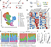 Figure 1
Study design. (A) Schematic overview of sample collection. PBMCs were collected from patients with colorectal cancer (CRC; n = 4) and glioblastoma (GBM; n = 3) before and after rhIL-7-hyFc administration. CRC patients received 2 doses. (B) Schematic illustration of the single-cell sequencing workflow. To assess the effects of rhIL-7-hyFc on peripheral T cells, sorted T cells were subjected to scRNA-seq and scTCR-seq. (C) Cell-type annotation of single cells. A total of 185,331 cells were first annotated as CD4+ or CD8+ T cells, and then further classified using canonical gene markers. (D) Expression of canonical markers in T cells. The expressions of well-established genes associated with a naive state, survival, memory, activation, cytotoxicity, inhibition, and proliferation were plotted to characterize each cluster. (E) Proportions of the CD8+ T cell (left) and CD4+ T cell (right) subsets per patient at baseline (W0) and 1 week after treatment (W1). (F and G) Proportion of proliferating CD8 (F) and CD4 (G) cluster per sample, based on scRNA-seq data from W0 and W1 (n = 7). Paired t test was used for comparison. **P < 0.01, ****P < 0.0001.
From PBMCs, T cells were isolated using a pan T cell isolation kit and were analyzed by scRNA-seq and single-cell TCR sequencing (scTCR-seq) (Figure 1B). These analyses yielded transcriptomic profiles for 185,331 cells across 7 patients and 4 time points and showed 13 distinct clusters (Figure 1C). Principal component analysis revealed minimal diversity among patients having different cancer types (Supplemental Figure 1, A and B). W1 was segregated from the other time points (Supplemental Figure 1C). Minimal batch effects were confirmed (Supplemental Figure 2). T cells were classified into CD4+ and CD8+ subsets, and further divided based on canonical markers, including CCR7, BCL2, IL7R, TNFRSF4, GZMB, HAVCR2, MKI67, and FOXP3 (Supplemental Figure 3A). CD8+ T cells formed 7 clusters: naive CD8, BCL2hi-CD8, memory CD8, activated CD8, cytotoxic CD8, exhausted CD8, and proliferating CD8. CD4+ T cells formed 6 clusters: naive CD4, BCL2–CD4 (BCL2hi-CD4), memory CD4, activated CD4, CD4 Treg, and proliferating CD4. These clusters were defined based on gene expression patterns including markers of a naive state (TCF7, SELL, LEF1, and CCR7), survival (BCL2), memory (IL7R), activation (TNFRSF4), cytotoxicity and effector function (GZMA, GZMB, GZMK, IFNG, and NKG7), inhibition (LAG3, TIGIT, HAVCR2, and CTLA4), and proliferation (STMN1 and MKI67) (Figure 1D).
To focus on the effects of the first rhIL-7-hyFc dose, we assessed T cell composition at W0 and W1. The T cell profiles exhibited significant shifts after rhIL-7-hyFc administration. Several clusters were markedly expanded at W1, including BCL2hi-CD8, activated CD8, cytotoxic CD8, proliferating CD8, BCL2hi-CD4, activated CD4, and proliferating CD4 (Figure 1E and Supplemental Figure 3B). In consensus with previous findings (28), sample-wise comparison of the proportion of proliferating or CD4+ T cells revealed a dramatic increase, 0.39% at baseline to 5.82% after treatment for CD8+ T cells and 0.39% to 9.0% for CD4+ T cells (Figure 1, F and G). The notable changes in cluster proportion represent rhIL-7-hyFc–induced changes in peripheral blood T cell dynamics.
Cell cycling and antiapoptotic features of peripheral blood T cells after rhIL-7-hyFc treatment. To examine rhIL-7-hyFc–induced features across T cell clusters, we performed differentially expressed gene (DEG) analysis between whole T cell transcriptomes from W0 and W1. This revealed 3,071 upregulated genes (notably including CORO1B, ISG15, IFITM3, PSMB, IL4R, CD55, ISG20, CD5, CD7, IFNGR2, CD74, CD69, CEACAM1, IFITM1, IL32, ICOS, GZMA, and GZMB) and 405 downregulated genes (notably including DUSP1, BTG2, BTG1, IL7R, CXCR4, BCL11B, TCF7, DUSP2, CCL3, IL5RA, and IKZF2) after treatment (Figure 2A).
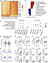 Figure 2
rhIL-7-hyFc–induced transcriptional changes in T cells. (A) Row-normalized heatmap of differentially expressed genes (DEGs) between W0 and W1. Representative genes are annotated. (B) Pathway enrichment analysis of DEGs. Pathways enriched at baseline (blue) and after rhIL-7-hyFc treatment (red) are shown. (C) GSEA of upregulated pathways after rhIL-7-hyFc treatment, among CD8+ and CD4+ T cells. (D) Module scores for the GO term “Regulation of T cell apoptotic process” were calculated and compared between W0 and W1. Unpaired t test was used for P value calculation. (E and F) Mean BCL2 expression per sample among CD8+ T cells and CD4+ T cells from scRNA-seq data (E) (n = 7) and from geometric mean fluorescence intensity (gMFI) measured by flow cytometry (F) (n = 12). (G) Flow cytometry–measured CD38 gMFI among CD8+ and CD4+ T cells, subdivided into total, naive, central memory, effector memory, and EMRA subsets (n = 9). Statistical testing was conducted using the paired Wilcoxon test (E–G). *P < 0.05, **P < 0.01, ***P < 0.001.
Gene ontology (GO) analysis of the upregulated genes revealed enrichment of cell cycle and cell division processes, which is consistent with the expected effects of IL-7 treatment (Figure 2B, red bars). On the other hand, downregulated genes were associated with developmental processes not related to T cell function, such as organ development and morphogenesis (Figure 2B, blue bars). These findings were supported by gene set enrichment analysis (GSEA), which revealed significant enrichment of lymphocyte activation and cell growth after rhIL-7-hyFc treatment (Figure 2C and Supplemental Figure 4, A and B).
We also analyzed the expression of cytotoxic genes, including GZMA, GZMB, and GZMK, on a per-cell basis within each cluster. While the cytotoxic CD8 cluster proportion did not markedly increase (Supplemental Figure 3B), per-cell expression of cytotoxic genes within this cluster was substantially higher at W1 compared to W0 (Supplemental Figure 4C). We further examined granzyme genes across all CD8+ T cells, per cluster, and per patient at W0 versus W1 (Supplemental Figure 4C). This analysis confirms that cytotoxic gene expression increased at W1 both at the bulk CD8+ level and within individual clusters, particularly the cytotoxic CD8 and exhausted CD8 subsets, and that this pattern was consistent across patients.
Due to the increased proportions of the BCL2hi-CD8 and BCL2hi-CD4 clusters after rhIL-7-hyFc treatment, we next examined apoptosis-related genes. We calculated module scores for genes involved in regulating T cell apoptosis, curated by GO biological process (GOBP). These module scores were significantly lower at W1 compared with W0, indicating reduced apoptotic features after rhIL-7-hyFc treatment (Figure 2D). Moreover, BCL2 mRNA levels were significantly higher at W1 across the CD8+ and CD4+ T cell clusters (Figure 2E). To validate BCL2 expression at the protein level, we performed flow cytometry analysis on PBMCs from an expanded cohort (n = 29), including 7 patients whose T cells had been analyzed by scRNA-seq. Flow cytometry revealed significantly increased BCL2 protein expression in total CD8+ and CD4+ T cells after rhIL-7-hyFc treatment (Figure 2F). In subset analysis, BCL2 expression was significantly increased in the naive subset (TN) and in all 4 subsets of CD8+ and CD4+ T cells (Supplemental Figure 4D). Thus, both single-cell and flow cytometry analyses revealed that rhIL-7-hyFc induced qualitative changes in T cells, including upregulation of cell cycle, activation of immune response, and induction of antiapoptosis.
Moreover, T cells treated with rhIL-7-hyFc exhibited increased activation, as evidenced by the increased populations of OX40+CD4+ and OX40+CD8+ T cells. Flow cytometry analysis (n = 9) also confirmed the upregulation of activation markers, such as CD38, HLA-DR, and OX40, particularly in the effector memory T (TEM) and CD45RA+ effector memory (TEMRA) cell populations (Figure 2G and Supplemental Figure 4, E and F).
Temporal determination of T cell state in response to rhIL-7-hyFc. We performed Monocle2 analysis to better understand dynamic changes of CD8+ and CD4+ T cell state induced by rhIL-7-hyFc. Proliferating T cell populations were excluded from trajectory construction. The trajectory of CD8+ T cells generated 3 branches, with the naive CD8+ T cell cluster as the starting point (Supplemental Figure 5A). Two prominent branches were observed at branch point 3 — with the left branch (state 1) representing the progression of memory CD8+ T cells into cytotoxic CD8+ T cells, and the right branch (state 2) illustrating the trajectory of BCL2hi-CD8+ into activated CD8+ T cells (Figure 3A). The state 2 branch ended at exhausted CD8+ T cells, although their relative frequency was negligible. Notably, state 1 was enriched with cells from time point W0, and state 2 was enriched with cells from W1 (Figure 3B).
 Figure 3
Pseudotime analysis of T cells after rhIL-7-hyFc treatment. (A and B) Pseudotime trajectory of CD8+ T cells was inferred using Monocle2. Cell annotation based on transcriptomic profile (A) and sampling time points (B) was mapped. Branch 3 divided state 1 versus 2. (C and D) Differentially expressed genes (DEGs) contributing to state bifurcation at branch 3 were identified by branch expression analysis modeling (BEAM) (C), and subjected to pathway enrichment analysis (D). Genes contributing to each state are marked with arrow. (E and F) Pseudotime trajectory of CD4+ T cells was inferred. Cell annotation based on transcriptomic profile (E) and sampling time points (F) was mapped. (G and H) DEGs contributing to CD4+ T cell state bifurcation were identified by BEAM (G), and analyzed by pathway enrichment (H).
To further investigate genes involved in the state decisions, we conducted branch expression analysis modeling (BEAM) analysis (Figure 3C). Pathway analysis of significant genes revealed that state 1 — mainly comprising CD8+ T cells from W0 — was enriched for TCR signaling and antigen presentation of MHC I (Figure 3D). State 2 — mainly comprising CD8+ T cells from W1 — was significantly enriched with terms including signaling by interleukins and cytokine signaling in immune system, reflecting the direct effects of rhIL-7-hyFc treatment. Additionally, state 2 exclusively showed upregulation of G1/S transition, suggesting increased cell proliferation.
Among CD4+ T cells, the trajectory construction yielded 2 paths from branch point 1, with pseudotime starting from the naive CD4+ T cell population (Supplemental Figure 5B). The upper branch (state 1) shows progression into memory CD4+ T cells and CD4+ Treg cells, while the lower arm (state 2) shows the trajectory of BCL2hi-CD4+ T into activated CD4+ T cells (Figure 3E). The 2 CD4+ T cell states were also differentially enriched across time points (Figure 3F).
We performed BEAM analysis to identify significant genes related to state decisions (Figure 3G). State 1 — mainly comprising CD4+ T cells from W0 — showed weaker enrichment of TCR signaling and cytokine signaling in the immune system, but stronger enrichment of MHC II antigen presentation and interferon signaling (Figure 3H). On the other hand, state 2 — mainly comprising CD4+ T cells from W1 — was highly enriched with terms representing cytokine and TCR signaling, as well as G1/S transition, indicating rhIL-7-hyFc–induced effects (Figure 3H). Thus, the trajectory analyses of CD8+ and CD4+ T cells revealed differential temporal enrichment patterns across cell states, characterized by strong enrichment of cytokine signaling pathways and G1/S phase transition, highlighting the immunomodulatory effects of rhIL-7-hyFc on peripheral blood T cells.
Hyporesponsiveness after repeated rhIL-7-hyFc treatment. In a recent phase Ib clinical trial (NCT03478995), it was reported that the second rhIL-7-hyFc administration at W3 failed to elicit a significant increase in T cell numbers (30). We investigated molecular mechanisms underlying this hyporesponsiveness. Consistent with the previous finding, scRNA-seq revealed distinct transcriptomic changes following the first administration (W1), while the second dose did not induce comparable transcriptomic changes (W4) (Figure 4A). The proportions of proliferating CD8+ and CD4+ T cell clusters were not increased after the second dose administration (Figure 4, B). This finding was further confirmed by flow cytometry analysis using PBMCs (n = 23), which revealed that the frequency of Ki-67+ T cells significantly increased at W1 compared with W0, but remained unchanged between W3 and W4 (Figure 4, C and D).
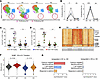 Figure 4
Hyporesponsiveness of T cells after repeated rhIL-7-hyFc administration. (A) UMAP of T cells from each time point, with proliferating T cells highlighted in red. (B and C) The frequency of proliferating CD8 (B) and CD4 (C) clusters at each time point was quantified using scRNA-seq data. Paired t tests were used for statistical comparison. (D) The proportions of Ki-67+ CD8+ (left) and CD4+ (right) T cells among peripheral T cells were measured across time points at different doses: 60–120 μg/kg (n = 6), 240–480 μg/kg (n = 5), 720–960 μg/kg (n = 6), and 1200–1700 μg/kg (n = 6). (E) Differentially expressed genes (DEGs) from W0 to W1 were projected across all time points using heatmap. (F) Comparison of module scores for the GO biological process “G0-to-G1 transition” between time points. Statistical differences were assessed using the Kolmogorov-Smirnov D value. (G) Comparison of immune activation–related pathway enrichment between the first and second rhIL-7-hyFc doses. *P < 0.05; ***P < 0.001; ****P < 0.0001.
Treatment-induced transcriptomic profile changes were also distinct between the first and second administrations. The DEGs identified at W4 were not similar to the patterns identified at W1 (Figure 4E). Rather, DEG analysis between W3 and W4 revealed genes having little relevance to T cells or their functionality (Supplemental Table 2). In accordance with these findings, the G0-to-G1 transition term, which includes genes required for exiting quiescence and entering the cell division phase, showed an increased module score from W0 to W1 (D = 0.44), but only a marginally increased module score between W3 and W4 (D = 0.16), as measured by the Kolmogorov-Smirnov D statistic (Figure 4F). Additionally, expression of the key antiapoptotic gene BCL2 was markedly increased at W1, but not at W4 (Supplemental Figure 6).
Finally, we compared the functional enrichment of upregulated genes across the 2 treatment periods. While W1 showed enrichment of T cell activation terms, such as response to cytokine or positive regulation of cytokine release, these terms were not enriched at W4 (Figure 4G). Overall, these findings indicate that repeated treatment failed to induce peripheral T cell proliferation or activation.
Hyporesponsiveness is not attributed to anti-drug antibodies or receptor modulation. To investigate mechanisms underlying the hyporesponsiveness observed after the second rhIL-7-hyFc treatment, we examined various factors that could influence the effects of rhIL-7-hyFc. We first hypothesized that rhIL-7-hyFc might elicit anti-drug antibodies (ADAs) that could neutralize the effect of the second dose. Therefore, we measured the ADA titers of serum samples obtained after the first administration (W3). Among 23 individuals, 14 were ADA-positive and 9 were ADA-negative. To further examine the effect of ADAs on responsiveness to rhIL-7-hyFc, we compared the frequency of Ki-67+ cells among CD8+ and CD4+ T cells between the ADA-negative group (n = 9) and individuals with a high ADA titer (>1:200) (n = 7). However, both patient groups showed a significantly decreased Ki-67+ frequency at W4 compared with W1 (Figure 5A), indicating that ADAs cannot fully explain the hyporesponsiveness observed upon the second administration.
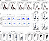 Figure 5
Impaired induction of T cell proliferation following repeated rhIL-7-hyFc administration. (A) Proliferative responses of CD8+ T cells (left) and CD4+ T cells (right) were measured in all samples (n = 23). The data were further divided into ADA-negative individuals (titer = 0; n = 9), and ADA-high individuals (>1:200; n = 7). (B) PBMCs collected at W0 and W3 (n = 11) were stained with CellTrace Violet (CTV), and stimulated for 120 hours with native IL-7 (10 ng/mL) or rhIL-7-hyFc (60 ng/mL). Representative flow cytometry plots for CD8+ T and CD4+ T cells (left), and the cumulative data regarding CTVlo T cells after stimulation (right), are shown. (C) The frequency of CD127+ T cells within the CD8+ (top) and CD4+ (bottom) subsets were assessed for W0 and W3 (n = 29). Statistical analyses were conducted with the Wilcoxon test. NS, not significant. *P < 0.05; **P < 0.01; ***P < 0.001; ****P < 0.0001.
Next, we treated PBMCs obtained at W0 and W3 with rhIL-7-hyFc or rhIL-7 ex vivo, and examined the frequency of CellTrace Violet–low (CTVlo) proliferating cells among CD8+ and CD4+ T cells. In this experiment, we could exclude any potential effect of ADAs, because the culture did not include autologous sera. We found that rhIL-7-hyFc treatment significantly increased the CTVlo cell frequency among CD8+ and CD4+ T cells obtained at both W0 and W3. However, the rhIL-7-hyFc–induced increase in the CTVlo frequency was significantly lower among CD8+ and CD4+ T cells obtained at W3 compared with those obtained at W0 (Figure 5B). Similar results were observed when PBMCs were treated with rhIL-7. These findings corroborate that the hyporesponsiveness is attributed to cell-intrinsic factors, and not ADAs.
We also hypothesized that the expression level of IL-7Rα (CD127) on the T cell membrane at W3 might be downregulated compared with W0. However, IL-7Rα expression was instead upregulated in all subsets of CD8+ T cells at W3 compared with W0, with the exception of CD8+ TCM cells (Figure 5C). All 4 subsets of CD4+ T cells exhibited the same result. These findings indicate that the observed hyporesponsiveness is not attributable to the downregulation of IL-7Rα.
Intracellular disruption of distal signaling accounts for IL-7 hyporesponsiveness. We next investigated whether the proximal signaling of IL-7Rα was attenuated in T cells obtained at W3 compared with those at W0. To this end, we treated PBMCs from W0 and W3 with rhIL-7-hyFc (n = 11) or rhIL-7 (n = 10), and examined the level of phosphorylated STAT5 (p-STAT5). Treatment with rhIL-7-hyFc or rhIL-7 significantly increased the p-STAT5 level in CD8+ T cells obtained at both W0 and W3 (Figure 6A). Importantly, the rhIL-7-hyFc–induced increase in p-STAT5 did not differ between CD8+ T cells obtained at W0 versus W3, and similar results were observed in CD4+ T cells. These data indicate that the proximal signaling of IL-7Rα was not disrupted in T cells obtained at W3.
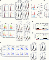 Figure 6
Altered cytokine signaling following repeated rhIL-7-hyFc treatment. (A) PBMCs collected at W0 and W3 were incubated for 120 hours with no stimulation or with stimulation using native IL-7 (10 ng/mL) or rhIL-7-hyFc (60 ng/mL). Subsequently, the frequency of p-STAT5+ cells among T cells was measured (n = 11 for rhIL-7-hyFc and n = 10 for IL-7). (B) PIM-1 kinase expression was assessed by gMFI in CD8+ (upper) and CD4+ (lower) T cells from W0 and W3 (n = 9). (C) PIM1 expression at the single-cell resolution is plotted for CD4+ (upper) and CD8+ (lower) T cells. (D) Induction of c-Myc was assessed in CD8+ (upper) and CD4+ (lower) T cells from W0 and W3 (n = 6). (E and F) GSEA was performed to measure enrichment of HALLMARK: MYC_TARGETS after the first dose (E) and the second dose (F). (G) PBMCs from W0 and W3 (n = 8) were stimulated for 120 hours with rhIL-2 (100 IU/mL) or rhIL-15 (10 ng/mL). The cumulative data regarding CTVlo T cell frequencies after stimulation are shown on the right. Statistical comparisons were performed using paired t tests. The control data for G are shared with Figure 5B, as both panels were derived from the same experiment designed to compare donor PBMC proliferative capacity following stimulation with rhIL-7 or rhIL-7-hyFc (Figure 5B) and rhIL-2 or rhIL-15 (G). NS, not significant. *P < 0.05; **P < 0.01; ***P < 0.001; ****P < 0.0001.
Next, we examined PIM-1 kinase, a downstream target of p-STAT5 (31) which is involved in cell cycle regulation. We performed flow cytometry to measure PIM-1 expression in CD8+ and CD4+ T cells obtained at W0 and W3 (n = 9), and found significantly lower PIM-1 levels at W3 compared with W0 (Figure 6B). This reduction in PIM1 expression was additionally confirmed by scRNA-seq data (Figure 6C). This low PIM-1 kinase expression at W3 may contribute to the IL-7 hyporesponsiveness.
We also analyzed the expression of STAT5 downstream target genes across T cell clusters in scRNA-seq data. We computed a STAT5 transcriptional activity score using the MSigDB HALLMARK_IL2_STAT5_SIGNALING gene set as a proxy for STAT5 downstream output. The STAT5 target gene module score was elevated at W1 compared with W0 across most clusters (Supplemental Figure 7A). In contrast, comparing W4 to W3, the STAT5 target gene score showed minimal change, indicating attenuated STAT5 transcriptional output following the second dose. PIM1 expression also showed a concordant pattern across T cell clusters (Supplemental Figure 7B).
We additionally examined the expression of c-Myc, a critical regulator of cell proliferation, which is downstream of PIM-1 (32, 33). PBMCs obtained at W0 and W3 were treated with rhIL-7-hyFc (n = 6) or rhIL-7 (n = 6) ex vivo. Either treatment led to significantly increased c-Myc expression in CD8+ T cells obtained at W0, but not in CD8+ T cells obtained at W3 (Figure 6D). A similar trend was observed among CD4+ T cells, although rhIL-7-hyFc significantly increased the c-Myc expression level in CD4+ T cells obtained at W3. GSEA conducted on scRNA-seq data confirmed that Myc-targeted genes were upregulated after first dose, but not after the second dose (Figure 6, E and F).
Our results indicated that the distal signaling of IL-7Rα, at the stage of PIM-1 kinase/c-Myc, was disrupted in T cells obtained at W3. Thus, we hypothesized that these T cells might also exhibit hyporesponsiveness to other common γ-chain cytokines that share the same distal signaling of PIM-1/c-Myc. To investigate this possibility, we treated PBMCs obtained at W0 and W3 with rhIL-2 or rhIL-15. Our results showed that rhIL-2 or rhIL-15 treatment significantly increased the CTVlo cell frequency among CD8+ T cells obtained at both W0 and W3 (Figure 6G). However, the rhIL-2– or rhIL-15–induced increase in the CTVlo frequency was significantly lower among CD8+ T cells obtained at W3 compared with those obtained at W0. Moreover, rhIL-2 or rhIL-15 treatment significantly increased the CTVlo cell frequency among CD4+ T cells obtained at W0, but not among CD4+ T cells obtained at W3. These data confirmed that T cells obtained at W3 also exhibited partial hyporesponsiveness to other common γ-chain cytokines, such as IL-2 and IL-15, supporting the notion that disruption in the distal signaling of PIM-1/c-Myc accounts for the hyporesponsiveness observed after the second rhIL-7-hyFc administration.


