
Research ArticleImmunologyNephrology
Open Access |  10.1172/jci.insight.197348
Innate allorecognition induces HSP70 in monocytes contributing to renal allograft rejection
Zeping Gui,1 Neda Feizi,1 Berkay Demirors,1 Canxiang Lin,1 Hehua Dai,1 Mouhamad Al Moussawy,1 Andrew Friday,1 Steven M. Sanders,1 Latha Halesha,1 Amanda L. Williams,1 Faruk Sacirbegovic,1 Parmjeet S. Randhawa,1,2 Khodor I. Abou-Daya,1 and Martin H. Oberbarnscheidt1,3
1Thomas E. Starzl Transplantation Institute, Department of Surgery, University of Pittsburgh School of Medicine and University of Pittsburgh Medical Center, Pittsburgh, Pennsylvania, USA.
2Division of Transplant Pathology, Department of Pathology, University of Pittsburgh School of Medicine, Pittsburgh, Pennsylvania, USA.
3Department of Immunology, University of Pittsburgh, Pittsburgh, Pennsylvania, USA.
Address correspondence to: Martin H. Oberbarnscheidt or Khodor I. Abou-Daya, 200 Lothrop Street BST 15th floor, Thomas E. Starzl Transplantation Institute, University of Pittsburgh, Pittsburgh, Pennsylvania, 15213, USA. Email: mho6@pitt.edu (MHO); kha17@pitt.edu (KIAD).
Authorship note: ZG and NF contributed equally to this work and have been designated as co–first authors. KIAD and MHO contributed equally to this work.
Find articles by Gui, Z. in: PubMed | Google Scholar
1Thomas E. Starzl Transplantation Institute, Department of Surgery, University of Pittsburgh School of Medicine and University of Pittsburgh Medical Center, Pittsburgh, Pennsylvania, USA.
2Division of Transplant Pathology, Department of Pathology, University of Pittsburgh School of Medicine, Pittsburgh, Pennsylvania, USA.
3Department of Immunology, University of Pittsburgh, Pittsburgh, Pennsylvania, USA.
Address correspondence to: Martin H. Oberbarnscheidt or Khodor I. Abou-Daya, 200 Lothrop Street BST 15th floor, Thomas E. Starzl Transplantation Institute, University of Pittsburgh, Pittsburgh, Pennsylvania, 15213, USA. Email: mho6@pitt.edu (MHO); kha17@pitt.edu (KIAD).
Authorship note: ZG and NF contributed equally to this work and have been designated as co–first authors. KIAD and MHO contributed equally to this work.
Find articles by Feizi, N. in: PubMed | Google Scholar
1Thomas E. Starzl Transplantation Institute, Department of Surgery, University of Pittsburgh School of Medicine and University of Pittsburgh Medical Center, Pittsburgh, Pennsylvania, USA.
2Division of Transplant Pathology, Department of Pathology, University of Pittsburgh School of Medicine, Pittsburgh, Pennsylvania, USA.
3Department of Immunology, University of Pittsburgh, Pittsburgh, Pennsylvania, USA.
Address correspondence to: Martin H. Oberbarnscheidt or Khodor I. Abou-Daya, 200 Lothrop Street BST 15th floor, Thomas E. Starzl Transplantation Institute, University of Pittsburgh, Pittsburgh, Pennsylvania, 15213, USA. Email: mho6@pitt.edu (MHO); kha17@pitt.edu (KIAD).
Authorship note: ZG and NF contributed equally to this work and have been designated as co–first authors. KIAD and MHO contributed equally to this work.
Find articles by Demirors, B. in: PubMed | Google Scholar
1Thomas E. Starzl Transplantation Institute, Department of Surgery, University of Pittsburgh School of Medicine and University of Pittsburgh Medical Center, Pittsburgh, Pennsylvania, USA.
2Division of Transplant Pathology, Department of Pathology, University of Pittsburgh School of Medicine, Pittsburgh, Pennsylvania, USA.
3Department of Immunology, University of Pittsburgh, Pittsburgh, Pennsylvania, USA.
Address correspondence to: Martin H. Oberbarnscheidt or Khodor I. Abou-Daya, 200 Lothrop Street BST 15th floor, Thomas E. Starzl Transplantation Institute, University of Pittsburgh, Pittsburgh, Pennsylvania, 15213, USA. Email: mho6@pitt.edu (MHO); kha17@pitt.edu (KIAD).
Authorship note: ZG and NF contributed equally to this work and have been designated as co–first authors. KIAD and MHO contributed equally to this work.
Find articles by Lin, C. in: PubMed | Google Scholar
1Thomas E. Starzl Transplantation Institute, Department of Surgery, University of Pittsburgh School of Medicine and University of Pittsburgh Medical Center, Pittsburgh, Pennsylvania, USA.
2Division of Transplant Pathology, Department of Pathology, University of Pittsburgh School of Medicine, Pittsburgh, Pennsylvania, USA.
3Department of Immunology, University of Pittsburgh, Pittsburgh, Pennsylvania, USA.
Address correspondence to: Martin H. Oberbarnscheidt or Khodor I. Abou-Daya, 200 Lothrop Street BST 15th floor, Thomas E. Starzl Transplantation Institute, University of Pittsburgh, Pittsburgh, Pennsylvania, 15213, USA. Email: mho6@pitt.edu (MHO); kha17@pitt.edu (KIAD).
Authorship note: ZG and NF contributed equally to this work and have been designated as co–first authors. KIAD and MHO contributed equally to this work.
Find articles by Dai, H. in: PubMed | Google Scholar
1Thomas E. Starzl Transplantation Institute, Department of Surgery, University of Pittsburgh School of Medicine and University of Pittsburgh Medical Center, Pittsburgh, Pennsylvania, USA.
2Division of Transplant Pathology, Department of Pathology, University of Pittsburgh School of Medicine, Pittsburgh, Pennsylvania, USA.
3Department of Immunology, University of Pittsburgh, Pittsburgh, Pennsylvania, USA.
Address correspondence to: Martin H. Oberbarnscheidt or Khodor I. Abou-Daya, 200 Lothrop Street BST 15th floor, Thomas E. Starzl Transplantation Institute, University of Pittsburgh, Pittsburgh, Pennsylvania, 15213, USA. Email: mho6@pitt.edu (MHO); kha17@pitt.edu (KIAD).
Authorship note: ZG and NF contributed equally to this work and have been designated as co–first authors. KIAD and MHO contributed equally to this work.
Find articles by Al Moussawy, M. in: PubMed | Google Scholar
1Thomas E. Starzl Transplantation Institute, Department of Surgery, University of Pittsburgh School of Medicine and University of Pittsburgh Medical Center, Pittsburgh, Pennsylvania, USA.
2Division of Transplant Pathology, Department of Pathology, University of Pittsburgh School of Medicine, Pittsburgh, Pennsylvania, USA.
3Department of Immunology, University of Pittsburgh, Pittsburgh, Pennsylvania, USA.
Address correspondence to: Martin H. Oberbarnscheidt or Khodor I. Abou-Daya, 200 Lothrop Street BST 15th floor, Thomas E. Starzl Transplantation Institute, University of Pittsburgh, Pittsburgh, Pennsylvania, 15213, USA. Email: mho6@pitt.edu (MHO); kha17@pitt.edu (KIAD).
Authorship note: ZG and NF contributed equally to this work and have been designated as co–first authors. KIAD and MHO contributed equally to this work.
Find articles by Friday, A. in: PubMed | Google Scholar
1Thomas E. Starzl Transplantation Institute, Department of Surgery, University of Pittsburgh School of Medicine and University of Pittsburgh Medical Center, Pittsburgh, Pennsylvania, USA.
2Division of Transplant Pathology, Department of Pathology, University of Pittsburgh School of Medicine, Pittsburgh, Pennsylvania, USA.
3Department of Immunology, University of Pittsburgh, Pittsburgh, Pennsylvania, USA.
Address correspondence to: Martin H. Oberbarnscheidt or Khodor I. Abou-Daya, 200 Lothrop Street BST 15th floor, Thomas E. Starzl Transplantation Institute, University of Pittsburgh, Pittsburgh, Pennsylvania, 15213, USA. Email: mho6@pitt.edu (MHO); kha17@pitt.edu (KIAD).
Authorship note: ZG and NF contributed equally to this work and have been designated as co–first authors. KIAD and MHO contributed equally to this work.
Find articles by
Sanders, S.
in:
PubMed
|
Google Scholar
|

1Thomas E. Starzl Transplantation Institute, Department of Surgery, University of Pittsburgh School of Medicine and University of Pittsburgh Medical Center, Pittsburgh, Pennsylvania, USA.
2Division of Transplant Pathology, Department of Pathology, University of Pittsburgh School of Medicine, Pittsburgh, Pennsylvania, USA.
3Department of Immunology, University of Pittsburgh, Pittsburgh, Pennsylvania, USA.
Address correspondence to: Martin H. Oberbarnscheidt or Khodor I. Abou-Daya, 200 Lothrop Street BST 15th floor, Thomas E. Starzl Transplantation Institute, University of Pittsburgh, Pittsburgh, Pennsylvania, 15213, USA. Email: mho6@pitt.edu (MHO); kha17@pitt.edu (KIAD).
Authorship note: ZG and NF contributed equally to this work and have been designated as co–first authors. KIAD and MHO contributed equally to this work.
Find articles by Halesha, L. in: PubMed | Google Scholar
1Thomas E. Starzl Transplantation Institute, Department of Surgery, University of Pittsburgh School of Medicine and University of Pittsburgh Medical Center, Pittsburgh, Pennsylvania, USA.
2Division of Transplant Pathology, Department of Pathology, University of Pittsburgh School of Medicine, Pittsburgh, Pennsylvania, USA.
3Department of Immunology, University of Pittsburgh, Pittsburgh, Pennsylvania, USA.
Address correspondence to: Martin H. Oberbarnscheidt or Khodor I. Abou-Daya, 200 Lothrop Street BST 15th floor, Thomas E. Starzl Transplantation Institute, University of Pittsburgh, Pittsburgh, Pennsylvania, 15213, USA. Email: mho6@pitt.edu (MHO); kha17@pitt.edu (KIAD).
Authorship note: ZG and NF contributed equally to this work and have been designated as co–first authors. KIAD and MHO contributed equally to this work.
Find articles by Williams, A. in: PubMed | Google Scholar
1Thomas E. Starzl Transplantation Institute, Department of Surgery, University of Pittsburgh School of Medicine and University of Pittsburgh Medical Center, Pittsburgh, Pennsylvania, USA.
2Division of Transplant Pathology, Department of Pathology, University of Pittsburgh School of Medicine, Pittsburgh, Pennsylvania, USA.
3Department of Immunology, University of Pittsburgh, Pittsburgh, Pennsylvania, USA.
Address correspondence to: Martin H. Oberbarnscheidt or Khodor I. Abou-Daya, 200 Lothrop Street BST 15th floor, Thomas E. Starzl Transplantation Institute, University of Pittsburgh, Pittsburgh, Pennsylvania, 15213, USA. Email: mho6@pitt.edu (MHO); kha17@pitt.edu (KIAD).
Authorship note: ZG and NF contributed equally to this work and have been designated as co–first authors. KIAD and MHO contributed equally to this work.
Find articles by Sacirbegovic, F. in: PubMed | Google Scholar
1Thomas E. Starzl Transplantation Institute, Department of Surgery, University of Pittsburgh School of Medicine and University of Pittsburgh Medical Center, Pittsburgh, Pennsylvania, USA.
2Division of Transplant Pathology, Department of Pathology, University of Pittsburgh School of Medicine, Pittsburgh, Pennsylvania, USA.
3Department of Immunology, University of Pittsburgh, Pittsburgh, Pennsylvania, USA.
Address correspondence to: Martin H. Oberbarnscheidt or Khodor I. Abou-Daya, 200 Lothrop Street BST 15th floor, Thomas E. Starzl Transplantation Institute, University of Pittsburgh, Pittsburgh, Pennsylvania, 15213, USA. Email: mho6@pitt.edu (MHO); kha17@pitt.edu (KIAD).
Authorship note: ZG and NF contributed equally to this work and have been designated as co–first authors. KIAD and MHO contributed equally to this work.
Find articles by Randhawa, P. in: PubMed | Google Scholar
1Thomas E. Starzl Transplantation Institute, Department of Surgery, University of Pittsburgh School of Medicine and University of Pittsburgh Medical Center, Pittsburgh, Pennsylvania, USA.
2Division of Transplant Pathology, Department of Pathology, University of Pittsburgh School of Medicine, Pittsburgh, Pennsylvania, USA.
3Department of Immunology, University of Pittsburgh, Pittsburgh, Pennsylvania, USA.
Address correspondence to: Martin H. Oberbarnscheidt or Khodor I. Abou-Daya, 200 Lothrop Street BST 15th floor, Thomas E. Starzl Transplantation Institute, University of Pittsburgh, Pittsburgh, Pennsylvania, 15213, USA. Email: mho6@pitt.edu (MHO); kha17@pitt.edu (KIAD).
Authorship note: ZG and NF contributed equally to this work and have been designated as co–first authors. KIAD and MHO contributed equally to this work.
Find articles by
Abou-Daya, K.
in:
PubMed
|
Google Scholar
|
1Thomas E. Starzl Transplantation Institute, Department of Surgery, University of Pittsburgh School of Medicine and University of Pittsburgh Medical Center, Pittsburgh, Pennsylvania, USA.
2Division of Transplant Pathology, Department of Pathology, University of Pittsburgh School of Medicine, Pittsburgh, Pennsylvania, USA.
3Department of Immunology, University of Pittsburgh, Pittsburgh, Pennsylvania, USA.
Address correspondence to: Martin H. Oberbarnscheidt or Khodor I. Abou-Daya, 200 Lothrop Street BST 15th floor, Thomas E. Starzl Transplantation Institute, University of Pittsburgh, Pittsburgh, Pennsylvania, 15213, USA. Email: mho6@pitt.edu (MHO); kha17@pitt.edu (KIAD).
Authorship note: ZG and NF contributed equally to this work and have been designated as co–first authors. KIAD and MHO contributed equally to this work.
Find articles by
Oberbarnscheidt, M.
in:
PubMed
|
Google Scholar
|
Published October 8, 2026 - More info
JCI Insight. 2026;11(19):e197348. https://doi.org/10.1172/jci.insight.197348.
© 2026 Gui et al. This work is licensed under the Creative Commons Attribution 4.0 International License. To view a copy of this license, visit http://creativecommons.org/licenses/by/4.0/.
Received: June 30, 2025; Accepted: August 13, 2026

-
Results
HSP70 is a key regulator downstream of monocyte allorecognition. To gain insight into transcriptional changes of monocytes after innate allorecognition, we analyzed published scRNA-seq datasets from mice (GSE147596) (9) and human kidney allograft biopsies (E-MTAB-12051) (10) to identify upregulated genes after monocyte allorecognition. Datasets were reprocessed.
The scRNA-seq dataset GSE147596 consists of splenic monocytes sorted from B6-Rag2−/− Il2rg−/− mice 1 week after either allogeneic or syngeneic injection of 20 million irradiated BALB, C3H, or B6 splenocytes (9). Four unique neighborhoods were visualized in the UMAP (Figure 1A). Initially, we classified these neighborhoods based on the expression of Ly-6C encoded by Ly6c2, revealing Ly-6Chi, Ly-6Cint, and Ly-6Clo subsets (Figure 1B). Further profiling for antigen presentation–related gene expression showed that the Ly-6Cint subset is transitioning to mo-DC, as it differentially expressed the highest levels of MHCII (H2-Ab1) and CD74 (Cd74) as well as an intermediate level of CD11c (Itgax) (11, 12). One of the Ly-6Clo subsets expressed the highest levels of CD36 (Cd36) and CD11c, supporting its transition to macrophages, given CD36’s role in phagocytosis.
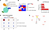 Figure 1
Inducible HSP70 is upregulated downstream of innate allorecognition. Analysis of our in-house published scRNA-seq dataset (GSE147596) of murine splenic monocytes. B6 Rag-gc-KO mice were immunized with allogeneic (Balb/c, C3H) or injected with syngeneic (B6) splenocytes and monocytes analyzed 1 week later as published (9). (A) UMAP of monocytes annotated by Ly-6C expression and antigen presentation gene signature. (B) Heatmap displaying expression of differentially expressed genes used to annotate monocytes. (C) Ingenuity analysis of upstream regulators differentially expressed after allostimulation. (D) Bubble plot displaying expression of Hspa1a by color scale and bubble size in monocyte subsets across groups. (E) STRING analysis of downstream targets of HSPA1A.
After classification of monocyte subtypes, we performed differential gene expression analysis comparing Ly-6Chi monocytes in Balb/c versus B6-injected groups. Differentially expressed genes (DEG) (|fold change > 1.5| and FDR < 0.01) were used to perform Qiagen’s Ingenuity Pathway Analysis (IPA) in order to pinpoint upstream regulators that are differentially expressed after allogeneic stimulation. Only 4 upstream regulators were shortlisted (Figure 1C). Hspa1a was one of the shortlisted upstream regulators, and its downstream targets included Ptk2, Tnf, Xbp1, and Cdk1. We validated that Hspa1a upregulation was not restricted to Ly-6Chi monocytes recognizing allogeneic Balb/c splenocytes, but we also found that it was upregulated in Ly-6Chi and Ly-6Cint transition to mo-DC subsets in Balb/c and C3H splenocyte–injected groups (Figure 1D). Notably, upregulation of Hspa1a was higher in the Balb/c as compared with the C3H group, consistent with our previous reports of the monocyte response to different allogeneic sources of injected splenocytes (8). This upregulation was absent in the Ly-6Clo transition to macrophage subset, supporting its specific involvement in monocyte to mo-DC differentiation. To pinpoint which gene products are directly interacting with HSPA1A, we queried Hspa1a’s downstream targets and the other upstream regulators in STRING, an online database for protein-protein interaction. Only XBP1 and TNF directly interact with HSPA1A, as highlighted in Figure 1E. In addition, XBP1 is known to be required for DC development and survival (13). To note, Xbp1 was upregulated in Ly-6Chi monocytes after allostimulation with a fold change of 1.7; however, it did not reach significance (P = 0.15 and FDR step up = 0.47). Nevertheless, HSPA1A might be interacting with XBP1 during monocyte to DC differentiation. To confirm that the DEG in Ly-6Chi monocytes after injection of irradiated Balb/c or C3H monocytes are specific to allostimulation, we performed bulk RNA-seq on BM-derived monocytes after nonspecific GM-CSF–driven monocyte to DC differentiation, comparing Hspa1a shRNA knockdown versus controls. Only 34 genes were both downregulated after shRNA knockdown and upregulated specifically after allostimulation by injection of irradiated Balb/c splenocytes (Supplemental Figure 1, A and B; supplemental material available online with this article; https://doi.org/10.1172/jci.insight.197348DS1). None of these genes are linked to monocyte differentiation to DCs, suggesting that the observed transcriptional changes in the Ly-6Chi subset are specific to innate allorecognition.
To investigate if similar transcriptional changes can be observed in human transplant recipients, we analyzed a publicly available scRNA-seq dataset generated from kidney allograft biopsies (BioStudies accession no, E-MTAB-12051) of patients with transplant rejection (R, n = 4) and no rejection (NR, n = 12) (10). We classified the dataset’s major cell types using the Automated Garnett Cell Classifier (14). Only 1 unique neighborhood’s cells were labeled as myeloid (Supplemental Figure 2A). In addition, the myeloid classified neighborhood was the only PTPRC-expressing (CD45-expressing) neighborhood that also abundantly expressed HSPA1A. We selected this neighborhood and performed a dimensionality reduction and projection using t-SNE. This revealed the heterogeneity of the myeloid population as 7 distinct neighborhoods (Figure 2A). The cells were then classified by the expression of known unique markers for myeloid subsets (Supplemental Figure 2B). This allowed us to label the neighborhoods as cDC1, cDC2, mo-DC, M1 macrophages (MΦ), M2 MΦ, classical monocytes, and nonclassical monocytes (Figure 2A). Mo-DC and nonclassical monocytes were only detected in the R group, and M2 MΦ were only observed in the NR group (Figure 2B). This is consistent with an ongoing robust alloresponse in the R group and a dominant reparative response in the NR group. Supplemental Figure 2A shows that HSPA1A expression in immune cells was absent in B cells, minimal in T cell subsets and prominent in myeloid cells. We then profiled the myeloid subsets for differential expression of HSPA1A while comparing the 2 patient groups. Classical monocytes and cDC1, which are known to activate T cells, upregulated HSPA1A expression while cDC2, and M2 MΦ downregulated it during rejection (Figure 2C). Moreover, HSPA1A transcripts were present in mo-DC, a myeloid subset exclusively found in kidney biopsies in the rejection group, and in M2 MΦ, a myeloid subset exclusively detected in the nonrejection group. Upregulation cannot be assessed for either subset, owing to their absence in the comparator condition. We confirmed that this transcriptional upregulation is reflected at the protein level by intracellular HSP70 staining of graft-infiltrating monocytes (Supplemental Figure 3).
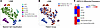 Figure 2
HSPA1A expression in myeloid cells in rejecting human kidney allografts. Analysis of myeloid cells infiltrating kidney transplant biopsies from published scRNA-seq dataset (E-MTAB-12051). (A) t-SNE of the myeloid cells with visualized classification from Supplemental Figure 2. (B) t-SNE of the myeloid cells highlighting their source from rejection or no rejection groups. (C) Heatmap visualizing expression of HSPA1A in the distinct myeloid subsets comparing rejection versus nonrejection groups. Color reflects HSPA1A expression: blue, low; red, high. For subsets present in both patient groups (cDC1, cDC2, classical monocytes, M1 macrophages), color represents relative expression between rejection and nonrejection. mo-DC (rejection only), M2 macrophages (nonrejection only) and nonclassical monocytes (rejection only) are present in a single group and have no comparator. For these, color reflects the level of transcript detected in that group and up- or downregulation cannot be inferred. Gray indicates subset absent from that group.
Blocking the HSP70 pathway diminishes innate allorecognition. To test the effect of blocking the HSP70 pathway on innate allorecognition, we used the allogeneic BM plug transplantation model, in which the intact BM cylinder from the femur of a donor mouse was transplanted under the kidney capsule of the recipient mouse (5, 8, 9). We used 2 different approaches. First, we blocked HSP70 by treating recipients with the HSP70 inhibitor deoxyspergualin (DSG) in recipients lacking all lymphoid cells (B6 Rag-gc–KO) to investigate the effect on innate allorecognition in the absence of adaptive immunity. DSG is a synthetic analogue of spergualin that engages the conserved C-terminal EEVD motif and the N-terminal nucleotide-binding domain of cytosolic HSP70 family members, inhibiting ATPase activity and downregulating Hspa1a expression (15–17). NOD BM plugs (Figure 3A) or Balb/c BM plugs (Figure 3B) were transplanted under the kidney capsule and procured on day 7 after transplantation. Flow cytometric analysis of the graft infiltrate demonstrated the presence of mo-DC in untreated recipients, both for the NOD and the Balb/c graft. Treatment of recipients with DSG, administered daily i.p., resulted in significant reduction of infiltration with mo-DC in the graft, indicating that blocking HSP70 with DSG impairs innate allorecognition by monocytes.
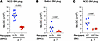 Figure 3
Blocking the HSP70 pathway reduces innate allorecognition. Bone marrow plugs from NOD or Balb/c donors were transplanted under the kidney capsule of recipient mice as indicated. BM plugs were removed and analyzed by flow cytometry on day 7 after transplantation. Mo-DC (lin–CD11b+F4/80–CD11c+) were enumerated. (A and B) NOD (P < 0.0001) and Balb/c BM plugs (P = 0.0087) transplanted to B6 Rag-gc-KO or B6 Rag-gc-KO recipients treated with deoxyspergualin (n = 6–12/group, n = 2 experiments). (C) Mo-DC infiltration of allogeneic NOD BM plugs in B6 WT and HSP70-KO recipients treated with anti-CD4, -CD8, -CD20 and -NK1.1 (P = 0.0087) (P < 0.05 considered significant, Mann-Whitney U test) (n = 6/group, N = 2 experiments).
We then investigated if innate allorecognition is impaired in HSP70-deficient WT recipients. NOD BM plugs were transplanted under the kidney capsule of either WT or HSP70-KO recipients and graft infiltrate analyzed by flow cytometry. HSP70-KO recipients displayed significantly reduced infiltration of BM plugs with mo-DC compared with WT recipients (Figure 3C), indicating that HSP70 is a downstream regulator of innate allorecognition.
Interfering with the HSP70 pathway improves renal transplant outcomes in mice. To investigate if the HSP70 pathway is relevant in a more clinically relevant transplantation setting, we utilized an allogeneic kidney transplant model in mice and transplanted allogeneic Balb/c kidneys to B6 recipients. This model results in slow, chronic rejection with long-term survival and histopathological hallmarks of chronic rejection. Recipients were either WT or constitutive HSP70-deficient mice. An additional group received treatment with DSG i.p. for 60 days. Syngeneic kidney transplants served as controls. Since the kidney transplants were life-sustaining, survival was monitored. Grafts were procured at day 120 after transplantation, and histopathology and the renal immune infiltrate were assessed. Survival curves of all groups were not significantly different, although some allografts were lost in the WT and HSP70 groups due to urinary obstruction (Figure 4A). Histopathological Banff scoring of kidney graft rejection showed significantly lower Banff scores in the HSP70-KO and DSG-treated groups, compared with the untreated WT control group (Figure 4B). Photomicrographs of kidney grafts showed a visually higher immune infiltrate in the WT group compared with the HSP70-KO and DSG-treated groups (Figure 4C). Quantitation of fibrosis in kidney grafts showed no significant differences across groups, but quantitation of the immune infiltrate was significantly lower in the HSP70-KO and DSG-treated groups compared with the untreated WT group and similar to syngeneic grafts (Figure 4B). Although chronic rejection of kidney grafts in this model is not dependent on donor-specific antibodies, we measured DSA in all groups to determine if HSP70 deficiency or blockade confers any differences in alloantibody production. Figure 4D shows no differences in DSA production between all groups, indicating that there is no direct or indirect effect of blocking HSP70 on DSA production. To further characterize the differences in immune infiltrate in the absence of HSP70, we analyzed the phenotype of the immune infiltrate of the grafts by multicolor spectral flow cytometry at day 120 (Supplemental Figure 4). Infiltrating monocytes were not different between groups, but there were significantly reduced numbers of mono-DC in the HSP70-KO group and significantly less mature (CD80+MHCII+) mono-DC in both the HSP70-KO and DSG-treated group. Allograft infiltration with CD8 and CD4 T cells was significantly reduced in HSP70-KO recipients but did not reach significance in the DSG-treated group. Overall, these findings suggest that blocking HSP70 blocks maturation of mono-DC and causes a reduction in graft-infiltrating T cells.
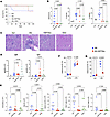 Figure 4
Interfering with the HSP70 pathway improves renal transplant outcomes. Balb/c kidneys were transplanted to B6 recipients as indicated. Syngeneic B6 KTx shown as controls (n = 2). Grafts were procured at day 120 after transplant (n = 4–8, N = 2–3 experiments). (A) Graft survival (log-rank test). (B) Quantitation of Banff score, fibrosis, and immune infiltrate of grafts is shown. (P < 0.05 considered significant, 1-way ANOVA). (C) Photomicrograph of H&E histology of indicated representative kidney graft. Scale bar: 100 μm. (D) MFI of serum donor-specific antibody measurements for IgM and IgG (n = 4–10, syn shown as baseline reference) (P < 0.05 was considered significant, 1-way ANOVA with Tukey’s multiple comparison test). (E) Quantitation of immune cell infiltrate in allografts (P < 0.05 was considered significant, 1-way ANOVA with Tukey’s multiple comparison test). (F and G) In vitro BMDC maturation assay. Maturation of B6 WT and HSP70-KO BMDC after LPS stimulation (F), and after coculture with syngeneic or allogeneic splenocytes (G) (P < 0.05 considered significant, 2-way ANOVA) (n = 6, N = 1 experiment).
To confirm that Hspa1a mRNA upregulation is reflected at the protein level and to localize HSP70 protein expression, we measured intracellular HSP70 by flow cytometry in lymphoreplete and HSP70-KO kidney transplant recipients at day 7 (Supplemental Figure 3). Graft-infiltrating monocytes from WT recipients expressed significantly higher intracellular HSP70 in kidney transplant and spleen, whereas HSP70 was absent in HSP70-KO recipients (Supplemental Figure 3B), confirming the HSP70-KO genotype. Within WT grafts HSP70 protein was expressed predominantly by myeloid subsets (Supplemental Figure 3C). Characterization of the graft immune infiltrate at day 7 showed significantly reduced T cell (P = 0.0004) and CD8 T cell (P = 0.0053) numbers in HSP70-KO compared with WT recipients (Supplemental Figure 3D), consistent with the reduced T cell infiltration observed in the chronic kidney transplant model (Figure 4E). To assess whether HSP70 deficiency alters allospecific T cell priming, we enumerated allospecific CD8 T cells using a pooled H-2K(d) tetramer panel. Allospecific tetramer+ CD8 T cell numbers did not differ between WT and HSP70-KO recipients in either the graft (P = 0.1000) or spleen (P = 0.2000) at day 7 (Supplemental Figure 3E), indicating that initial allospecific CD8 priming is intact in the absence of HSP70 and that the reduced intragraft T cell infiltrate reflects a graft-local rather than a systemic priming defect. Overall immune subset composition in the spleen of naive WT and HSP70-KO mice was similar (Supplemental Figure 3F), whereas NK and monocyte numbers were significantly reduced, and macrophage numbers increased, in HSP70-KO mice 7 days after allosensitization, consistent with a predominantly myeloid-restricted effect (Supplemental Figure 3F).
To further test the role of the HSP70 pathway in innate allorecognition, we generated BM-derived immature DC in vitro. Immature BMDC from either B6 WT or B6 HSP70-KO mice were cocultured with congenic B6 syngeneic or Balb/c allogeneic splenocytes for 5 days. Flow cytometric analysis for mature (CD80+MHCII+) BMDC showed similar maturation of BMDC in both WT and HSP70-KO groups after LPS stimulation (Figure 4F). B6 BMDC matured after allogeneic stimulation to a similar extent than LPS stimulated WT BMDC. Maturation of HSP70-KO BMDC was significantly impaired after allogeneic stimulation (P ≤ 0.0001) compared with WT BMDC (Figure 4G). These data show not only that the HSP70 pathway plays a role in maturation of monocyte-derived DC after allorecognition but also demonstrates that pathways causing maturation of BMDC after LPS stimulation are not dependent on HSP70.


