
Clinical Research and Public HealthClinical ResearchReproductive biology
Open Access |  10.1172/jci.insight.202443
Programmed cycle-induced endometrial perturbations do not independently influence angiogenic imbalance or hypertensive disorders in pregnancy
David Huang,1,2 Emily Flynn,3 Brittany Davidson,3 Juan C. Irwin,2 Mohammad Naser,3 Ana Laura Almonte,4,5 Jennifer Qin,6 Yue Song,6 Fleurdeliza B. Rabara,1 Rebecca Wong,1 Lydia B. Zablotska,7 Mitchell P. Rosen,1 Torsten Wittmann,3 Gabriela K. Fragiadakis,3,8 Alexis J. Combes,3,9 Marina Sirota,2,4 Marcelle I. Cedars,1,2 and Linda C. Giudice1,2
1Division of Reproductive Endocrinology & Infertility, Department of Obstetrics, Gynecology & Reproductive Sciences,
2Center for Reproductive Sciences,
3CoLabs,
4Bakar Computational Health Sciences Institute,
5Biomedical Informatics Program,
6Department of Obstetrics, Gynecology & Reproductive Sciences,
7Department of Epidemiology and Biostatistics,
8Division of Rheumatology, Department of Medicine, and
9Department of Pathology, Division of Gastroenterology, Department of Medicine, Bakar ImmunoX Initiative, UCSF, San Francisco, California, USA.
Address correspondence to: David Huang, 499 Illinois St., 6th Floor, San Francisco, California 94107, USA. Email: David.Huang@UCSF.edu.
Find articles by Huang, D. in: PubMed | Google Scholar
1Division of Reproductive Endocrinology & Infertility, Department of Obstetrics, Gynecology & Reproductive Sciences,
2Center for Reproductive Sciences,
3CoLabs,
4Bakar Computational Health Sciences Institute,
5Biomedical Informatics Program,
6Department of Obstetrics, Gynecology & Reproductive Sciences,
7Department of Epidemiology and Biostatistics,
8Division of Rheumatology, Department of Medicine, and
9Department of Pathology, Division of Gastroenterology, Department of Medicine, Bakar ImmunoX Initiative, UCSF, San Francisco, California, USA.
Address correspondence to: David Huang, 499 Illinois St., 6th Floor, San Francisco, California 94107, USA. Email: David.Huang@UCSF.edu.
Find articles by Flynn, E. in: PubMed | Google Scholar
1Division of Reproductive Endocrinology & Infertility, Department of Obstetrics, Gynecology & Reproductive Sciences,
2Center for Reproductive Sciences,
3CoLabs,
4Bakar Computational Health Sciences Institute,
5Biomedical Informatics Program,
6Department of Obstetrics, Gynecology & Reproductive Sciences,
7Department of Epidemiology and Biostatistics,
8Division of Rheumatology, Department of Medicine, and
9Department of Pathology, Division of Gastroenterology, Department of Medicine, Bakar ImmunoX Initiative, UCSF, San Francisco, California, USA.
Address correspondence to: David Huang, 499 Illinois St., 6th Floor, San Francisco, California 94107, USA. Email: David.Huang@UCSF.edu.
Find articles by Davidson, B. in: PubMed | Google Scholar
1Division of Reproductive Endocrinology & Infertility, Department of Obstetrics, Gynecology & Reproductive Sciences,
2Center for Reproductive Sciences,
3CoLabs,
4Bakar Computational Health Sciences Institute,
5Biomedical Informatics Program,
6Department of Obstetrics, Gynecology & Reproductive Sciences,
7Department of Epidemiology and Biostatistics,
8Division of Rheumatology, Department of Medicine, and
9Department of Pathology, Division of Gastroenterology, Department of Medicine, Bakar ImmunoX Initiative, UCSF, San Francisco, California, USA.
Address correspondence to: David Huang, 499 Illinois St., 6th Floor, San Francisco, California 94107, USA. Email: David.Huang@UCSF.edu.
Find articles by Irwin, J. in: PubMed | Google Scholar
1Division of Reproductive Endocrinology & Infertility, Department of Obstetrics, Gynecology & Reproductive Sciences,
2Center for Reproductive Sciences,
3CoLabs,
4Bakar Computational Health Sciences Institute,
5Biomedical Informatics Program,
6Department of Obstetrics, Gynecology & Reproductive Sciences,
7Department of Epidemiology and Biostatistics,
8Division of Rheumatology, Department of Medicine, and
9Department of Pathology, Division of Gastroenterology, Department of Medicine, Bakar ImmunoX Initiative, UCSF, San Francisco, California, USA.
Address correspondence to: David Huang, 499 Illinois St., 6th Floor, San Francisco, California 94107, USA. Email: David.Huang@UCSF.edu.
Find articles by Naser, M. in: PubMed | Google Scholar
1Division of Reproductive Endocrinology & Infertility, Department of Obstetrics, Gynecology & Reproductive Sciences,
2Center for Reproductive Sciences,
3CoLabs,
4Bakar Computational Health Sciences Institute,
5Biomedical Informatics Program,
6Department of Obstetrics, Gynecology & Reproductive Sciences,
7Department of Epidemiology and Biostatistics,
8Division of Rheumatology, Department of Medicine, and
9Department of Pathology, Division of Gastroenterology, Department of Medicine, Bakar ImmunoX Initiative, UCSF, San Francisco, California, USA.
Address correspondence to: David Huang, 499 Illinois St., 6th Floor, San Francisco, California 94107, USA. Email: David.Huang@UCSF.edu.
Find articles by Almonte, A. in: PubMed | Google Scholar
1Division of Reproductive Endocrinology & Infertility, Department of Obstetrics, Gynecology & Reproductive Sciences,
2Center for Reproductive Sciences,
3CoLabs,
4Bakar Computational Health Sciences Institute,
5Biomedical Informatics Program,
6Department of Obstetrics, Gynecology & Reproductive Sciences,
7Department of Epidemiology and Biostatistics,
8Division of Rheumatology, Department of Medicine, and
9Department of Pathology, Division of Gastroenterology, Department of Medicine, Bakar ImmunoX Initiative, UCSF, San Francisco, California, USA.
Address correspondence to: David Huang, 499 Illinois St., 6th Floor, San Francisco, California 94107, USA. Email: David.Huang@UCSF.edu.
Find articles by Qin, J. in: PubMed | Google Scholar
1Division of Reproductive Endocrinology & Infertility, Department of Obstetrics, Gynecology & Reproductive Sciences,
2Center for Reproductive Sciences,
3CoLabs,
4Bakar Computational Health Sciences Institute,
5Biomedical Informatics Program,
6Department of Obstetrics, Gynecology & Reproductive Sciences,
7Department of Epidemiology and Biostatistics,
8Division of Rheumatology, Department of Medicine, and
9Department of Pathology, Division of Gastroenterology, Department of Medicine, Bakar ImmunoX Initiative, UCSF, San Francisco, California, USA.
Address correspondence to: David Huang, 499 Illinois St., 6th Floor, San Francisco, California 94107, USA. Email: David.Huang@UCSF.edu.
Find articles by Song, Y. in: PubMed | Google Scholar
1Division of Reproductive Endocrinology & Infertility, Department of Obstetrics, Gynecology & Reproductive Sciences,
2Center for Reproductive Sciences,
3CoLabs,
4Bakar Computational Health Sciences Institute,
5Biomedical Informatics Program,
6Department of Obstetrics, Gynecology & Reproductive Sciences,
7Department of Epidemiology and Biostatistics,
8Division of Rheumatology, Department of Medicine, and
9Department of Pathology, Division of Gastroenterology, Department of Medicine, Bakar ImmunoX Initiative, UCSF, San Francisco, California, USA.
Address correspondence to: David Huang, 499 Illinois St., 6th Floor, San Francisco, California 94107, USA. Email: David.Huang@UCSF.edu.
Find articles by Rabara, F. in: PubMed | Google Scholar
1Division of Reproductive Endocrinology & Infertility, Department of Obstetrics, Gynecology & Reproductive Sciences,
2Center for Reproductive Sciences,
3CoLabs,
4Bakar Computational Health Sciences Institute,
5Biomedical Informatics Program,
6Department of Obstetrics, Gynecology & Reproductive Sciences,
7Department of Epidemiology and Biostatistics,
8Division of Rheumatology, Department of Medicine, and
9Department of Pathology, Division of Gastroenterology, Department of Medicine, Bakar ImmunoX Initiative, UCSF, San Francisco, California, USA.
Address correspondence to: David Huang, 499 Illinois St., 6th Floor, San Francisco, California 94107, USA. Email: David.Huang@UCSF.edu.
Find articles by Wong, R. in: PubMed | Google Scholar
1Division of Reproductive Endocrinology & Infertility, Department of Obstetrics, Gynecology & Reproductive Sciences,
2Center for Reproductive Sciences,
3CoLabs,
4Bakar Computational Health Sciences Institute,
5Biomedical Informatics Program,
6Department of Obstetrics, Gynecology & Reproductive Sciences,
7Department of Epidemiology and Biostatistics,
8Division of Rheumatology, Department of Medicine, and
9Department of Pathology, Division of Gastroenterology, Department of Medicine, Bakar ImmunoX Initiative, UCSF, San Francisco, California, USA.
Address correspondence to: David Huang, 499 Illinois St., 6th Floor, San Francisco, California 94107, USA. Email: David.Huang@UCSF.edu.
Find articles by Zablotska, L. in: PubMed | Google Scholar
1Division of Reproductive Endocrinology & Infertility, Department of Obstetrics, Gynecology & Reproductive Sciences,
2Center for Reproductive Sciences,
3CoLabs,
4Bakar Computational Health Sciences Institute,
5Biomedical Informatics Program,
6Department of Obstetrics, Gynecology & Reproductive Sciences,
7Department of Epidemiology and Biostatistics,
8Division of Rheumatology, Department of Medicine, and
9Department of Pathology, Division of Gastroenterology, Department of Medicine, Bakar ImmunoX Initiative, UCSF, San Francisco, California, USA.
Address correspondence to: David Huang, 499 Illinois St., 6th Floor, San Francisco, California 94107, USA. Email: David.Huang@UCSF.edu.
Find articles by Rosen, M. in: PubMed | Google Scholar
1Division of Reproductive Endocrinology & Infertility, Department of Obstetrics, Gynecology & Reproductive Sciences,
2Center for Reproductive Sciences,
3CoLabs,
4Bakar Computational Health Sciences Institute,
5Biomedical Informatics Program,
6Department of Obstetrics, Gynecology & Reproductive Sciences,
7Department of Epidemiology and Biostatistics,
8Division of Rheumatology, Department of Medicine, and
9Department of Pathology, Division of Gastroenterology, Department of Medicine, Bakar ImmunoX Initiative, UCSF, San Francisco, California, USA.
Address correspondence to: David Huang, 499 Illinois St., 6th Floor, San Francisco, California 94107, USA. Email: David.Huang@UCSF.edu.
Find articles by Wittmann, T. in: PubMed | Google Scholar
1Division of Reproductive Endocrinology & Infertility, Department of Obstetrics, Gynecology & Reproductive Sciences,
2Center for Reproductive Sciences,
3CoLabs,
4Bakar Computational Health Sciences Institute,
5Biomedical Informatics Program,
6Department of Obstetrics, Gynecology & Reproductive Sciences,
7Department of Epidemiology and Biostatistics,
8Division of Rheumatology, Department of Medicine, and
9Department of Pathology, Division of Gastroenterology, Department of Medicine, Bakar ImmunoX Initiative, UCSF, San Francisco, California, USA.
Address correspondence to: David Huang, 499 Illinois St., 6th Floor, San Francisco, California 94107, USA. Email: David.Huang@UCSF.edu.
Find articles by
Fragiadakis, G.
in:
PubMed
|
Google Scholar
|

1Division of Reproductive Endocrinology & Infertility, Department of Obstetrics, Gynecology & Reproductive Sciences,
2Center for Reproductive Sciences,
3CoLabs,
4Bakar Computational Health Sciences Institute,
5Biomedical Informatics Program,
6Department of Obstetrics, Gynecology & Reproductive Sciences,
7Department of Epidemiology and Biostatistics,
8Division of Rheumatology, Department of Medicine, and
9Department of Pathology, Division of Gastroenterology, Department of Medicine, Bakar ImmunoX Initiative, UCSF, San Francisco, California, USA.
Address correspondence to: David Huang, 499 Illinois St., 6th Floor, San Francisco, California 94107, USA. Email: David.Huang@UCSF.edu.
Find articles by
Combes, A.
in:
PubMed
|
Google Scholar
|
1Division of Reproductive Endocrinology & Infertility, Department of Obstetrics, Gynecology & Reproductive Sciences,
2Center for Reproductive Sciences,
3CoLabs,
4Bakar Computational Health Sciences Institute,
5Biomedical Informatics Program,
6Department of Obstetrics, Gynecology & Reproductive Sciences,
7Department of Epidemiology and Biostatistics,
8Division of Rheumatology, Department of Medicine, and
9Department of Pathology, Division of Gastroenterology, Department of Medicine, Bakar ImmunoX Initiative, UCSF, San Francisco, California, USA.
Address correspondence to: David Huang, 499 Illinois St., 6th Floor, San Francisco, California 94107, USA. Email: David.Huang@UCSF.edu.
Find articles by
Sirota, M.
in:
PubMed
|
Google Scholar
|
1Division of Reproductive Endocrinology & Infertility, Department of Obstetrics, Gynecology & Reproductive Sciences,
2Center for Reproductive Sciences,
3CoLabs,
4Bakar Computational Health Sciences Institute,
5Biomedical Informatics Program,
6Department of Obstetrics, Gynecology & Reproductive Sciences,
7Department of Epidemiology and Biostatistics,
8Division of Rheumatology, Department of Medicine, and
9Department of Pathology, Division of Gastroenterology, Department of Medicine, Bakar ImmunoX Initiative, UCSF, San Francisco, California, USA.
Address correspondence to: David Huang, 499 Illinois St., 6th Floor, San Francisco, California 94107, USA. Email: David.Huang@UCSF.edu.
Find articles by Cedars, M. in: PubMed | Google Scholar
1Division of Reproductive Endocrinology & Infertility, Department of Obstetrics, Gynecology & Reproductive Sciences,
2Center for Reproductive Sciences,
3CoLabs,
4Bakar Computational Health Sciences Institute,
5Biomedical Informatics Program,
6Department of Obstetrics, Gynecology & Reproductive Sciences,
7Department of Epidemiology and Biostatistics,
8Division of Rheumatology, Department of Medicine, and
9Department of Pathology, Division of Gastroenterology, Department of Medicine, Bakar ImmunoX Initiative, UCSF, San Francisco, California, USA.
Address correspondence to: David Huang, 499 Illinois St., 6th Floor, San Francisco, California 94107, USA. Email: David.Huang@UCSF.edu.
Find articles by Giudice, L. in: PubMed | Google Scholar
Published June 23, 2026 - More info
JCI Insight. 2026;11(15):e202443. https://doi.org/10.1172/jci.insight.202443.
© 2026 Huang et al. This work is licensed under the Creative Commons Attribution 4.0 International License. To view a copy of this license, visit http://creativecommons.org/licenses/by/4.0/.
Received: December 2, 2025; Accepted: June 11, 2026

-
Results
Global differences in transcriptomic signature and cellular topography in PC endometrium.
To rigorously isolate the effects of PC endometrial preparation on the global WOI endometrial environment, we first compared PC (n = 7) to NC (n = 9) WOI endometrium using single-nucleus transcriptomic analysis (snRNA-seq). The samples were processed in a single batch using tissue from patients with unexplained infertility and normal endometrial cavity evaluation. Additional relevant clinical data of these 16 patients and their hormonal exposures are summarized (Supplemental Tables 1 and 2).
Single nuclei were extracted from WOI endometrial biopsy samples and sequenced using the 10x Genomics platform (Figure 1A). After quality control, a total of 93,064 nuclei were retained for final analyses: 44,230 nuclei (47.5%) from NC endometrium and 48,834 nuclei (52.5%) from PC endometrium samples. Five major cell types were identified by cell lineage marker genes (Supplemental Figure 1): epithelial (glandular, luminal, and ciliated subtypes; total n = 36,762), stromal fibroblasts (n = 46,354), immune (macrophage, uterine natural killer, and B cell; total n = 8,878), endothelial (n = 840), and endocervical (n = 230) (Figure 1B). We were not able to confidently map T cells in this dataset, which could be secondary to the low number of T cells in the secretory phase (26), and/or lower sensitivity for this cell type intrinsic to snRNA processing (27). Global transcriptomic differences were observed between PC and NC WOI endometrium using UMAP dimensionality reduction (Figure 1C). The degree of transcriptomic changes associated with PC preparation was not uniformly observed across all endometrial cell types. Transcriptomic signatures were markedly altered in glandular epithelial and stromal fibroblast cells, with clear clustering on UMAP by endometrial preparation method in these 2 populations (Figure 1C). Proportion of variance component analysis estimated that 30.0% of variance in principal component (PC) expression space among glandular epithelium is explained by endometrial preparation method, and 16.0% in stromal fibroblasts. Ciliated epithelium also showed distinct transcriptomic signatures in PC compared with NC, with 11.7% of variance in PC-expression space explained by endometrial preparation method (Supplemental Figure 2A). Other cell types showed less discernible clustering between NC and PC, all with less than 5.0% of variance in PC-expression space attributed to endometrial preparation method (Supplemental Figure 2, B–F).
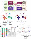 Figure 1
snRNA-seq of WOI human endometrium in NC and PC settings. (A) Study methodology combining single-nucleus transcriptomics (left) and a prospective clinical cohort study (right) to examine differences between NC and PC endometrium at both molecular and clinical levels. (B) Uniform manifold approximation and projection (UMAP) visualization of snRNA data from a total of 16 individuals and 93,064 nuclei after quality control measures and cell type annotation. (C) UMAP visualization of WOI endometrium transcriptomes by endometrial preparation method (NC and PC). (D) Bar plot depicting the cellular compositions of NC and PC endometrium. (E) Beeswarm plot showing distribution of log-fold change (FC) of various cell type neighborhoods by endometrial preparation method. Differentially abundant neighborhoods with |logFC| > 2.5 and Spatial FDR < 0.1 are colored.
We next examined the cellular composition of WOI NC and PC endometrium. The relative proportions of identified cell types by endometrial preparation method are shown in Figure 1D. The epithelial/stromal ratio was higher in PC (ratio: 1.30) compared with NC (ratio: 0.40) endometrium, potentially in the setting of supraphysiologic estradiol exposure, which is similar to that observed in endometrium following ovarian hyperstimulation (28). Strikingly, the percentage of uterine NK cells was significantly lower in PC endometrium (1.71%) compared with NC endometrium (10.4%). To examine differential abundance at increased granularity, we applied miloR to the data to identify and assess patterns in cellular neighborhoods by cell type in NC and PC endometrium (Figure 1E) (29). We identified 1,507 and 1,869 neighborhoods that were significantly enriched (spatial FDR <0.1) in PC and NC, respectively. Similar to the coarse cell type results, only 3 uterine NK cell neighborhoods were significantly enriched in PC endometrium, compared with 99 uterine NK cell neighborhoods enriched in NC endometrium (binomial test P < 0.001). Concordant with the observation of a higher epithelial/stromal ratio in PC endometrium, 964 glandular epithelial neighborhoods were significantly enriched in PC endometrium, compared with 364 in NC (P < 0.001). Similarly, 123 and 2 luminal epithelial neighborhoods were significantly enriched in PC and NC endometrium, respectively (P < 0.001). Neighborhood enrichment patterns in other cell types were not significantly different by endometrial preparation method (all P > 0.05). These findings suggest that PC endometrium exhibits substantial differences in cellular composition from NC endometrium.
Differentially regulated genes and biological pathways in PC endometrium.
We identified differentially expressed genes (DEGs) by cell type using DESeq2 after pseudobulking to limit false-positive discovery and identify shared per patient effect (Figure 2A). The number of significant DEGs (up- or downregulated in PC endometrium) are shown in Figure 2B. The highest number of significant DEGs (Padj < 0.05) were observed in glandular epithelial (682 upregulated; 979 downregulated) and stromal fibroblast populations (108 upregulated; 168 downregulated). In glandular epithelium (Figure 2C), several genes were notably downregulated in PC: LIF, PAEP, GPX3, COMP, and DPP4 (log2FC < –1.0, Padj < 0.001). These genes were reported to participate in implantation and endometrial decidualization and thought to reflect endometrial receptivity (30–33). PAEP and GPX3 were also significantly downregulated in PC stromal fibroblasts compared with those from NC endometrium (Figure 2C). THBS1, RARRES1, and DEPP-1, which are also genes with proposed roles in decidualization and implantation (32, 34, 35), were also significantly downregulated in PC stromal fibroblasts. These observations suggest that the PC endometrial preparation method may exert cell type–specific effects on postulated key regulators of endometrial receptivity and decidualization, which may influence the early maternal-fetal interface environment.
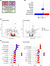 Figure 2
Transcriptomic differences and differentially regulated pathways between natural cycle and programmed cycle window of implantation endometrium. (A) Study design to identify differentially expressed genes (DEGs) in programmed cycle (PC) compared with natural cycle (NC) WOI endometrium. (B) Number of significant DEGs (|Log2FC| > 0.1 and Padj < 0.05, DESeq2) by cell type comparing PC to NC endometrium. (C) Volcano plots depicting the distribution of DEGs by Log2FC value (comparing PC to NC) and –Log10 Padj (DESeq2) in Glandular Epithelial and Stromal Fibroblast cell types. The most significant DEGs are depicted in red using the dashed lines, which indicate |Log2FC| > 1.0 and Padj < 0.001 (DESeq2). (D) Ridgeline plot demonstrating differentially regulated biological pathways from gene set enrichment analysis using cell type–specific DEGs in glandular epithelial and stromal fibroblasts (Padj values from gene set enrichment analysis).
Given the ongoing debate on optimal endometrial preparation method for fertility treatment, we also examined the relative expressions of markers from a gene panel identified through a meta-analysis (34 epithelial- and 22 stromal-specific markers), whose upregulation is thought to represent the meta-signature of human endometrial receptivity (30). We found that many receptivity markers had differential expression between NC and PC. Eighteen epithelial receptivity markers had significantly lower expression (log2FC < –1.0, Padj < 0.05) in PC endometrium in our dataset (Supplemental Figure 3). In stromal fibroblasts, 3 markers (GPX3, PAEP, and GBP2) also had significantly lower expression in PC. No receptivity markers in this panel were significantly upregulated in a PC endometrium.
Furthermore, CXCL14 — a highly conserved homeostatic chemokine — was also significantly downregulated in PC endometrium. CXCL14 has been shown to modulate chemotaxis, as well as differentiation and activation of various immune cell types (36). CXCL14, critically regulated by progesterone, has been shown to exert a chemoattractive effect on uterine NK cells in the endometrium (37). The significantly lower expression of CXCL14 may explain our finding that uterine NK cells were markedly less abundant in PC endometrium (Figure 1E).
To contextualize the DEGs between PC and NC endometrium, gene set enrichment analyses (GSEA) were performed to identify differentially regulated biological pathways in the setting of exogenous hormone administration and the absence of a corpus luteum (Figure 2D). A total of 156 pathways in glandular epithelium and 615 pathways in stromal fibroblasts were significantly enriched (Padj < 0.05) by endometrial preparation method. Gene ontology pathways that were significantly downregulated in PC glandular epithelium were related to protein localization, response to oxidative stress, wound healing, cell migration, and smooth muscle cell proliferation. Concordant with the observed DEGs, embryo implantation also emerged as downregulated in PC from functional enrichment analysis. In PC glandular epithelium, we identified upregulated pathways related to neuron development, sterol biosynthesis, and detection of chemical stimulus. Similarly, detection of chemical stimulus involved in sensory perception was also identified using the upregulated DEGs in PC stromal fibroblasts. The downregulated DEGs in PC stromal fibroblasts heavily mapped to biological pathways related to vascular development, angiogenesis, and extracellular matrix organization, which may have implications for angiogenic balance and remodeling of the endometrium in preparation for implantation and placentation. Negative regulation of cell migration was also seen in functional enrichment analysis from the downregulated DEGs in PC stromal fibroblasts.
Cell-cell interactome of NC and PC endometrium in the WOI.
Given the substantial differences in the transcriptomic landscape between NC and PC WOI endometrium, we used CellChat to analyze cell-cell communication patterns of various biological pathways using differentially expressed ligand-receptor pairs (38). Global communication patterns showed that the number of cell-cell interactions are preserved in PC compared with NC endometrium. While there was a higher absolute number of interactions in PC endometrium, the interaction strength appeared higher in NC endometrium (Figure 3A).
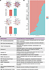 Figure 3
Cell-cell interactome in human window of implantation endometrium in natural cycle and programmed cycle. (A) Interaction net count and weight plots in natural cycle (NC) and programmed cycle (PC) window of implantation (WOI) endometrium. (B) Relative information flow analysis of biological pathways that demonstrated significant differential enrichment (paired Wilcoxon test P < 0.05) between NC and PC WOI endometrium. (C) Summary table describing the relevant function of significantly enriched pathways between NC and PC endometrium in reproductive biology
To better understand PC-associated changes in the endometrial microenvironment, a bar plot was first used to visualize the relative information flow of signaling pathways that demonstrated significant differences (P < 0.05) in relative strength in NC versus PC (Figure 3B). We observed a significant shift in signaling dynamics of 62 pathways between the 2 endometrial preparation methods: 41 showed greater relative information flow in NC, and 21 showed greater relative information flow in PC. A comparative global heatmap of overall signaling patterns in NC and PC WOI endometrium by cell type is also provided (Supplemental Figure 4).
Several of these signaling pathways have been proposed with important functions in reproduction (Figure 3B). For example, signaling pathways involved in endometrial decidualization and stromal homeostasis were more prominent in NC, such as SPP1, THBS, and FGF. Pathways that may have important roles in embryo implantation (SPP1, CSF, SEMA4, and NRG) were also significantly enriched in NC endometrium. Inflammatory modulation (IL-6, IL-10, CD40, and IFN-I) and vascular remodeling (FGF, ANGPT) pathways at the maternal-fetal interface also demonstrated greater signaling activity in NC. On the other hand, pathway involving VEGI — an antiangiogenic factor — was more prominent in PC. The BMP pathway — which has been characterized as an important pathway in endometrial decidualization — was also significantly enriched in PC endometrium. A summary table describing the relevant function of these pathways in endometrial biology is provided in Figure 3C (34, 39–46).
We further examined select pathways relevant to endometrial decidualization, inflammatory response, and angiogenesis between the 2 endometrial environments (Figure 4). The number of interactions in SPP1 and THBS pathways were decreased in PC compared with NC endometrium. On the other hand, BMP signaling was more prominent in PC endometrium. These observations highlight altered epithelial-stromal communication patterns that may affect decidualization and embryo implantation between endometrial preparation methods (Figure 4A). Signaling patterns were also altered in inflammatory pathways (IL-10, IL-6, and IFN-I), with less interaction numbers and strength in a PC microenvironment (Figure 4B). The complex interplay of these immunomodulators at the maternal-fetal interface may hold implications for maternal tolerance of the embryo as well as affecting trophoblastic invasion. Lastly, concordant with the functional enrichment analysis using stromal fibroblast DEGs, specific vasculogenesis/angiogenesis-related pathways were differentially signaled in NC versus PC (Figure 4C). Stromal fibroblast signaling in VEGF, ANGPTL, and FGF pathways appeared attenuated in PC endometrium. Together, these inferred cell-cell communication patterns suggest potentially perturbed signaling in key biological pathways relevant to reproduction and pregnancy success in a PC endometrial environment.
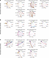 Figure 4
Cell-cell communication patterns of biological pathways relevant to reproduction by endometrial preparation method. (A) Circle plot visualization of inferred cell-cell communication network of pathways related to decidualization (SPP1, THBS, and BMP). (B) Circle plot visualization of inferred cell-cell communication network of pathways related to inflammatory cytokines (IL-10, IL-6, and INF-I). (C) Circle plot visualization of inferred cell-cell communication network of pathways related to angiogenesis and vascular remodeling (VEGF, FGF, and ANGPTL).
Angiogenic balance in pregnancies from NC and PC endometrium.
We separately performed an independent, prospective clinical cohort study in 548 patients (Figure 1A; clinical metadata in Supplemental Table 3) to investigate our findings of potentially perturbed biological pathways related to decidualization and angiogenesis, as well as the postulated increased risk of HDP with PC endometrial preparation. Critically, we evaluated obstetric outcomes in pregnancies established in NC or PC endometrium while adjusting for ART use and health parameters related to underlying infertility — both of which could independently contribute to obstetric complications. This methodology helps pinpoint specific risk(s) attributed to endometrial preparation method.
First, we examined serum levels of 2 maternal serum angiogenic markers, sFlt-1 and PlGF, in pregnancies conceived via 3 broad categories of mode of conception: (a) non-ART embryo implanted in a NC endometrium, (b) ART-derived embryo in a NC endometrium, and (c) ART-derived embryo in a PC endometrium (Figure 5A). We specifically measured early pregnancy levels of these 2 analytes to see if angiogenic imbalance is observed in the initial stages of establishing the maternal-fetal interface. As shown in Figure 5B., sFlt-1 concentrations did not significantly differ by mode of conception in early gestation, from 6–7 weeks through 18–20 weeks (all P > 0.05). Similarly, PlGF concentrations also did not differ significantly by mode of conception, regardless of ART use or endometrial preparation method. We then graphed serum sFlt-1/PlGF ratios to probe the state of angiogenic balance in these gestational periods (Figure 5C). Serum sFlt-1/PlGF ratios did not significantly differ by mode of conception at 6–7 weeks, 8–10 weeks, 11–13 weeks, or 18–20 weeks (all P > 0.05). sFlt-1/PlGF ratios were high in very early pregnancy but gradually and substantially shifted toward lower ratios by 18–20 weeks. This is likely secondary to a marked increase in PlGF at 18- to 20-week gestational age (GA) in all modes of conception (Figure 5B).
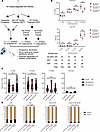 Figure 5
Angiogenic balance and incidences of hypertensive disorders of pregnancy by mode of conception. (A) Study design of a prospective clinical cohort to investigate the effect of PC endometrium on angiogenic balance and risk of HDP, adjusting for underlying infertility and use of ART. (B) Serum levels of angiogenic markers (sFlt1 and PlGF; mean ± SEM) by mode of conception across various gestational periods. Kruskal-Wallis test used. (C) Ratios of sFlt-1:PlGF by mode of conception and gestational period. Kruskal-Wallis test used. (D) Incidences of HDP (composite), preeclampsia (PreE), preeclampsia with severe features (PreE with SF), and preterm preeclampsia with severe features (Preterm PreE with SF; disease diagnosis < 37 weeks GA) by mode of conception (P value by χ2 test).
Incidences of HDP by mode of conception.
Incidences of HDP diagnoses were subsequently analyzed in this prospective cohort of 548 singleton pregnancies: 208 conceived without ART in a NC endometrium (non-ART+NC), 83 conceived with an ART-derived embryo in a NC endometrium (ART+NC), and 257 conceived with an ART-embryo in a PC endometrium (ART+PC). We focused on specific diagnoses along the spectrum of this clinical syndrome: any HDP (which includes gestational hypertension), preeclampsia, preeclampsia with severe features, and preterm preeclampsia with severe features (defined as diagnosis at < 37 weeks GA). All diagnoses were adjudicated by OB/GYN-trained clinicians using the criteria set forth by the American College of Obstetricians and Gynecologists (47).
Baseline characteristics of this cohort are provided in Supplemental Table 3. Female patients who conceived with ART+NC or ART+PC were significantly older than those who conceived via non-ART+NC. The median (interquartile range [IQR]) age of female patients was 39 (range, 36–43) in ART+PC, 38 (range, 35–40) in ART+NC, and 36 (range, 34–38) in non-ART+NC (P < 0.01). Distribution of primarily infertility diagnoses was also significantly different among the 3 groups (P < 0.01). The proportion of nulliparity was significantly different, with the non-ART+NC group having the highest percentage of nulliparous patients (71.2%) (P < 0.01). The distribution of body mass index (BMI), history of prior miscarriage, and history of hypertension or diabetes diagnoses prior to pregnancy did not differ among the 3 groups (all P > 0.10).
We compared the incidences of various HDP diagnoses by mode of conception (Figure 5D). Composite HDP incidences did not differ significantly by mode of conception: 20.7% in non-ART +NC, 16.9% in ART+NC, and 20.6% in ART+PC (P = 0.73). We then analyzed if HDP diagnoses of greater severity, or earlier manifestation, would differ by mode of conception. The incidence of preeclampsia was 12.5% in non-ART+NC, 10.8% in ART+NC, and 13.2% in ART+PC (P = 0.85). Preeclampsia with severe features developed in 7.7% of patients in non-ART+NC, 7.2% in ART+NC, and 8.9% in ART+PC (P = 0.83). Lastly, the incidence of preterm preeclampsia with severe features were 5.8% in non-ART+NC, 3.6% in ART+NC, and 5.8% in ART+PC (P = 0.72). ART+PC also had similar rates of hypertensive disorders as patients in the non-ART+NC group. Finally, multivariable log-binomial regression was performed to estimate adjusted relative risk and 95% CI of HDP diagnoses by mode of conception, adjusting for age, BMI, nulliparity, and primary infertility diagnosis. Compared with non-ART+NC conceptions, there was no significantly increased RR of any of the HDP diagnoses in either ART+NC or ART+PC conceptions (Supplemental Table 4).


