Gastroenterology
Abstract
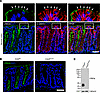
Multisystemic Smooth Muscle Dysfunction Syndrome (MSMDS) is a rare disorder caused by ACTA2 mutations, including the R179H variant, which alters actin filament stability and dynamics and smooth muscle contractility. While cardiovascular complications dominate its clinical presentation, gastrointestinal (GI) dysfunction significantly impacts quality of life. To investigate the structural, functional, and cellular basis of gut dysmotility in MSMDS, we reviewed clinical data from 24 MSMDS patients and studied the ACTA2 R179H mouse model Patients exhibited severe gut dysmotility, with 75% requiring medication for chronic constipation. ACTA2 mutant mice displayed cecal and colonic dilatation, reduced intestinal length, and disrupted colonic migrating motor complexes (CMMCs). Delayed whole-gut transit and impaired contractile responses to electrical and pharmacological stimulation were observed. Transcriptomic analysis revealed significant actin cytoskeleton-related gene changes in smooth muscle cells, and immune profiling identified increased lymphocytic infiltration. Despite functional abnormalities, there were no obvious changes in the enteric nervous system. These findings establish ACTA2 mice as a robust model for studying GI pathology in MSMDS, elucidating the role of smooth muscle dysfunction in gut dysmotility. This model provides a foundation for developing targeted therapies aimed at restoring intestinal motility by directly addressing actin cytoskeletal disruptions in smooth muscle cells.
Authors
Ahmed A. Rahman, Rhian Stavely, Leah C. Ott, Christopher Y. Han, Kensuke Ohishi, Ryo Hotta, Alan J. Burns, Sabyasachi Das, Emily Da Cruz, Diana Tambala, Mark E. Lindsay, Patricia L. Musolino, Allan M. Goldstein

Abstract
The tumor microenvironment plays a key role in cancer progression and therapy resistance, with cancer-associated fibroblasts (CAFs) contributing to desmoplasia, extracellular matrix (ECM) remodeling, and elevated interstitial fluid pressure, all of which hinder drug delivery. We investigated fibroblast activation protein–targeted (FAP-targeted) near-infrared photoimmunotherapy (NIR-PIT) as a strategy to improve drug penetration in CAF-rich tumors. In clinical esophageal cancer samples, FAP expression strongly correlated with increased collagen I, hyaluronic acid, and microvascular collapse. CAF-rich 3D spheroids demonstrated elevated ECM deposition and significantly impaired drug uptake compared with CAF-poor models. FAP-targeted NIR-PIT selectively reduced CAFs, reduced ECM components, and restored drug permeability. In vivo, FAP-targeted NIR-PIT enhanced the accumulation of panitumumab and Abraxane in CAF-rich tumors and improved antitumor efficacy when combined with chemotherapy. These findings highlight FAP-targeted NIR-PIT as a promising therapeutic approach to remodel the tumor stroma and overcome drug resistance in desmoplastic solid tumors.
Authors
Seitaro Nishimura, Kazuhiro Noma, Tasuku Matsumoto, Yasushige Takeda, Tatsuya Takahashi, Hijiri Matsumoto, Kento Kawasaki, Hotaka Kawai, Tomoyoshi Kunitomo, Masaaki Akai, Teruki Kobayashi, Noriyuki Nishiwaki, Hajime Kashima, Takuya Kato, Satoru Kikuchi, Shunsuke Tanabe, Toshiaki Ohara, Hiroshi Tazawa, Yasuhiro Shirakawa, Peter L. Choyke, Hisataka Kobayashi, Toshiyoshi Fujiwara
Abstract
Induction of heme oxygenase-1 (HO-1/Hmox1) is broadly considered cytoprotective, but the role of colonic epithelial HO-1 in colitis-associated tumorigenesis is poorly defined. HO-1 catabolizes heme, releasing ferrous iron, a key driver of oxidative stress and lipid peroxidation. We observed that colonic epithelial HO-1 is induced during colitis and tumorigenesis. We also found that HO-1 is upregulated in ferroptosis-inducing conditions in murine and human colonic epithelial organoids, and correlated with lipid peroxidation and ferroptosis markers in colonic tumors. In colonic epithelial organoids exposed to heme, deletion of Hmox1 amplified a compensatory oxidative stress and detoxification transcriptional program, likely reflecting unresolved oxidative and non-oxidative toxicity from heme. In vivo, epithelial HO-1 deficient mice developed significantly fewer and smaller tumors compared to littermate controls in a colitis-associated tumorigenesis model, despite similar inflammatory injury. Tumors from knockout mice exhibited reduced iron levels, decreased lipid peroxidation, lower oxidative DNA damage, and decreased proliferation. Single-cell RNA sequencing of tumor epithelial cells revealed a shift from a proliferative to a stress-adaptive program with loss of HO-1. These findings identify epithelial HO-1 as a context-dependent regulator of tumorigenesis: protective against acute heme toxicity, but promoting iron-dependent oxidative damage and proliferation in the setting of chronic inflammation.
Authors
Rosemary C. Callahan, Jillian C. Curry, Geetha Bhagavatula, Alyse W. Staley, Rachel E.M. Schaefer, Faiz Minhajuddin, Liheng Zhou, Rane M. Neuhart, Shaikh M. Atif, David J. Orlicky, Ian M. Cartwright, Mark E. Gerich, Calen A. Steiner, Arianne L. Theiss, Caroline H.T. Hall, Sean P. Colgan, Joseph C. Onyiah
Abstract
Pancreatic ductal adenocarcinoma (PDAC) has a dismal prognosis and current therapies show limited efficacy. Ligands and receptors of the TIGIT axis were analyzed using multicolor flow cytometry of tumor and blood samples, immunohistochemistry from primary tumors, and single-cell RNA sequencing from primary tumors and liver metastasis from patients with various stages of PDAC. The effect of soluble and plate-bound Nectin-4 on T cell function was tested in vitro. Further, patient-derived PDAC organoids were treated with the standard of care therapies FOLFIRINOX, gemcitabine plus paclitaxel, or the antibody-drug conjugate enfortumab vedotin. TIGIT expression was increased on tumor-infiltrating conventional and regulatory T cells compared with T cells from matched blood. Nectin-4, but not CD155 expression was associated with poor outcome. Nectin-4 was exclusively expressed by tumor cells and correlated with low immune infiltration. Notably, Nectin-4 inhibited T cell effector cytokine production in vitro. Targeting Nectin-4 with the antibody-drug conjugate enfortumab vedotin inhibited tumor growth in multiple patient-derived PDAC organoids. Collectively, our data underscores Nectin-4 as a novel therapeutic target and provides the rationale to test this agent in PDAC patients.
Authors
Max Heiduk, Carolin Beer, Sarah Cronjaeger, Emily A. Kawaler, Ulrich Sommer, Franziska Baenke, David Digomann, Loreen Natusch Bufe, Charlotte Reiche, Jessica Glück, Franziska Hoffmann, Sungsik Kim, Daniel E. Stange, Diane M. Simeone, Jürgen Weitz, Lena Seifert, Adrian M. Seifert
Abstract
Chronic liver injury results in activation of quiescent Hepatic Stellate Cells (qHSCs) into Collagen Type I-producing activated HSCs that make liver fibrotic. We identified ETS1/2 (E26 transformation-specific transcription factors 1/2) as lineage-specific transcription factors regulating HSC phenotypes. Here we investigated the role of ETS1/2 in HSCs in liver fibrosis using toxic liver injury models and 3D human liver spheroids. Liver fibrosis was induced in wild-type and HSC-specific Ets1 (Ets1ΔHSC) and Ets2 (Ets2ΔHSC) knockout mice by administration of carbon tetrachloride for 6 weeks, following cessation of liver injury for 2 weeks. Liver fibrosis was more severe in Ets1ΔHSC, and to lesser extent in Ets2ΔHSC, compared to wild-type mice. Regression of liver fibrosis was suppressed only in Ets1ΔHSC, indicating Ets1 as the predominant isoform maintaining quiescent-like phenotype in HSCs. Similar results were obtained in a MASH model using 3D human liver spheroids. Knockdown of ETS1 in human HSCs caused upregulation of fibrogenic genes in MASH human liver spheroids and prevented fibrosis regression. ETS1 regulated the qHSC phenotype via CRTC2/PGC1α/PPARγ pathway. Knockdown of CRTC2 (cAMP response element-binding protein (CREB)-regulated transcription co-activator 2) abrogated PPARγ responses and facilitated HSC activation. These findings suggest that ETS1 may represent a therapeutic target for anti-fibrotic therapy.
Authors
Wonseok Lee, Xiao Liu, Sara Brin Rosenthal, Charlene Miciano, Sadatsugu Sakane, Kanani Hokutan, Debanjan Dhar, Hyun Young Kim, David A. Brenner, Tatiana Kisseleva
Abstract
The gastrointestinal epithelium depends on the apical junctional complex (AJC), composed of tight and adherens junctions, to regulate barrier function. Here, we identify the apical polarity protein Crumbs homolog 3 (CRB3) as an important regulator of AJC assembly and barrier function in intestinal epithelium. Using primary murine colonic epithelial cells (colonoids) from inducible, conditional Crb3-knockout (Crb3ERΔIEC) and control (Crb3fl/fl) mice, we show that CRB3 deficiency compromised barrier function that was associated with a hypercontractile perijunctional actomyosin network and impaired AJC assembly. Loss of CRB3 exacerbated proinflammatory cytokine–induced AJC remodeling, leading to increased intestinal permeability. Crb3ERΔIEC cells exhibited increased RhoA activity and junctional tension, which could be reversed by ROCK-II or myosin II inhibition, restoring junctional architecture. Mechanistically, CRB3A interacts with the actin cytoskeletal linker protein, Merlin (NF2) via its FERM-binding domain, and NF2 knockdown phenocopied CRB3 loss, suggesting their cooperative role in AJC assembly. These findings establish CRB3 and NF2 signaling as key regulators of perijunctional actomyosin contractility and AJC organization during both de novo junctional assembly and inflammation-induced remodeling. This work defines a CRB3- and NF2-dependent pathway by which epithelial cells regulate mechanical tension to coordinate barrier assembly during homeostasis and junctional remodeling under inflammatory stress.
Authors
Shuling Fan, Saranyaraajan Varadarajan, Vicky Garcia-Hernandez, Sven Flemming, Arturo Raya-Sandino, Ben Margolis, Charles A. Parkos, Asma Nusrat
Abstract
Genetic variants in lipid metabolism influence the risk of developing metabolic dysfunction-associated steatotic liver disease (MASLD), cirrhosis, and end-stage liver disease (ESLD). The mechanisms by which these variants drive disease are poorly understood. Because of the PNPLA3-I148M variant's strong correlation with all stages of the MASLD spectrum and the lack of tractable therapeutic targets, we sought to understand its impact on cellular function and liver metabolism. Primary human hepatocytes (HAH) and iPSC-derived hepatocytes (iHeps) from healthy individuals possessing the PNPLA3-I148M mutation were characterized for changes in lipid metabolism, cellular stress, and survival. Using lipidomics, metabolomics, stable isotope tracing, and flux propensity analysis, we created a comprehensive metabolic profile of the changes associated with the PNPLA3-I148M variant. Functional analysis showed that the presence of the PNPLA3-I148M variant increased endoplasmic reticulum stress, mitochondrial dysfunction, and peroxisomal β-oxidation, ultimately leading to cell death via ferroptosis. Nutritional interventions, ferroptosis-specific inhibitors, and genetic approaches modulating GPX4 activity in PNPLA3-I148M HAH and iHeps decreased programmed cell death. Our findings indicate that therapies targeting ferroptosis in patients carrying the PNPLA3-I148M variant could affect the development of MASLD and ESLD and highlight the utility of iPSC-based models for the study of genetic contributions to hepatic disorders.
Authors
Rodrigo M. Florentino, Olamide Animasahun, Nils Haep, Minal Nenwani, Kehinde Omoloja, Leyla Nurcihan Altay, Abhinav Achreja, Kazutoyo Morita, Takashi Motomura, Ricardo Diaz-Aragon, Lanuza AP Faccioli, Yiyue Sun, Zhenghao Liu, Zhiping Hu, Bo Yang, Fulei Wuchu, Ajay Shankaran, Miya Paserba, Annalisa M. Baratta, Shohrat Arazov, Zehra N. Kocas-Kilicarslan, Noah Meurs, Jaideep Behari, Edgar N. Tafaleng, Jonathan Franks, Alina Ostrowska, Takahiro Tomiyama, Kyohei Yugawa, Akinari Morinaga, Zi Wang, Kazuki Takeishi, Dillon C. Gavlock, Mark Miedel, D. Lansing Taylor, Ira J. Fox, Tomoharu Yoshizumi, Deepak Nagrath, Alejandro Soto-Gutierrez
Abstract
Pancreatic ductal adenocarcinoma (PDAC) is a rapidly metastasizing cancer characterized by a dense desmoplastic stroma comprised of extracellular matrix (ECM) proteins, which complicates treatment. Upon stimulation, pancreatic stellate cells (PSCs) differentiated into cancer-associated fibroblasts (CAFs) that are the source of ECM and cytokines in PDAC. We previously reported that mechanical stress activates PSCs and induces fibrosis through mechanical ion channel PIEZO1-mediated TRPV4 channel activation, but its role in PDAC remains unclear. Here we report that pathological activation of PIEZO1 differentiated human PSCs into an inflammatory CAF phenotype that expresses chemoresistance and cancer stemness markers CD10 and GPR77. In an orthotopic PDAC model, TRPV4 knockout mice exhibited a significant reduction in tumor size, circulating inflammatory cytokines, tissue inhibitor of metalloproteinases-1 (TIMP1), and pre-metastatic niche markers, serum amyloid A (SAA) proteins. A similar trend was observed in mice lacking functional PIEZO1 in PSCs. The livers of TRPV4 knockout mice exhibited fewer cancer cell microlesions, lacked macro tumors, produced lower levels of inflammatory protein S100A8, and developed fewer inflammatory cell clusters. In orthotopic and genetically engineered models of PDAC, these mice also had improved survival, suggesting that blocking TRPV4 channels may be a promising therapeutic target for PDAC.
Authors
Joelle M.-J. Romac, Sandip M. Swain, Nidula Mullappilly, Bandana Bindhani, Rodger A. Liddle

Abstract
Clinical trials have identified 2 distinct eosinophilic esophagitis (EoE) treatment phenotypes: those that show proton pump inhibitor (PPI) responsiveness (PPI-R) and those that show PPI unresponsiveness (PPI-UR). Comprehensive clinical, endoscopic, and RNA-Seq analyses of patients with EoE prior to and following PPI therapy have not previously been performed to our knowledge. We showed that clinical, endoscopic, and histologic evaluation of esophageal biopsies from pediatric PPI-R and PPI-UR individuals with EoE prior to PPI therapy (diagnosis) were indistinguishable. RNA-Seq analyses revealed common immune and inflammatory transcriptional signatures in both PPI-R EoE and PPI-UR EoE esophageal biopsy samples at diagnosis and distinct signatures enriched for processes related to neuropeptide signaling and cell cycle and division. PPI therapy induced histologic, endoscopic, and transcriptional remission in PPI-R EoE, but not in PPI-UR EoE. Persistent disease in PPI-UR EoE was associated with the presence of Th2 inflammatory and dedifferentiated esophageal epithelial transcriptomic signatures, while PPI-R EoE revealed genes enriched in cellular responses to LPS, host defense against viruses, and type I IFN signaling. In silico analyses identified common and unique EoE disease gene drivers in PPI-R and PPI-UR EoE. These studies indicate that the 2 EoE phenotypes have unique transcriptomic elements that underlie the molecular nature of PPI-R and PPI-UR EoE disease.
Authors
Somdutta Chakraborty, Ankit Sharma, Sahiti Marella, Christian F. Rizza, Patrick A. O’Brien, Varsha Ganesan, Gila Idelman, Susie Min, Mayee Chen, Talaya McCright-Gill, Nancy Gonzalez, Alexandros D. Polydorides, Paul S. Foster, Simon P. Hogan, Mirna Chehade
Abstract
Intestinal epithelial barrier-integrity is essential for human health, and its disruption induces and exacerbates intestinal inflammatory disorders. While the epithelial cytoskeleton is critical for maintaining gut barrier-integrity, the role of septins- a family of GTP-binding, cytoskeletal proteins- is largely unknown. This highlights an important knowledge gap as dysfunction of septins, and specifically septin 9 (SEPT9), is associated with intestinal pathologies. We determined that SEPT9 localizes to the apical junctions of intestinal epithelial cells (IECs), overlapping with both tight and adherens junctions. IEC-specific ablation of SEPT9 in mice resulted in leaky gut, due to mislocalization of junctional proteins, and increased susceptibility to experimental colitis. Consistently, SEPT9 expression was significantly reduced in intestinal mucosa of inflammatory bowel disease (IBD) patients. Using affinity-purification mass spectrometry, super-resolution imaging, and genetic knockout, we determined that SEPT9 interacts with and is necessary to recruit non-muscle myosin IIC (NMIIC) to the IEC peri-junctional actomyosin belt. Loss of NMIIC also caused IEC barrier disruption. In summary, SEPT9 regulates intestinal barrier-integrity by supporting the assembly of tight and adherens junctions through NMIIC recruitment to the actomyosin belt. The septin cytoskeleton safeguards the intestinal mucosa during acute inflammation, and its disruption in IBD suggests a loss of this protective function.
Authors
Nayden G. Naydenov, Gaizun Hu, Dominik Robak, Atif Zafar, Khosiyat Makhmudova, Susana Lechuga, Yuta Ohno, Naseer Sangwan, Saikat Bandyopadhyay, Ryan Musich, Erin Jeffery, Lei Sun, Armando Marino-Melendez, Florian Rieder, Gloria Sheynkman, Andrei I. Ivanov, Seham Ebrahim
No posts were found with this tag.


